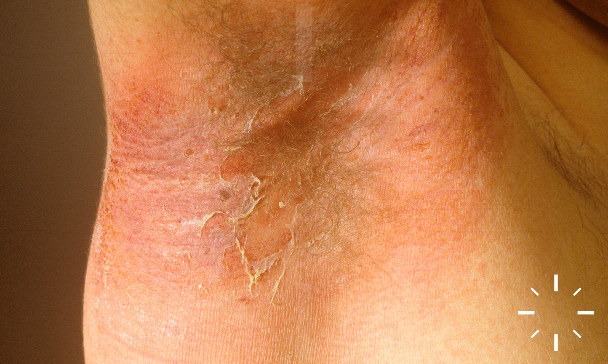
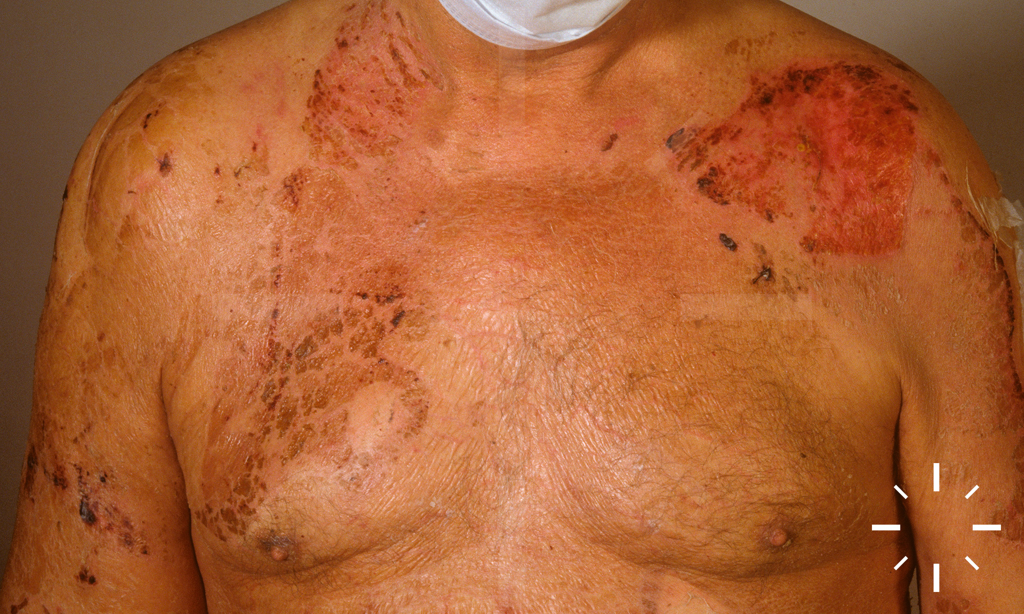
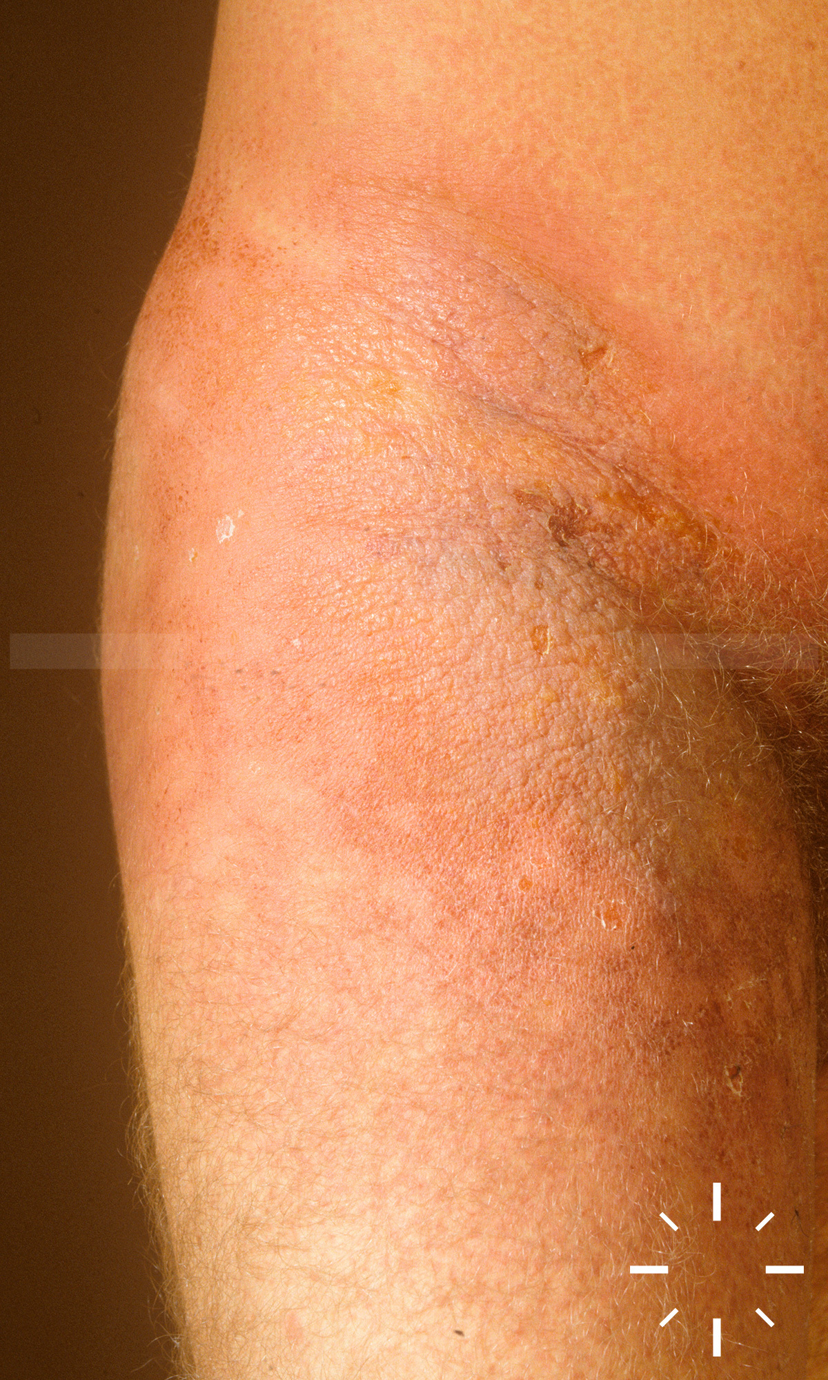
Staphylococcal scaled skin syndrome (SSSS)

Zuletzt aktualisiert: 2022-11-16

Autor(en): Anzengruber F., Navarini A.

ICD11: EA50.2

Lyell, 1956.

Staphylogenes Lyell-Syndrom.

  • Zählt zu den Staphylokokken-Toxin bedingten Dermatosen, welche zu einer Ablösung im Str. granulosum führt und letal sein kann
  • Erreger: Staphylococcus aureus spp.

  • Insbesondere sind Säuglinge (<3 Monaten) und ältere Kleinkinder betroffen
  • Selten Auftreten im Erwachsenenalter (bei Immunschwäche oder Niereninsuffizienz)

Der auslösende Staphylococcus aureus gehöhrt meist der Lyso(phagen)gruppe II, insbesondere Typ 3A, 3C, 55 und 71, an. Die von ihnen produzierten exfoliative Toxine ETA, ETB und ETD produzieren Blasen durch Spaltung des humanen und murinen Desmogleins 1.

  • Red. Allgemeinzustand und febrile Temperaturen, ggf. purulenter Rhinitis, Otitis, Pharyngitis oder Konjunktivitis
  • Initial ist ein rasch progredientes, skarlatiniformes Exanthem zu sehen. Nach 1-2 Tagen kommt es zum Auftreten großer, schlaffer, leicht rupturierender Blasen am ganzen Körper. Das klinische Bild kann einer Verbrennung 2. Grades ähneln. Das Nikolski-Zeichen ist positiv. Im Verlauf kommt es zu einer Austrocknung der Blasendecke, welche folglich groblamellös abschuppt. Die Schleimhaut kann in seltenen Fällen beteiligt sein. Mit einer erneuten Epithelisierung ist bei frühzeitigem Therapiebeginn innerhalb 1Woche zu rechnen.

  • Klinik
  • Bakt.-Abstrich
  • Fokussuche (Blutkulturen, U-Status, HNO-Konsil, Inspektion des Bauchnabels)

Akantholytischer Spalt im Stratum granulosum im Sinne einer subkornealen Blase, keine Zellnekrosen, kaum Entzündungszeichen, (DD: medikamentös ausgelöstes Lyell-Syndrom, wo sich die Spaltbildung subepidermal findet.

  • Sepsis
  • Pneumonie

Mit einer Reepithelisierung kann bei sofortigem Beginn einer adequaten Therapie gerechnet werden. Potentiell kann das Lyell Syndrom auch letal sein.

  1. Staphylococcal Toxin-Mediated Scalded Skin and Toxic Shock Syndromes.  Deadly Dermatologic Diseases: Springer Science + Business Media:133-6.
  2. Staphylococcal scalded skin syndrome.  SpringerReference: Springer Science + Business Media.
  3. Cribier B, Piemont Y, Grosshans E. Staphylococcal scalded skin syndrome in adults. Journal of the American Academy of Dermatology 1994;30:319-24.
  4. Dobson CM, King CM. Adult staphylococcal scalded skin syndrome: histological pitfalls and new diagnostic perspectives. British Journal of Dermatology 2003;148:1068-9.
  5. Elias PM. Staphylococcal Scalded Skin Syndrome. Arch Dermatol 1977;113:207.
  6. Gemmell CG. Staphylococcal scalded skin syndrome. Journal of Medical Microbiology 1995;43:318-27.
  7. Hanakawa Y, Schechter NM, Lin C, et al. Molecular mechanisms of blister formation in bullous impetigo and staphylococcal scalded skin syndrome. Journal of Clinical Investigation 2002;110:53-60.
  8. Has C, Misery L, David L, Cambazard F. Recurring Staphylococcal Scalded Skin Syndrome-like Bullous Mastocytosis: The Utility of Cytodiagnosis and the Rapid Regression with Steroids. Pediatric Dermatology 2002;19:220-3.
  9. Ladhani S, Evans RW. Staphylococcal scalded skin syndrome. Archives of Disease in Childhood 1998;78:85-8.
  10. Ladhani SMPCLJRWS. Staphylococcal scalded skin syndrome: exfoliative toxin A (ETA) induces serine protease activity when combined with A431 cells. Acta Paediatrica 1999;88:776-9.
  11. Melish ME, Glasgow LA. The Staphylococcal Scalded-Skin Syndrome. New England Journal of Medicine 1970;282:1114-9.
  12. Na. TOXIN IN BULLOUS IMPETIGO AND STAPHYLOCOCCAL SCALDED-SKIN SYNDROME TARGETS DESMOGLEIN 1. The Pediatric Infectious Disease Journal 2001;20:471.
  13. Nagasaka T, Nishifuji K, Ota T, Whittock NV, Amagai M. Defining the pathogenic involvement of desmoglein 4 in pemphigus and staphylococcal scalded skin syndrome. Journal of Clinical Investigation 2004;114:1484-92.
  14. Patel GK, Finlay AY. Staphylococcal Scalded Skin Syndrome. American Journal of Clinical Dermatology 2003;4:165-75.
  15. Stanley JR, Amagai M. Pemphigus, Bullous Impetigo, and the Staphylococcal Scalded-Skin Syndrome. New England Journal of Medicine 2006;355:1800-10.
  16. Ito Y, Funabashi Yoh M, Toda K, Shimazaki M, Nakamura T, Morita E. Staphylococcal scalded-skin syndrome in an adult due to methicillin-resistant Staphylococcus aureus. J Infect Chemother 2002;8:256-61.
  17. Chi CY, Wang SM, Lin HC, Liu CC. A clinical and microbiological comparison of Staphylococcus aureus toxic shock and scalded skin syndromes in children. Clin Infect Dis 2006;42:181-5.
  18. Pielop, J. (2016). Vesiculobullous and pustular lesions in the newborn. Uptodate.com. Retrieved 13 September 2015, from http://www.uptodate.com/contents/vesiculobullous-and-pustular-lesions-in-the-newborn?source=search_result&search=Staphylococcal+scaled+skin+syndrome&selectedTitle=1~7