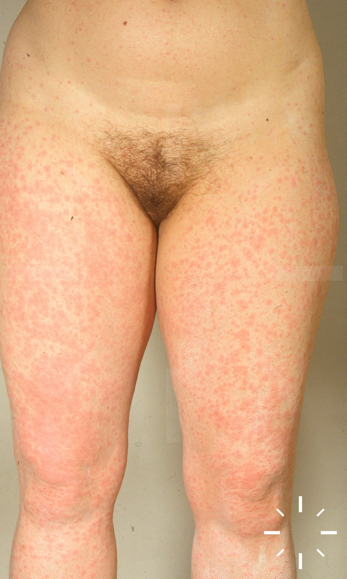
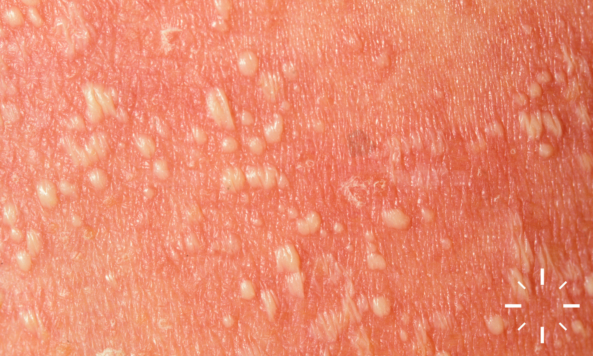
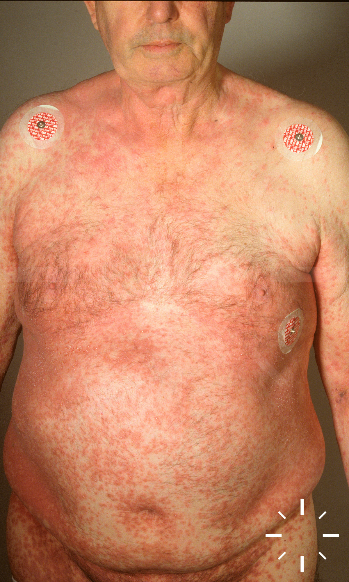
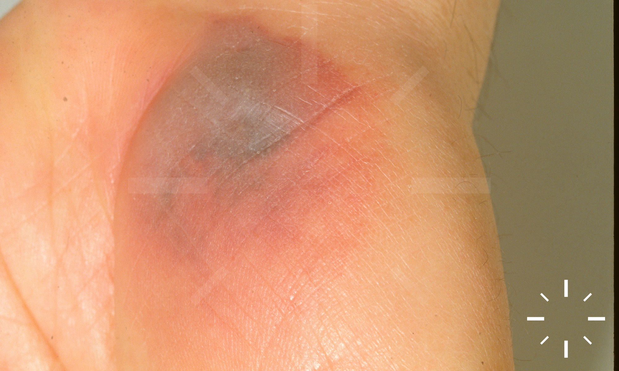
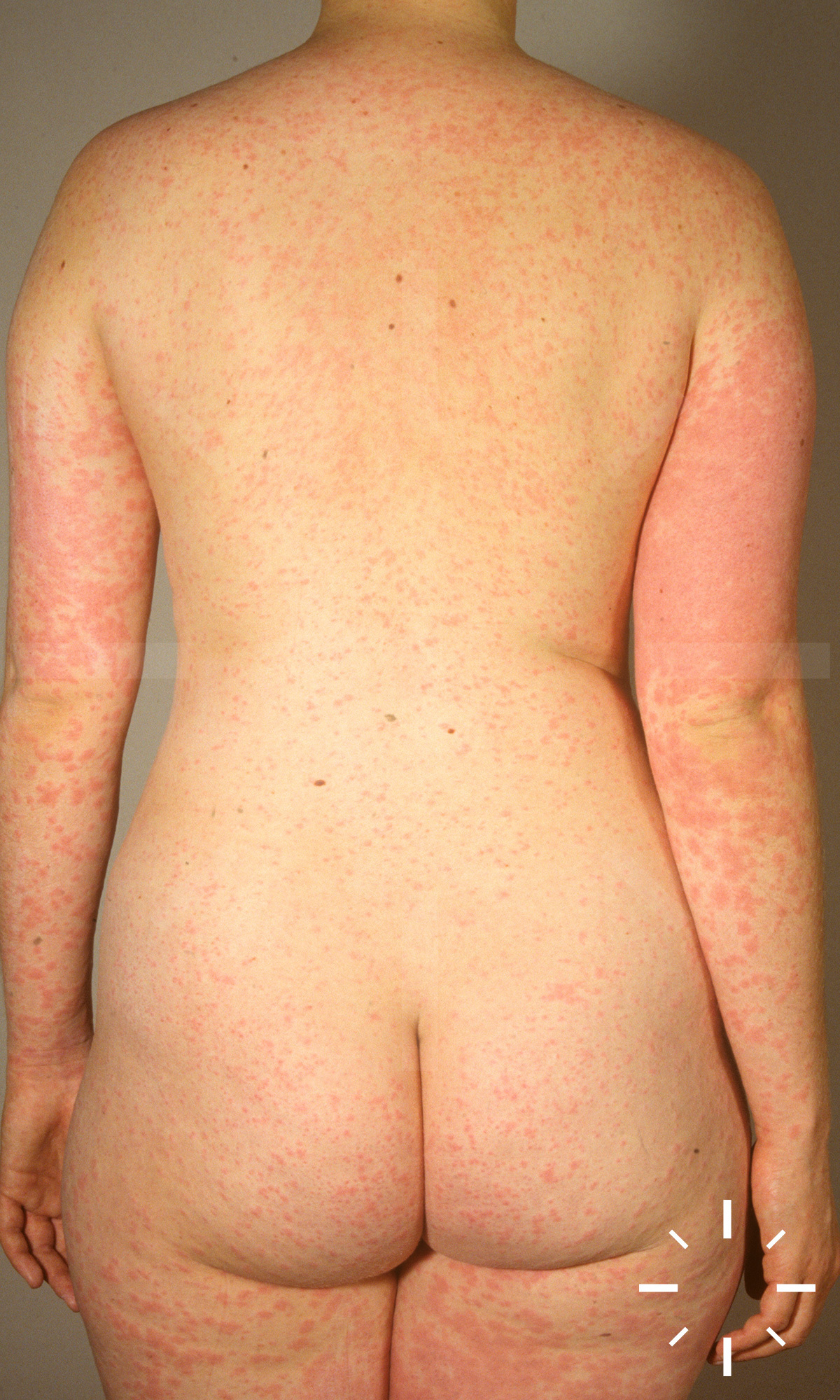
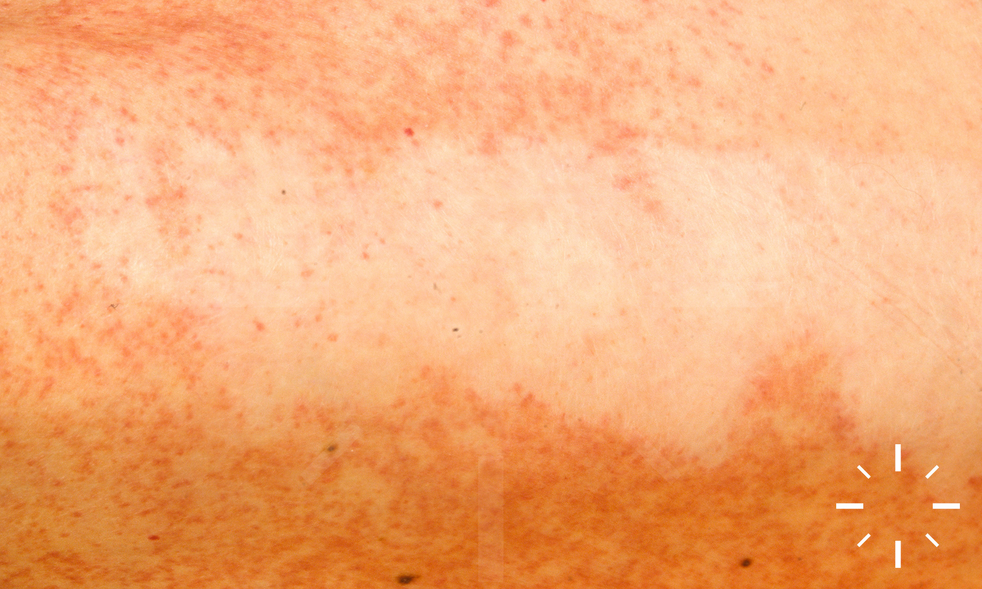
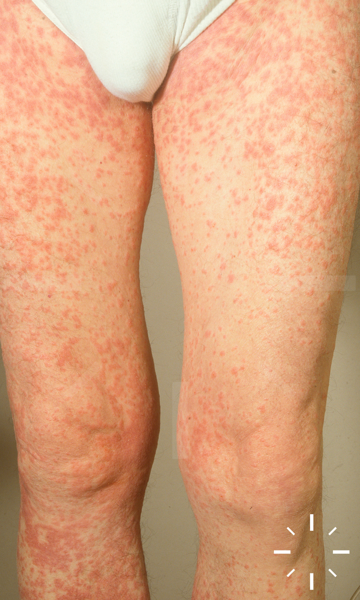
Stevens Johnson Syndrom und toxische epidermale Nekrolyse

Zuletzt aktualisiert: 2022-11-16

Autor(en): Anzengruber F.

ICD11: EB13.2

  • Stevens Johnson Syndrom: Stevens & Johnson, 1922
  • Toxisch epidermale Nekrolyse : Lyell, 1956

Stevens Johnson Syndrom, SJS, Toxisch epidermale Nekrolyse, TEN, Lyell-Syndrom, Epidermolysis necroticans combustiformis, Epidermolysis acuta toxica.

Seltne, ernsthafte, potenziell letale, mukokutane Erkrankung, welche meist durch die Einnahme von Arzneimitteln ausgelöst wird. Es kommt zu einer Abklösung der Epidermis und zum Auftreten von Nekrosen. Stevens Johnson Syndrom (SJS) und die toxisch epidermale Nekrolyse (TEN) entsprichen einem Krankheitsbild. In 100% gibt es eine Cheilitis und Stomatitis. Unterschiedlich ist die flächenmässige Ausbreitung an der restlichen Haut.

  • Einteilung
    • Stevens Johnson Syndrom: < 10% der Körperoberfläche, Schleimhäute sind betroffen (Augen, Mundhöhle, genital).
    • SJS/TEN overlap syndrome: > 10% bis < 30% der Körperoberfläche, Schleimhäute sind betroffen (Augen, Mundhöhle, genital).
    • TEN: > 30% der Körperoberfläche, Schleimhäute sind betroffen (Augen, Mundhöhle, genital).

  • Inzidenz (für SJS, SJS/TEN und TEN): 2-7/1.000.000/per annum
  • SJS:TEN = ca. 3:1
  • 100-fach erhöhte Inzidenz bei HIV-positiven Patienten
  • Frauen > Männer

  • Häufig auslösende Medikamente: Mnemonic SATAN
    • Sulfonamid-Antibiotika: Sulfamethoxazol (Bactrim), Sulfasalazine
    • Allopurinol
    • Tetracycline & andere Antibiotika: Amoxicillin, Cephalosporine, Chinolone, etc.
    • Antikonvulsiva: Carbamazepin, Lamotrigin, Phenytoin, etc.
    • NSAID
    • Virostatika (HAART): Nevirapin
  • Infektionen
    • Mycoplasma pneumoniae
    • Cytomegalovirus infections
  • Impfungen
  • Kontrastmittel
  • Nahrungsmittel
  • Knochenmarktransplantationen
  • Radiotherapie
  • Prädispositionierende Faktoren
    • HIV-Infektion
    • Genetik:
      • HLA: HLA-B*1502, HLA-A*3101, B*5801, HLA-B*1502, HLA-A*3101, HLA-B*5801.
      • Polymorphismen

  • Prodromalstadium:
    • Verschlechterter AZ, Fieber
    • Influenza-ähnliche Symptome
      • Fakultativ:
        • Photophobie
        • Konjunktivaler Pruritus
        • Schmerzen beim Schlucken
        • Myalgien
        • Arthralgien
        • Purulente Rhinitis
        • Gelenksbeschwerden
        • Generalisierte Lymphadenopathie
        •  Leber- und Milzbeteiligung
  • Mukokutane Symptome
    • Treten meist nach 1-3, jedoch manchmal nach bis zu 14 Tagen auf.
    • Läsionen beginnen meist im Gesicht oder am Rumpf bevor sie sich auf andere Körperlokalisationen ausbreiten.
    • Das Kapillitium ist nicht betroffen, jedoch zeigt sich auch in manchen Fällen eine palmoplantare Beteiligung.
    • Die Dauer der mukokutanen Symptome ist mit 8-12 Tagen angegeben.
  • Schleimhaut:
    • In 90% der Fälle ist die Mundschleimhaut betroffen.
      • Erosionen und Ulzerationen.
      • Hämorrhagische Krusten an den Lippen.
    • Die Augen (Konjunktivits, teilweise purulent) sind fast immer betroffen.
      • Verkrustete Läsionen an Ober- und Unterlidern.
      • Hornhautulkus, Panophthalmitis, Photophobie, Synechiaen, Symblepharon.
    • Genitale Erosionen.
      • Synechien und Stenosen können sich im Bereich der Labien, der Harnröhre ausbilden.
      • Urethritis, Harnverhalt, Cystitis.
    • Bei fast allen Patienten kann eine Beteiligung des Pharynx beobachtet werden.
    • Seltener können auch die Trachea, Bronchien, Ösophagus und Darm beteiligt sein.
  • Kutane Manifestation:
    • Epidermisablösungen, Erosionen und teils Ulzera
    • Kokardenförmige Plaques und Makulae
    • Oftmals skarlatiniformes, stammbetontes Exanthem
    • Ausbildung von Vesikeln und Bullae
      • Nikolski-Phänomen I (Blasen lassen sich auf gesunder Haut auslösen bzw. nicht blasige Haut lässt sich verschieben bei seitlichem Druck)
      • Nikolski-Phänomen II (schon bestehende Blasen lassen sich auf seitlichen Druck verschieben)
      • Asboe-Hansen Zeichen (die Blase expandiert auf Druck nach lateral
    • Grob-lammelläre Abschuppung/ die Haut schält sich flächig ab
    • Nekrosen
    • Ggf. Onycholyse
    • Eine Reepithelialisierung erfolgt nach wenigen Tagen
  • Labor
    • Gesteigerte BSG, CRP, Leukozytose
    • Eiweiselektrophorese (Gesteigerte α- & γ-Globulinfraktion)
    • Anämie (ca. 15%)
    • Lymphopenie (häufig)
    • Neutropenie
      • Kommt bei ca. 1/3 aller Patienten vor
      • Oft durch systemische Glukokortikoide maskiert
      • Korrelliert mit einer schlechten Prognose
    • Eosinophilie (20%)
    • Transaminasen erhöht (ca. 15%)
    • Proteinurie und Hämaturie (5%)
    • Bakteriämie (ca. 27%)
    • Kennzeichen für einen schweren Verlauf:
      • Harnstoff >10 mmol/L
      • Glukose >14 mmol/L
      • ALT (bis zum 2-3-fachen Wert)

  • Anamnese (Medikamentenanamnese)
  • Klinische Bild
  • Biopsie
    • Dermatopathologie-Schnellschnitt! Wichtig um eine Unterscheidung vom SSSS zu machen. Hat somit eine ausgeprägte Therapiekonsequenz!
    • Direkte Immunfluoreszenz
  • Mittels dem „algorithm of drug causality for epidermal necrolysis” (ALDEN)
  • Labor
    • Labor täglich: BB, kleiner Gerinnungsstatus, Chemogramm inkl. CRP, Harnstoff

      und Bicarbonat, Blutkulturen bei Bedarf, Serumelektrophorese

    • Initial einmalig: IgA quantitativ im Serum und ggf. HIV

  • Bakt. Abstriche von Haut und Schleimhaut, Blutkulturen, Kulturen von ggf. Harnkathetern und PEG-Sonden sollten alle 2 Tage wiederholt werden!
  • SCORTEN: Abschätzen des Schweregrades mittels SCORTEN (Severity-of-illness) Score* (bei Score >2 Verbrennungs-IPS Verlegung (USZ) organisieren). Täglich wiederholen.
  • Abgrenzung zu: EEMM, SSSS, bullöse Autoimmundermatosen, andere schwere Arzneimittelreaktionen
  • SJS/TEN: Disseminiert, generalisiert, Schleimhaut
    • Initial makulös (dolent!), Target-Läsionen, Blasen, „verbrühte Haut“

    • Kann innere Organe betreffen: Pneumonitis, Gastrointestinaltrakt

    • Fieber, Reduktion des Allgemeinzustands

    • Lymphopenie

    • Histologie: Epidermale Nekrose

    • Beginn nach Medikamenteneinnahme im Falle von Neusensibilisierungen: 1 – 4 Wochen ohne Vorsensibilisierung

  • AGEP: Disseminiert, generalisiert, initial evtl. beugenbetont. Erythrodermie, „Stecknadel- Pusteln“, evtl. mit Konfluenz und fein- lamellärer Abschuppung.
    • Kann innere Organe mitbetreffen (v.a. Hepatopathie, Nephropathie).
    • Häufig Fieber > 39°C. Neutrophilie.
    • Histologie subkorneale Pusteln. 
    • Beginn nach Medikamenteneinnahme im Falle von Neusensibilisierungen: 5 – 7 Tage ohne Sensibilisierung, 1 -2 Tage mit Sensibilisierung (1 Tag für Antibiotika)
  • DHS/DRESS: Generalisiert, zentrofaziale Schwellung
    • Polymorphe Exantheme inkl. vereinzelte Pusteln und Blasen möglich, seltene Verläufe ohne Exanthem

    • Hepatopathie, Pneumonitis, Lymphadenopathie, Nephritis, Kardiopathie. 

    • Fieber, Reduktion des Allgemeinzustands

    • Eosinophilie, atypische Lymphozyten, Zytopenien

    • Histologie Lymphozyten-Infiltrate.

    • Beginn nach Medikamenteneinnahme im Falle von Neusensibilisierungen: 2 – 6 Wochen ohne Vorsensibilisierung

  • GBFDE (Generalisiertes bullöses fixes Arzneimittelexanthem): Generalisiert, Schleimhaut, asymmetrisch verteilt

    • Livide Maculae und Plaques, Blasen, dazwischen intakte Haut

    • Innere Organe nicht betroffen, leichtes Fieber möglich. Normales Blutbild. 

    • Histologie epidermale Nekrose

    • Beginn nach Medikamenteneinnahme im Falle von Neusensibilisierungen: 5 – 7 Tage ohne Sensibilisierung, 1 -2 Tage mit Sensibilisierung (1 Tag für Antibiotika)


Subepidermaler Blasenspalt, nekrotische Keratinozyten in der Epidmermis am Blasendach, sonst unverändertes Str. Corneum, ↑ Eosinophile in  der Epidermis, Stachelzellen mit ballonierter Degeneration. Im Korium lymphohistiozytäres Infiltrat, vor allem perivaskulär angeordnet.

  • Hypoalbuminämie
  • Elektrolytentgleisungen
  • Hypovolämischer Schock
  • Insulin-Resistenz
  • Katabolische Stoffwechsellage
  • Multiorganversagen
  • Bakterielle Infektionen
  • Septischer Schock (häufig S. aureus und P. aeruginosa)
  • Pneumonie bzw. interstitielle Pneumonitis
  • Nekrose von Ösophagus, Dünndarm und Colon mit Perforation
  • Im Bereich der Schleimhäut zeigen sich meist schmerzhafte Erosionen
  • Schätzung des Körperoberflächenbefalls:
    • 1 Handfläche des Patienten entspricht ungefähr 1% der Körperoberfläche bei Kindern und Erwachsenen
    • Neunerregel: Regel zur Messung der Körperoberfläche
      • Kopf ≙ 9% der Körperoberfläche
      • 1 Arm ≙ 9% der Körperoberfläche
      • Brust & Bauch ≙ 9% & 9% (= 18%) der Körperoberfläche
      • 1 Bein ≙ 18% (!) der Körperoberfläche
      • Genital ≙ 1% der Körperoberfläche

  • SCORTEN (severity of illness score for toxic epidermal necrolysis)
    • Skala, die die Mortaliätswahrscheinlichkeit voraussagen soll.
  • Mortalität (für SJS, SJS/TEN und TEN): ca. 30%.
  • Mortalität (für SJS): 10%
  • Mortalität (TEN): >30%
     
SCORTEN (severity of illness score for toxic epidermal necrolysis) nach Bastuji-Garin
Alter ≥ 40 Jahre 1
Bestehende Neoplasie? Ja 1
Körperoberfläche betroffen >10% 1
Herzfrequenz ≥120/Minute 1
Serum Harnstoff >10 mmol/L 1
Serum Glukose >14 mmol/L 1
Serum Bicarbonat >20 mmol/L 1


  1. Wirz E., Scherer K., Häusermann P. Dermatologie USB Standards (internes Dokument, teils verbatim), 09.11.2017
  2. Arvalo JM, Lorente JA, Gonzalez-Herrada C , Jimenez-Reyes J. Treatment of Toxic Epidermal Necrolysis with Cyclosporin A. The Journal of Trauma: Injury, Infection, and Critical Care 2000;48:473-8.
  3. Assier H. Erythema Multiforme With Mucous Membrane Involvement and Stevens-Johnson Syndrome Are Clinically Different Disorders-Reply. Arch Dermatol 1996;132:711.
  4. Auquier-Dunant A, Mockenhaupt M, Naldi L, Correia O, Schröder W , Roujeau J-C. Correlations Between Clinical Patterns and Causes of Erythema Multiforme Majus, Stevens-Johnson Syndrome, and Toxic Epidermal Necrolysis. Arch Dermatol 2002;138.
  5. Bachot N, Revuz J , Roujeau J-C. Intravenous Immunoglobulin Treatment for Stevens-Johnson Syndrome and Toxic Epidermal Necrolysis. Arch Dermatol 2003;139.
  6. Bastuji-Garin S. Clinical Classification of Cases of Toxic Epidermal Necrolysis, Stevens-Johnson Syndrome, and Erythema Multiforme. Arch Dermatol 1993;129:92.
  7. Brown KM, Silver GM, Halerz M, Walaszek P, Sandroni A , Gamelli RL. Toxic Epidermal Necrolysis: Does Immunoglobulin Make a Difference? Journal of Burn Care & Rehabilitation 2004;25:81-8.
  8. Chung W-H, Hung S-I, Yang J-Y, Su S-C, Huang S-P, Wei C-Y et al. Granulysin is a key mediator for disseminated keratinocyte death in Stevens-Johnson syndrome and toxic epidermal necrolysis. Nature Medicine 2008;14:1343-50.
  9. Côté B. Clinicopathologic Correlation in Erythema Multiforme and Stevens-Johnson Syndrome. Arch Dermatol 1995;131:1268.
  10. Dietrich A, Kawakubo Y, Rzany B, Mockenhaupt M, Simon JC , Schopf E. Low N-acetylating capacity in patients with Stevens-Johnson syndrome and toxic epidermal necrolysis*. Experimental Dermatology 1995;4:313-6.
  11. Elias PM , Levy SW. Bullous impetigo. Occurrence of localized scalded skin syndrome in an adult. Arch Dermatol 1976;112:856-8.
  12. Fagot J-P, Mockenhaupt M, Bouwes-Bavinck J-N, Naldi L, Viboud C , Roujeau J-C. Nevirapine and the risk of Stevens–Johnson syndrome or toxic epidermal necrolysis. AIDS 2001;15:1843-8.
  13. Fouchard N, Bertocchi M, Roujeau J-C, Revuz J, Wolkenstein P , Bastuji-Garin S. SCORTEN: A Severity-of-Illness Score for Toxic Epidermal Necrolysis. Journal of Investigative Dermatology 2000;115:149-53.
  14. Garcia-Doval I, LeCleach L, Bocquet H, Otero X-L , Roujeau J-C. Toxic Epidermal Necrolysis and Stevens-Johnson Syndrome. Arch Dermatol 2000;136.
  15. Gubinelli E, Canzona F, Tonanzi T, Raskovic D , Didona B. Toxic epidermal necrolysis successfully treated with etanercept. The Journal of Dermatology 2009;36:150-3.
  16. Halebian PH, Corder VJ, Madden MR, Finklestein JL , Shires GT. Improved Burn Center Survival of Patients with Toxic Epidermal Necrolysis Managed without Corticosteroids. Annals of Surgery 1986;204:503-12.
  17. Halevy S, Ghislain P-D, Mockenhaupt M, Fagot J-P, Bouwes Bavinck JN, Sidoroff A et al. Allopurinol is the most common cause of Stevens-Johnson syndrome and toxic epidermal necrolysis in Europe and Israel. Journal of the American Academy of Dermatology 2008;58:25-32.
  18. Hebert AA , Bogle MA. Intravenous immunoglobulin prophylaxis for recurrent Stevens-Johnson syndrome. Journal of the American Academy of Dermatology 2004;50:286-8.
  19. Hess Schmid M , Elsner P. Ungewöhnliche hämorrhagische Variante eines Stevens-Johnson-Syndroms bei einem HIV-infizierten Patienten. Der Hautarzt 1999;50:52-5.
  20. Hewitt J , Ormerod AD. Toxic epidermal necrolysis treated with cyclosporin. Clin Exp Dermatol 1992;17:264-5.
  21. Hunger RE, Hunziker T, Buettiker U, Braathen LR , Yawalkar N. Rapid resolution of toxic epidermal necrolysis with anti-TNF-α treatment. Journal of Allergy and Clinical Immunology 2005;116:923-4.
  22. Kamanabroo D. Plasmapheresis in Severe Drug-Induced Toxic Epidermal Necrolysis. Arch Dermatol 1985;121:1548.
  23. Kardaun SH , Jonkman MF. Dexamethasone Pulse Therapy for Stevens-Johnson Syndrome/Toxic Epidermal Necrolysis. Acta Derm Venereol 2007;87:144-8.
  24. Laffitte E, Nenadov Beck M, Hofer M, Hohl D , Panizzon RG. Severe Stevens-Johnson syndrome induced by contrast medium iopentol (ImagopaqueR). Br J Dermatol 2004;150:376-8.
  25. Leckie MJ. Stevens-Johnson syndrome in association with hydroxychloroquine treatment for rheumatoid arthritis. Rheumatology 2002;41:473-4.
  26. Lonjou C, Borot N, Sekula P, Ledger N, Thomas L, Halevy S et al. A European study of HLA-B in Stevens???Johnson syndrome and toxic epidermal necrolysis related to five high-risk drugs. Pharmacogenetics and Genomics 2008;18:99-107.
  27. Lyell A. TOXIC EPIDERMAL NECROLYSIS: AN ERUPTION RESEMBLING SCALDING OF THE SKIN. Br J Dermatol 1956;68:355-61.
  28. Mockenhaupt M, Viboud C, Dunant A, Naldi L, Halevy S, Bavinck JNB et al. Stevens–Johnson Syndrome and Toxic Epidermal Necrolysis: Assessment of Medication Risks with Emphasis on Recently Marketed Drugs. The EuroSCAR-Study. Journal of Investigative Dermatology 2008;128:35-44.
  29. Nassif A, Bensussan A, Bachot N, Bagot M, Boumsell L, Roujeau J-C et al. Drug Specific Cytotoxic T-Cells in the Skin Lesions of a Patient with Toxic Epidermal Necrolysis. Journal of Investigative Dermatology 2002;118:728-33.
  30. Ng CY, Yeh YT, Wang CW, Hung SI, Yang CH, Chang YC et al. Impact of the HLA-B58:01 Allele and Renal Impairment on Allopurinol-Induced Cutaneous Adverse Reactions. J Invest Dermatol 2016.
  31. Paul C, Wolkenstein P, Adle H, Wechsler J, Garchon HJ, Revuz J et al. Apoptosis as a mechanism of keratinocyte death in toxic epidermal necrolysis. Br J Dermatol 1996;134:710-4.
  32. Pereira FA, Mudgil AV , Rosmarin DM. Toxic epidermal necrolysis. Journal of the American Academy of Dermatology 2007;56:181-200.
  33. Prins C. Treatment of Toxic Epidermal Necrolysis With High-Dose Intravenous Immunoglobulins. Arch Dermatol 2003;139:26.
  34. Redondo P, Felipe I, Pena A, Aramendia JM , Vanaclocha V. Drug-induced hypersensitivity syndrome and toxic epidermal necrolysis. Treatment with N-acetylcysteine. Br J Dermatol 1997;136:645-6.
  35. Revuz J , Roujeau JC. [Lyell's staphylococcal syndrome or bullous impetigo?]. Nouv Presse Med 1980;9:1097.
  36. Revuz JE , Roujeau JC. Advances in toxic epidermal necrolysis. Seminars in Cutaneous Medicine and Surgery 1996;15:258-66.
  37. Roujeau J-C. Genetic Susceptibility to Toxic Epidermal Necrolysis. Arch Dermatol 1987;123:1171.
  38. Roujeau J-C. Toxic Epidermal Necrolysis (Lyell Syndrome). Arch Dermatol 1990;126:37.
  39. Roujeau J-C, Chosidow O, Saiag P , Guillaume J-C. Toxic epidermal necrolysis (Lyell syndrome). Journal of the American Academy of Dermatology 1990;23:1039-58.
  40. Roujeau J-C, Kelly JP, Naldi L, Rzany B, Stern RS, Anderson T et al. Medication Use and the Risk of Stevens–Johnson Syndrome or Toxic Epidermal Necrolysis. New England Journal of Medicine 1995;333:1600-8.
  41. Rzany B, Hering O, Mockenhaupt M, Schroder W, Goerttler E, Ring J et al. Histopathological and epidemiological characteristics of patients with erythema exudativum multiforme major, Stevens-Johnson syndrome and toxic epidermal necrolysis. Br J Dermatol 1996;135:6-11.
  42. Rzany B, Mockenhaupt M, Baur S, Schröder W, Stocker U, Mueller J et al. Epidemiology of erythema exsudativum multiforme majus, Stevens-Johnson syndrome, and toxic epidermal necrolysis in Germany (1990–1992): Structure and results of a population-based registry. Journal of Clinical Epidemiology 1996;49:769-73.
  43. Saiag P, Caumes E, Chosidow O, Revuz J , Roujeau J-C. Drug-induced toxic epidermal necrolysis (Lyell syndrome) in patients infected with the human immunodeficiency virus. Journal of the American Academy of Dermatology 1992;26:567-74.
  44. Sassolas B, Haddad C, Mockenhaupt M, Dunant A, Liss Y, Bork K et al. ALDEN, an algorithm for assessment of drug causality in Stevens-Johnson Syndrome and toxic epidermal necrolysis: comparison with case-control analysis. Clin Pharmacol Ther 2010;88:60-8.
  45. Schneck J, Fagot J-P, Sekula P, Sassolas B, Roujeau JC , Mockenhaupt M. Effects of treatments on the mortality of Stevens-Johnson syndrome and toxic epidermal necrolysis: A retrospective study on patients included in the prospective EuroSCAR Study. Journal of the American Academy of Dermatology 2008;58:33-40.
  46. Schöpf E. Toxic Epidermal Necrolysis and Stevens-Johnson Syndrome. Arch Dermatol 1991;127:839.
  47. Shay E, Kheirkhah A, Liang L, Sheha H, Gregory DG , Tseng SCG. Amniotic Membrane Transplantation as a New Therapy for the Acute Ocular Manifestations of Stevens-Johnson Syndrome and Toxic Epidermal Necrolysis. Survey of Ophthalmology 2009;54:686-96.
  48. Stern RS. Improving the Outcome of Patients With Toxic Epidermal Necrolysis and Stevens-Johnson Syndrome. Arch Dermatol 2000;136.
  49. Ting W, Stone MS, Racila D, Scofield RH , Sontheimer RD. Toxic epidermal necrolysis-like acute cutaneous lupus erythematosus and the spectrum of the acute syndrome of apoptotic pan-epidermolysis (ASAP): a case report, concept review and proposal for new classification of lupus erythematosus vesiculobullous skin lesions. Lupus 2004;13:941-50.
  50. Tougeron-Brousseau B, Delcampe A, Gueudry J, Vera L, Doan S, Hoang-Xuan T et al. Vision-Related Function After Scleral Lens Fitting in Ocular Complications of Stevens-Johnson Syndrome and Toxic Epidermal Necrolysis. American Journal of Ophthalmology 2009;148:852-9.e2.
  51. Trent JT, Kirsner RS, Romanelli P , Kerdel FA. Analysis of Intravenous Immunoglobulin for the Treatment of Toxic Epidermal Necrolysis Using SCORTEN. Arch Dermatol 2003;139.
  52. Trent JT, Ma F, Kerdel F, Fien S, French LE, Romanelli P et al. Dose of intravenous immunoglobulin and patient survival in SJS and toxic epidermal necrolysis. Expert Review of Dermatology 2007;2:299-303.
  53. Watanabe H, Koide R , Iijima M. Toxic Epidermal Necrolysis Arising as a Sequela of Drug-induced Hypersensitivity Syndrome. Acta Dermato Venereologica 2012;92:214-5.
  54. Wetter DA, Davis MDP, Yiannias JA, Gibson LE, Dahl MV, El-Azhary RA et al. Effectiveness of Intravenous Immunoglobulin Therapy for Skin Disease Other Than Toxic Epidermal Necrolysis: A Retrospective Review of Mayo Clinic Experience. Mayo Clinic Proceedings 2005;80:41-7.
  55. Wolff K , Tappeiner G. Treatment of Toxic Epidermal Necrolysis. Arch Dermatol 2003;139.
  56. Wolkenstein P, Carri??re Vr, Charue D, Bastuji-Garin S, Revuz J, Roujeau J-C et al. A slow acetylator genotype is a risk factor for sulphonamide-induced toxic epidermal necrolysis and Stevens-Johnson syndrome. Pharmacogenetics 1995;5:255-8.
  57. Wolkenstein P, Chosidow O, Flechet M-L, Robbiola O, Paul M, Dume L et al. Patch testing in severe cutaneous adverse drug reactions, including Stevens-Johnson syndrome and toxic epidermal necrolysis. Contact Dermatitis 1996;35:234-6.
  58. Wolkenstein P, Latarjet J, Roujeau J-C, Duguet C, Boudeau S, Vaillant L et al. Randomised comparison of thalidomide versus placebo in toxic epidermal necrolysis. The Lancet 1998;352:1586-9.
  59. Wolkenstein PE, Roujeau JC , Revuz J. Drug-Induced Toxic Epidermal Necrolysis. Clinics in Dermatology 1998;16:399-408.
  60. Bastuji-Garin S et al. (2000) SCORTEN: a severity-of-illness score for toxic epidermal necrolysis. J Invest Dermatol 115: 149-153
  61. Nirken, M. (2016). Stevens-Johnson syndrome and toxic epidermal necrolysis: Pathogenesis, clinical manifestations, and diagnosis. Uptodate.com. Retrieved 22 May 2016, from http://www.uptodate.com/contents/stevens-johnson-syndrome-and-toxic-epidermal-necrolysis-pathogenesis-clinical-manifestations-and-diagnosis?source=search_result&search=stevens-johnson-syndrom&selectedTitle=1%7E150
  62. High, W. (2016). Stevens-Johnson syndrome and toxic epidermal necrolysis: Management, prognosis, and long-term sequelae. Uptodate.com. Retrieved 22 May 2016, from http://www.uptodate.com/contents/stevens-johnson-syndrome-andtoxic-epidermal-necrolysis-management-prognosis-and-long-term-sequelae?source=search_result&search=stevens-johnsonsyndrom&selectedTitle=2%7E150