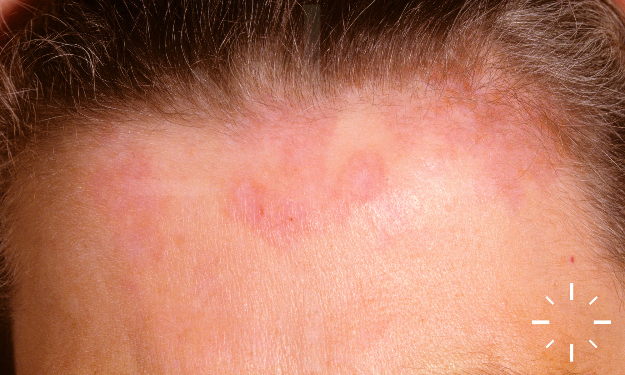
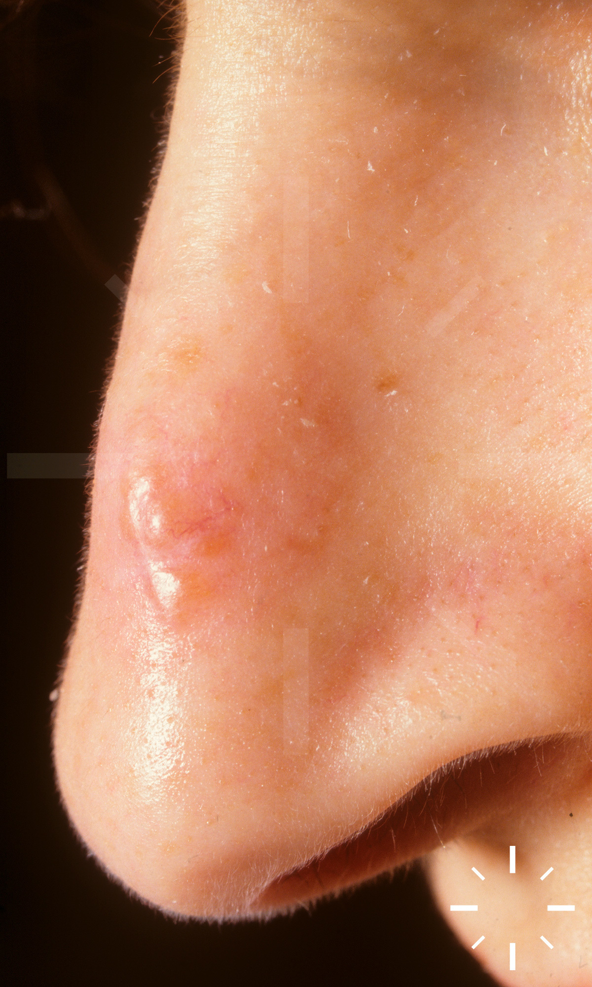
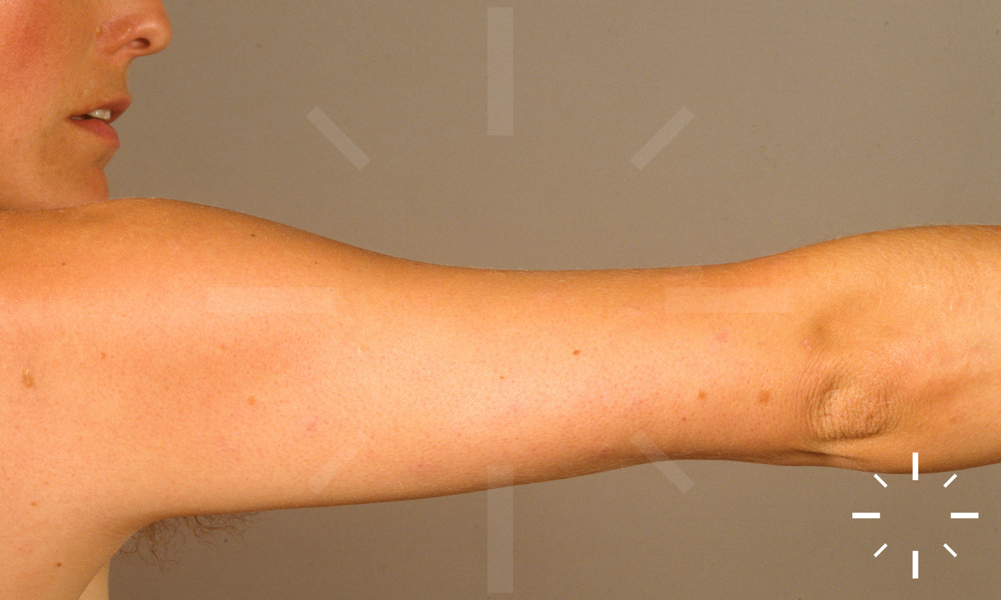
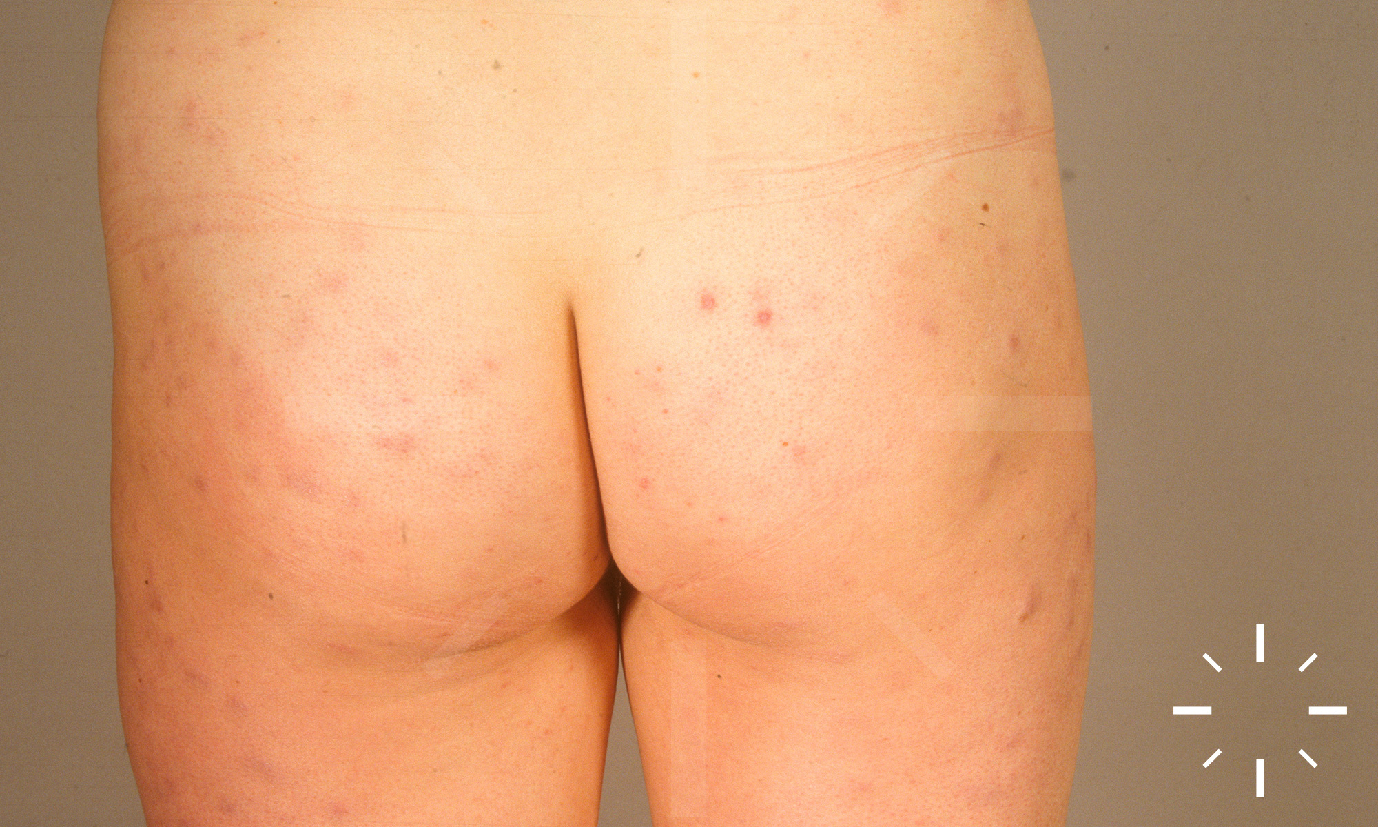
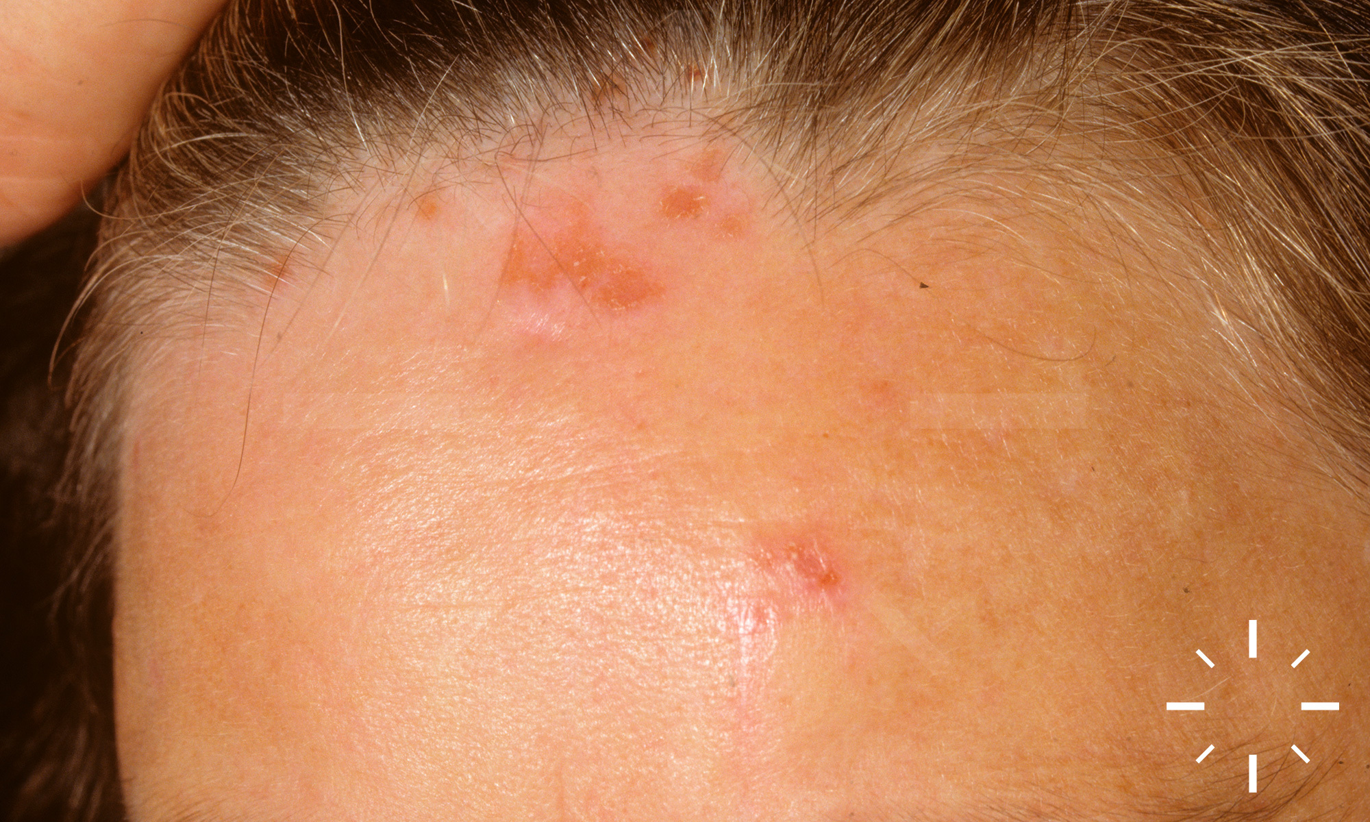
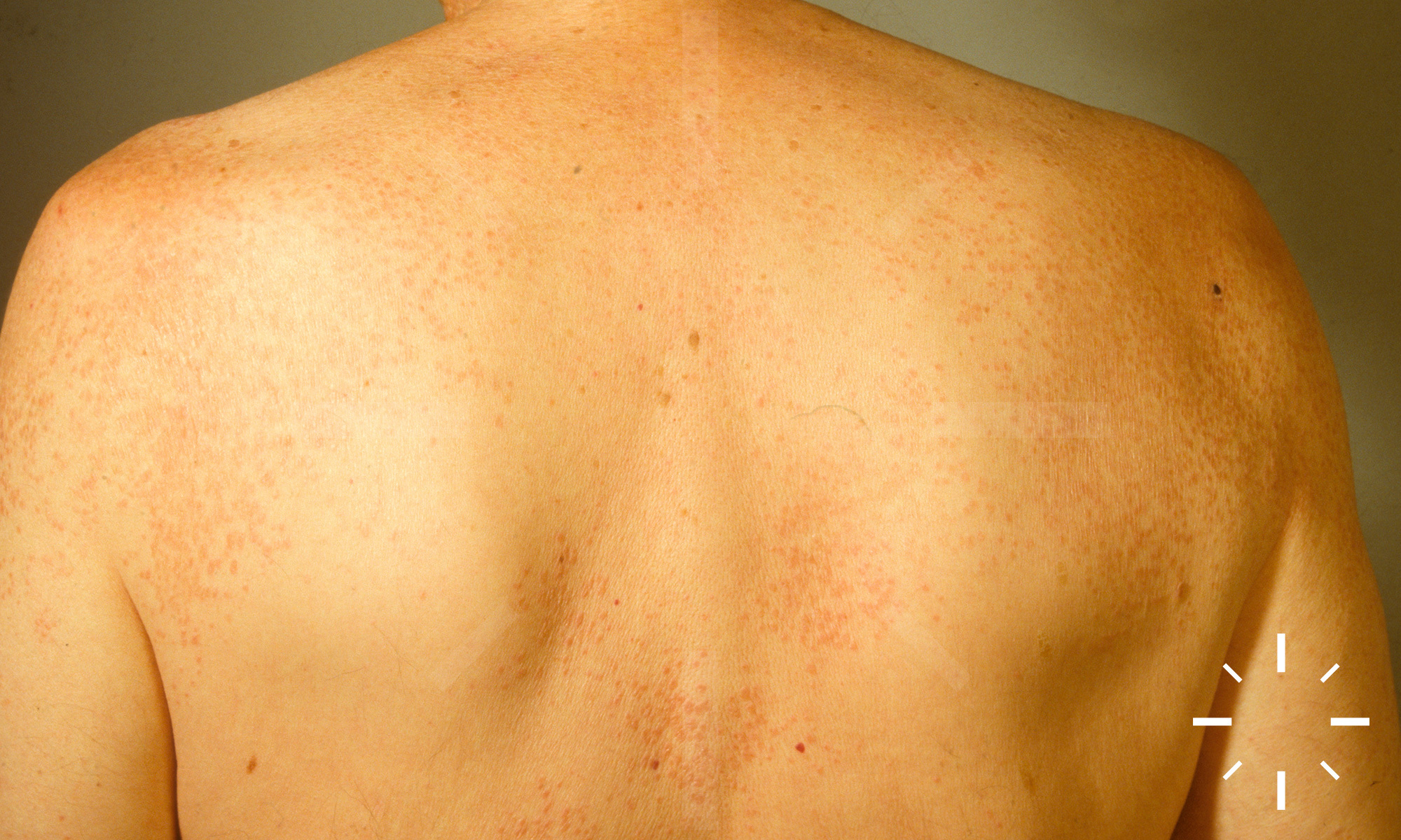
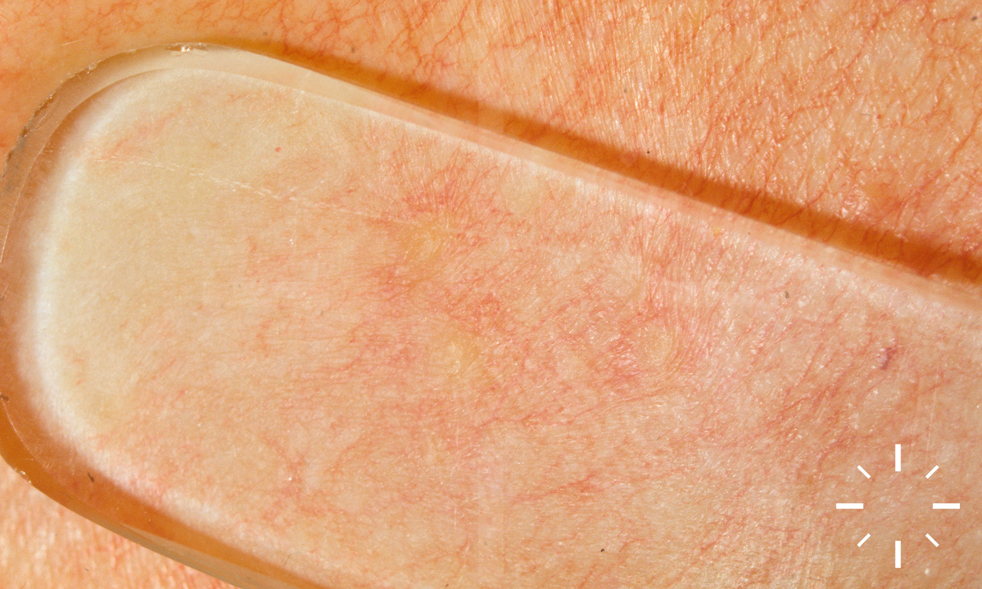
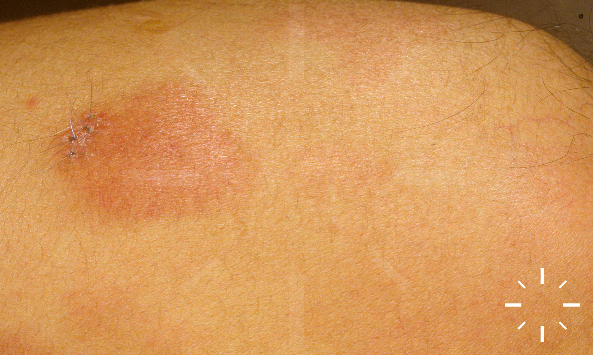
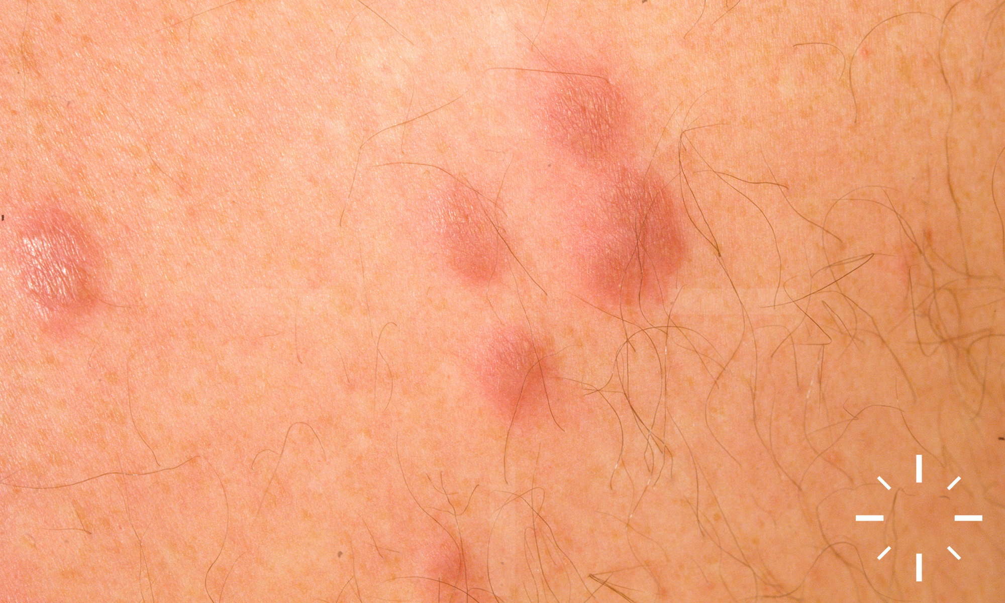
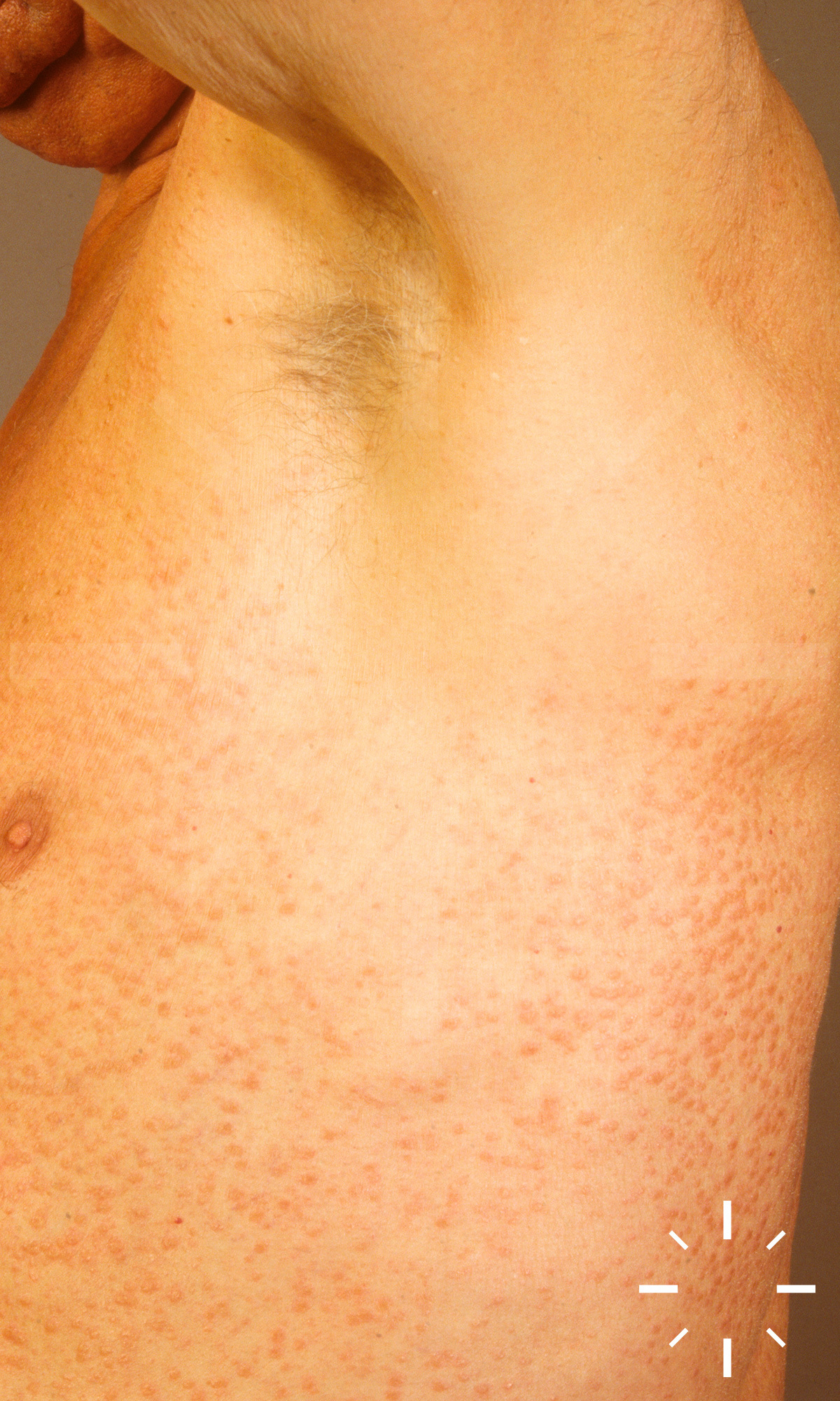
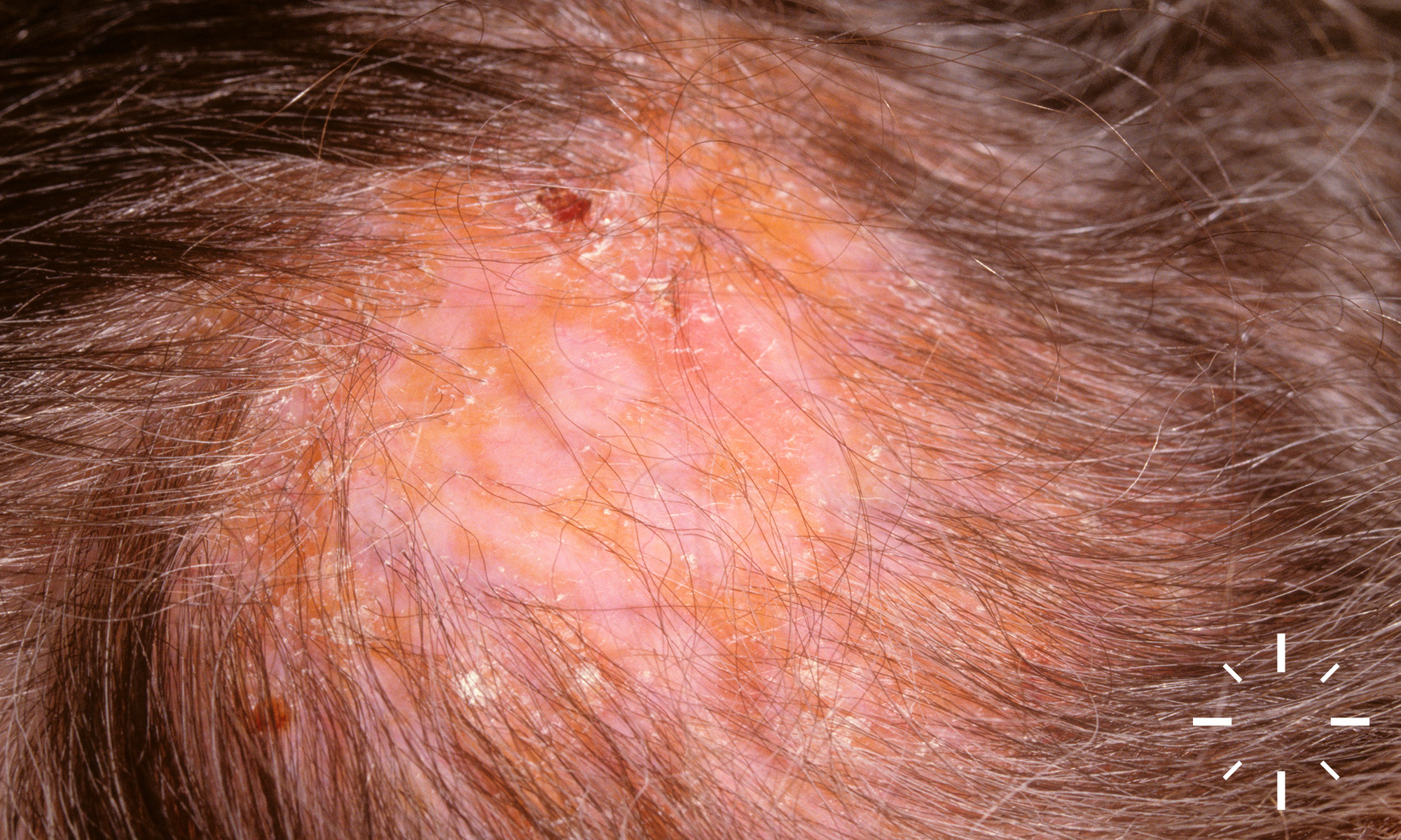
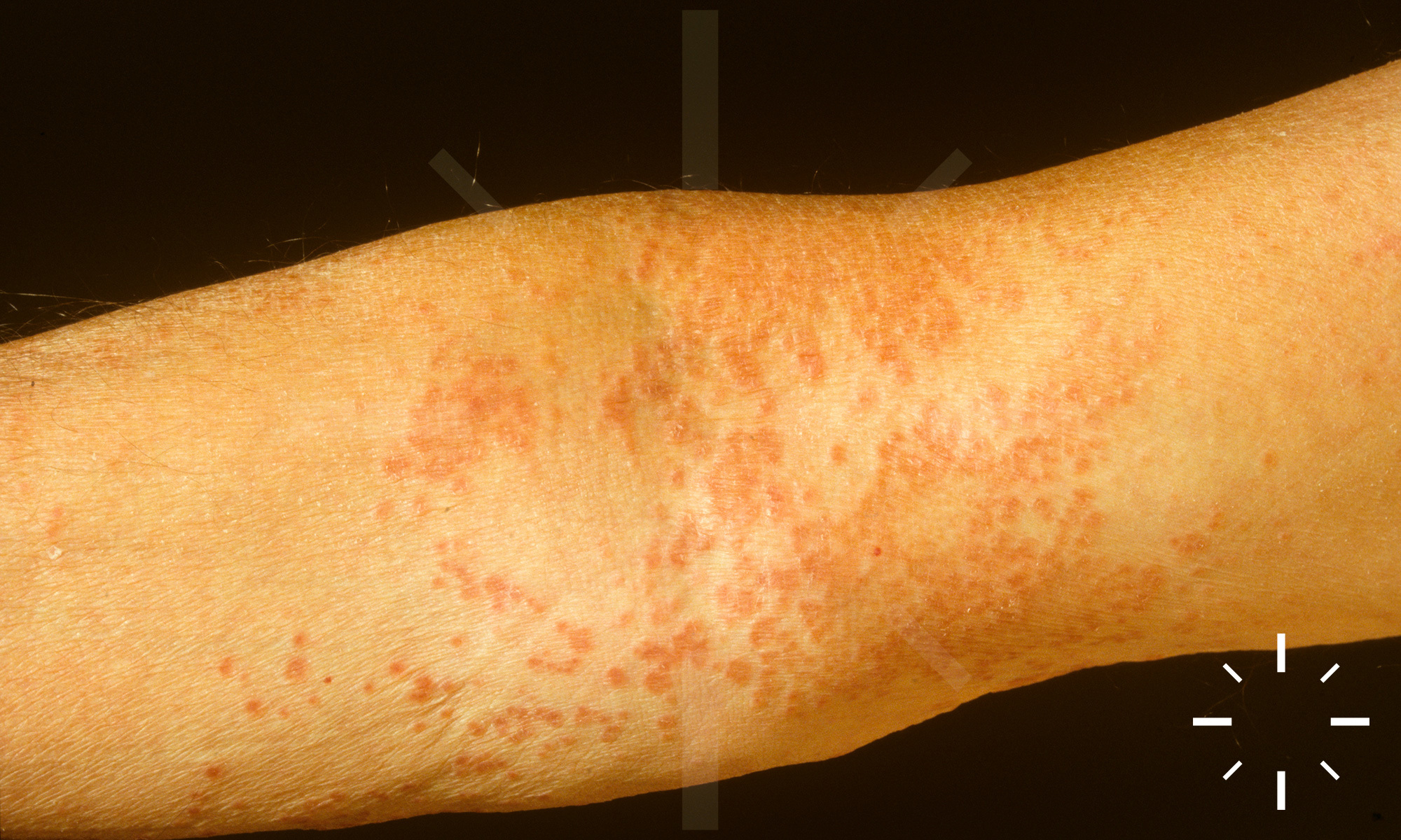
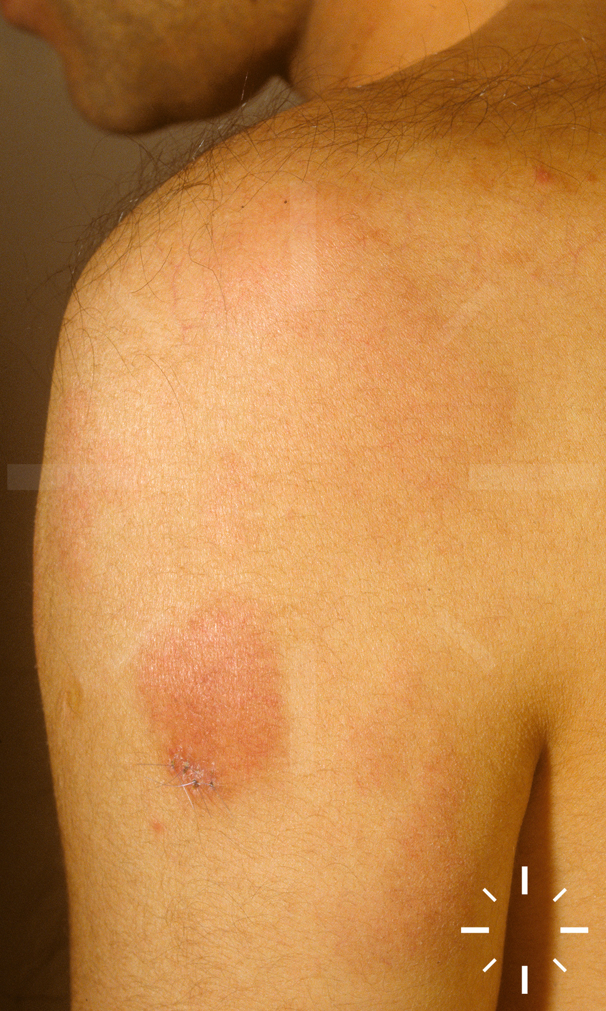
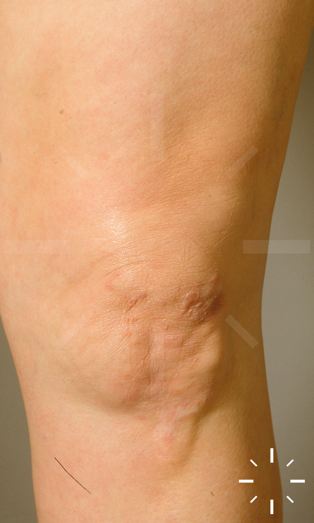
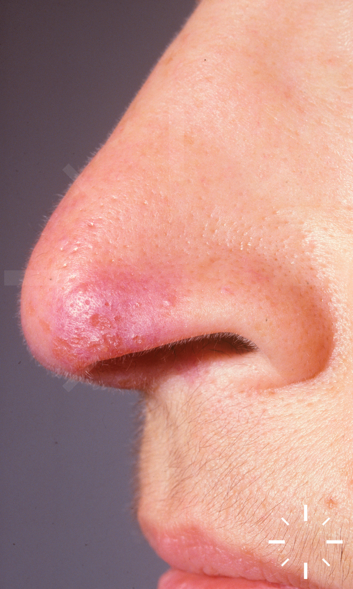
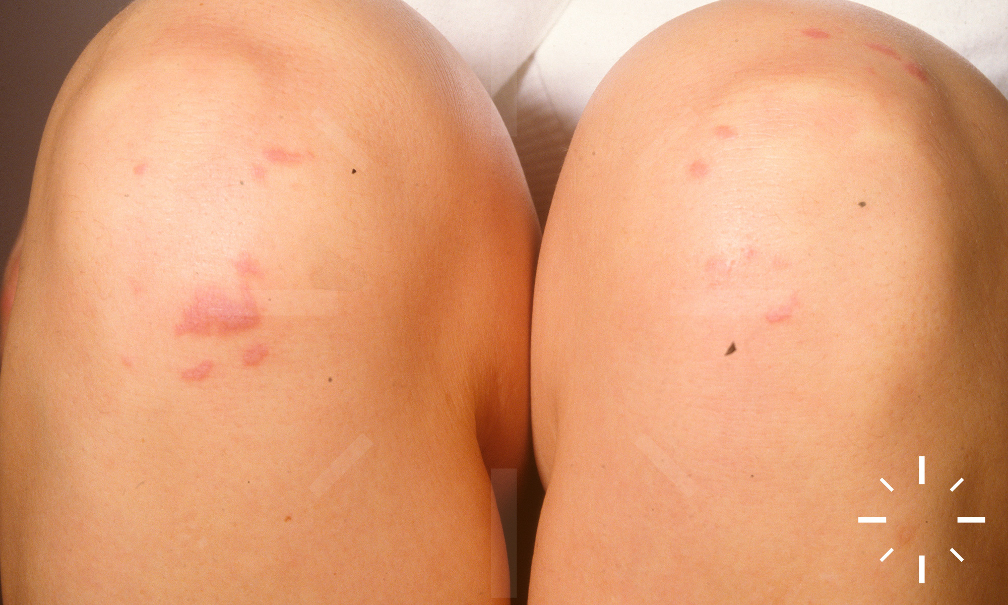
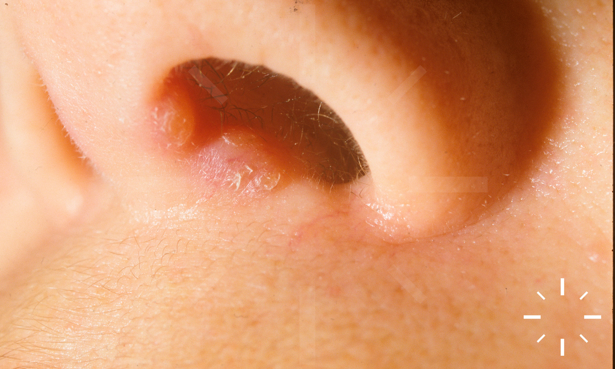
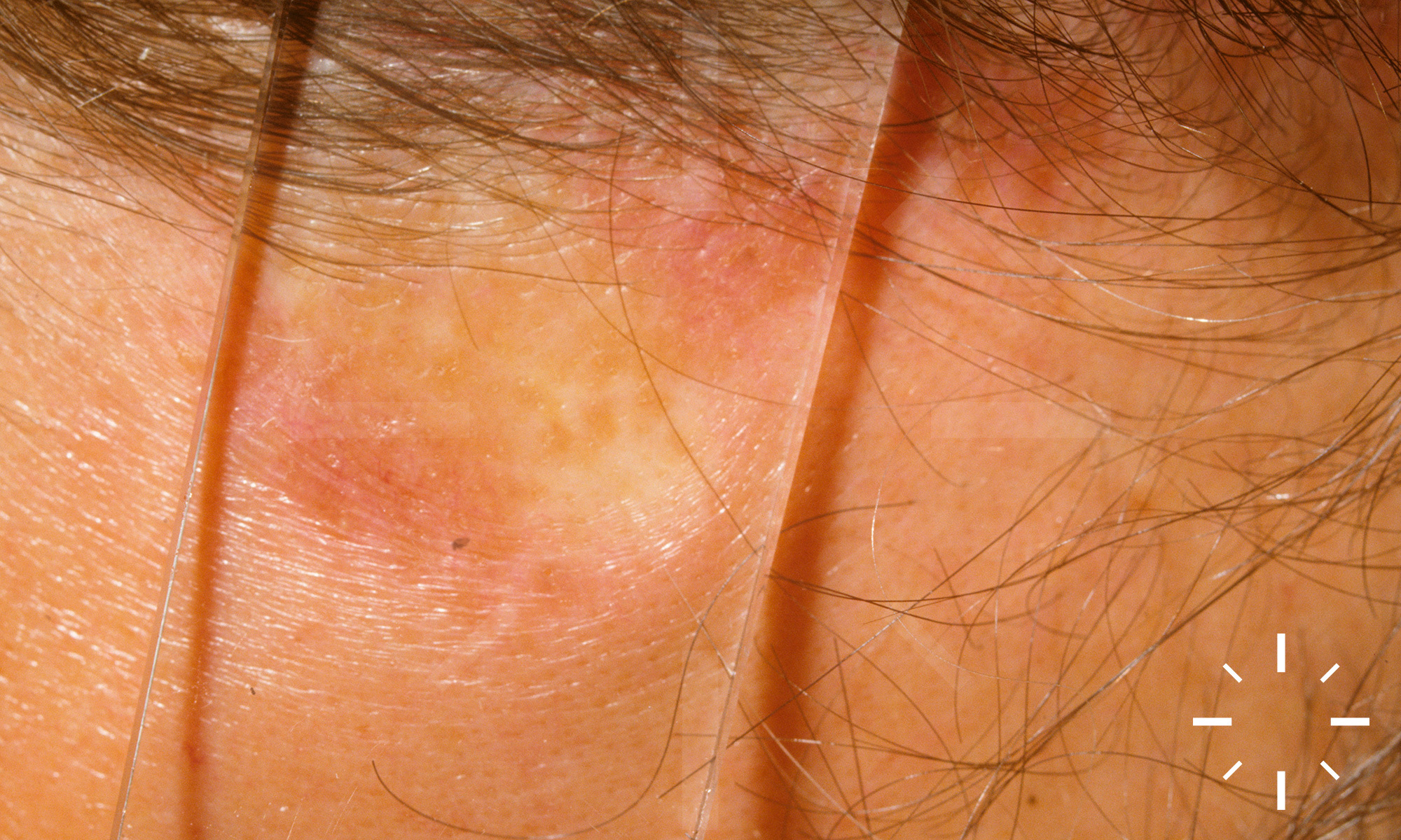
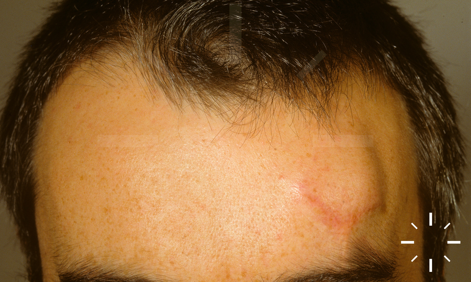
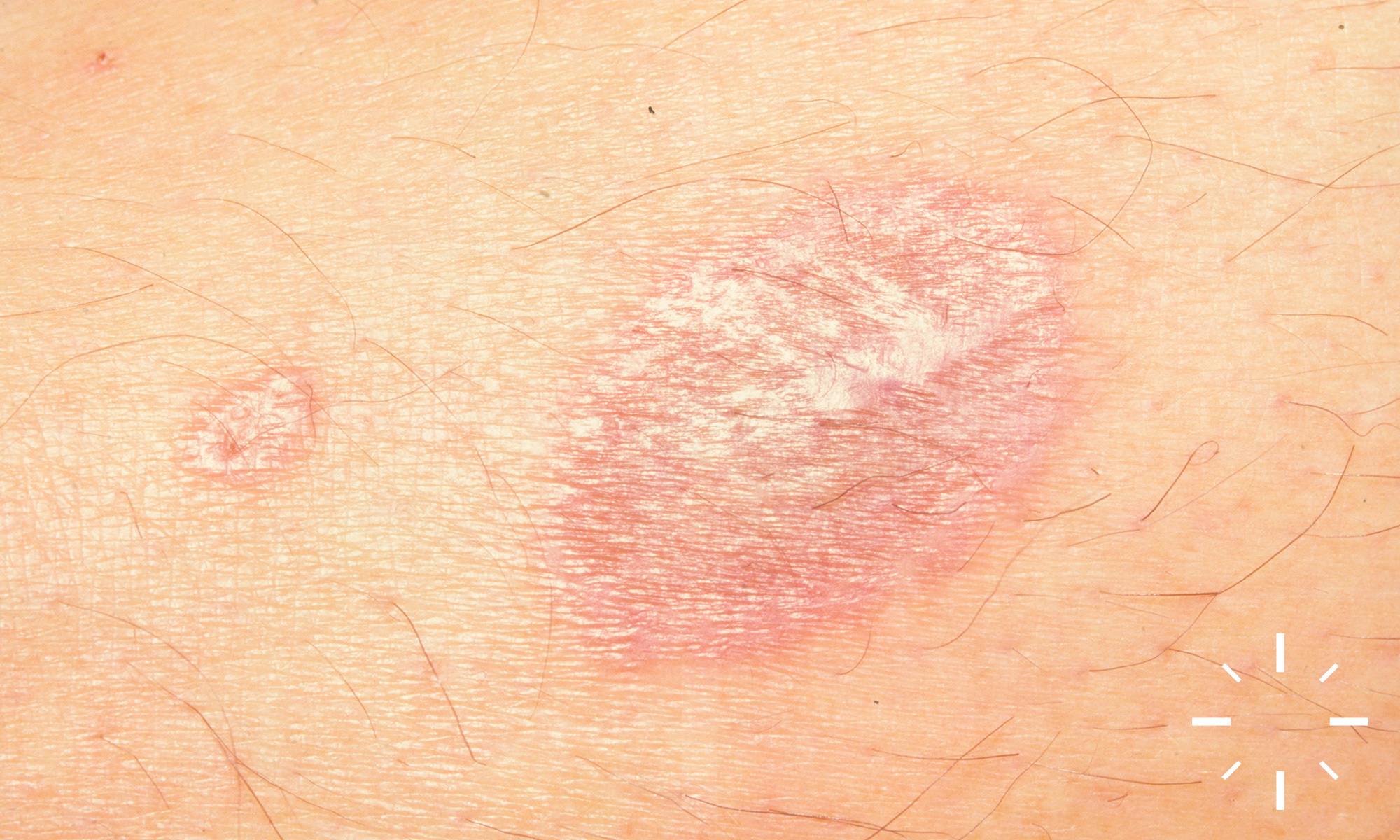
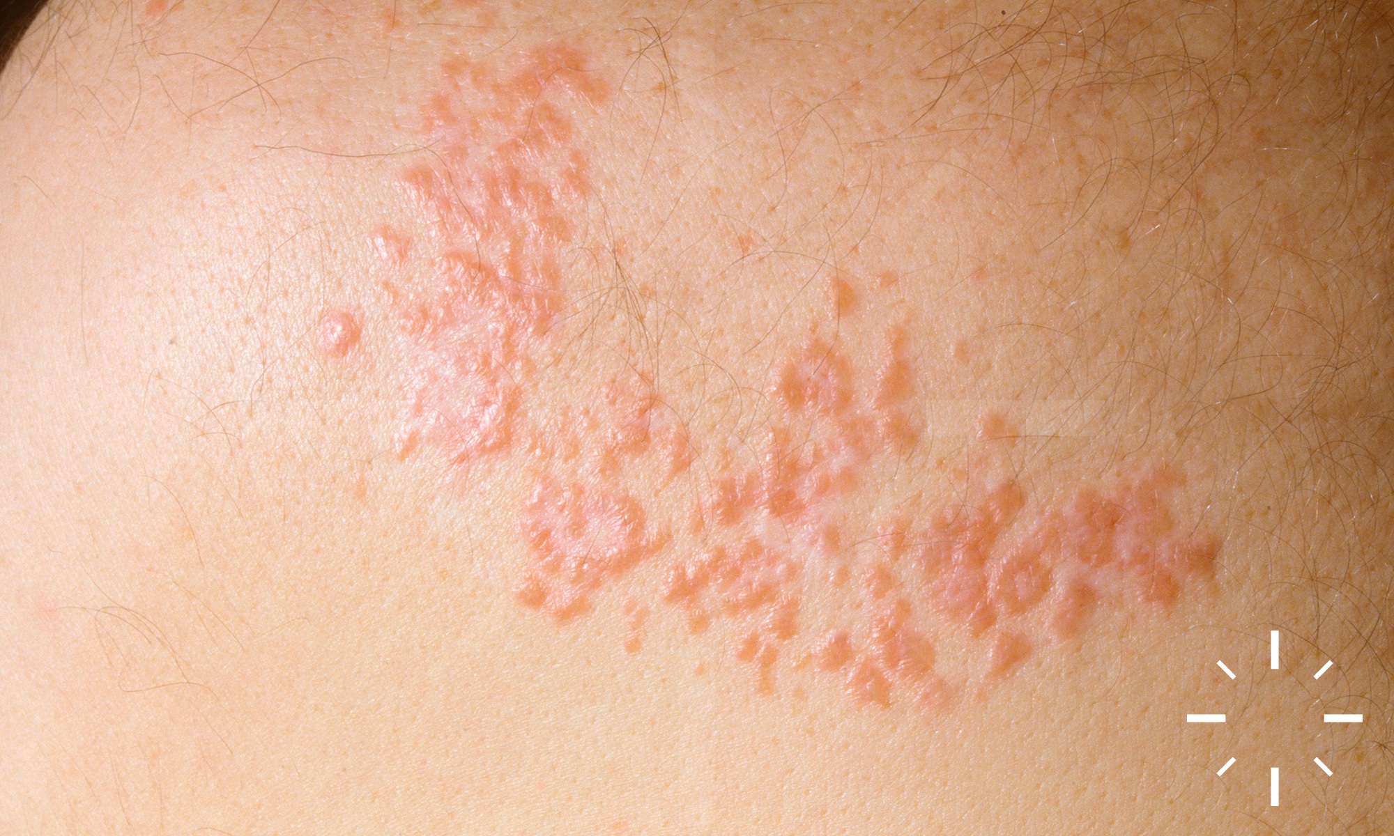
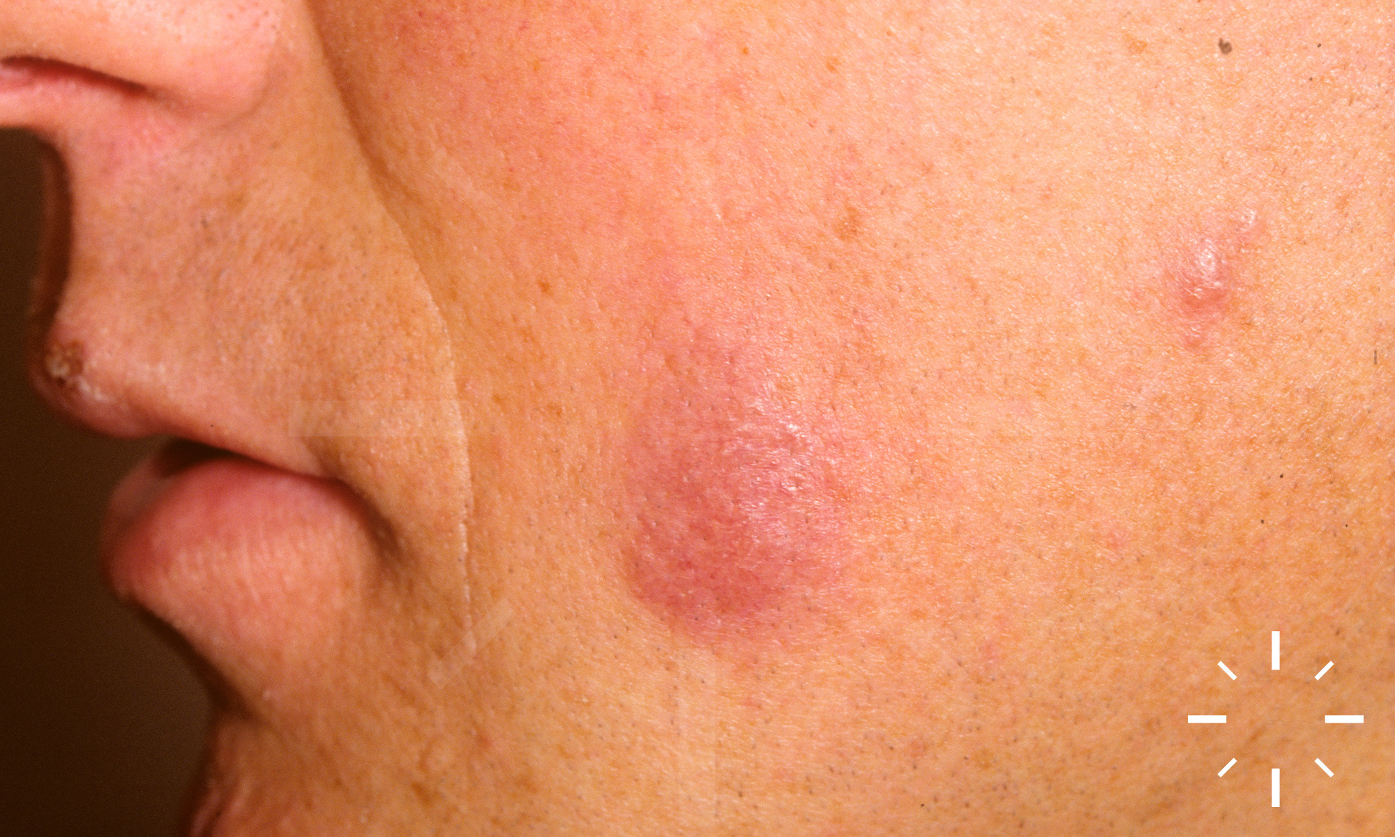
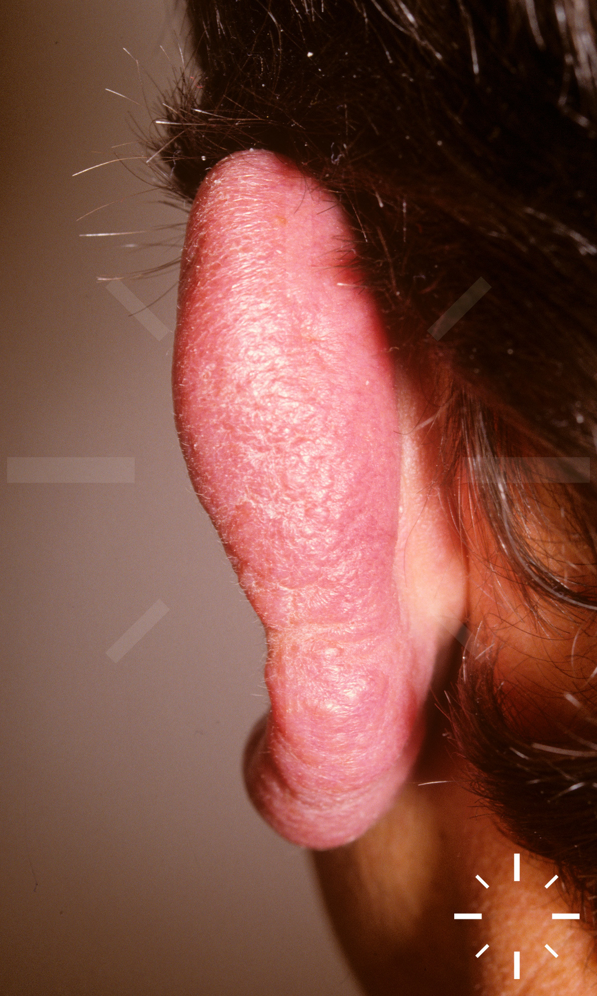
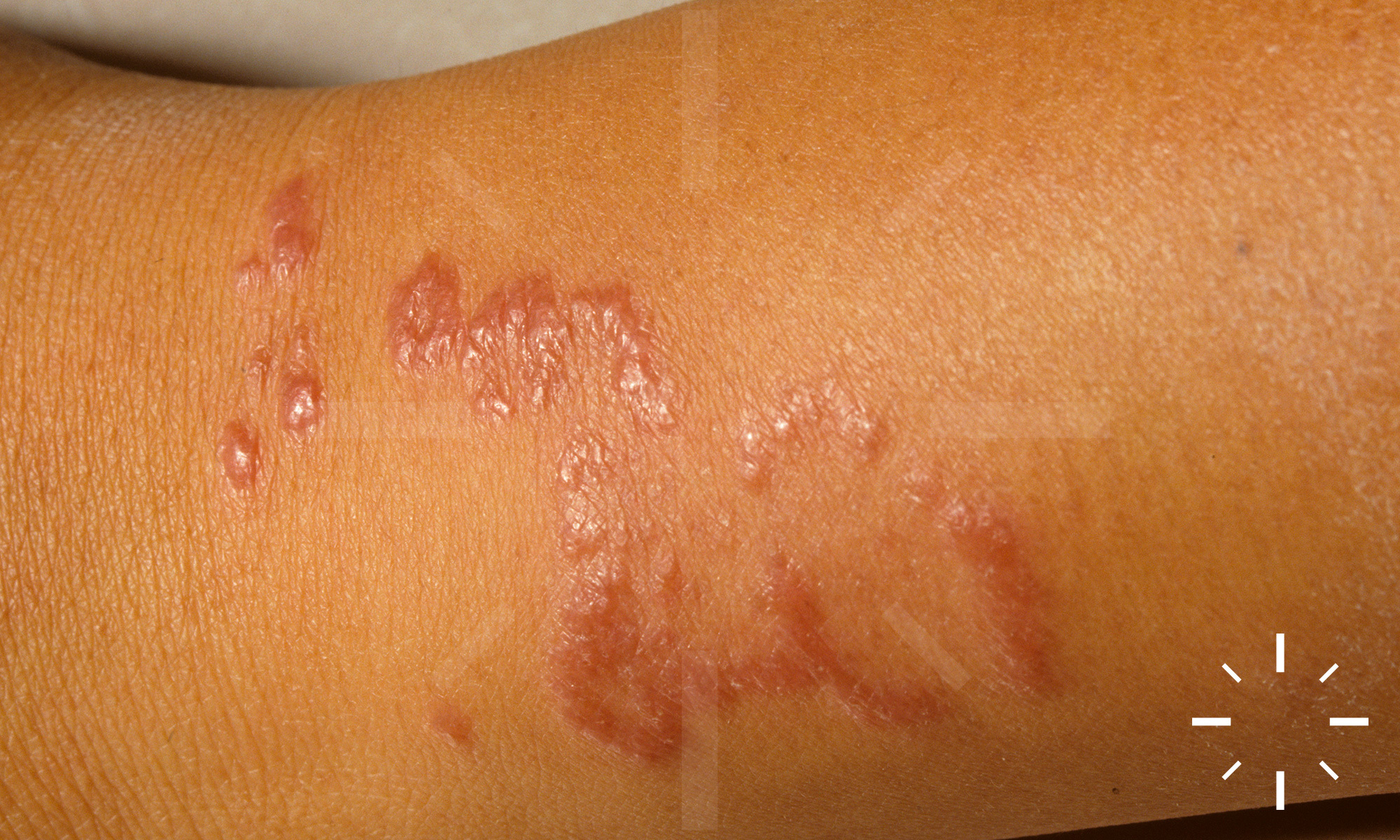
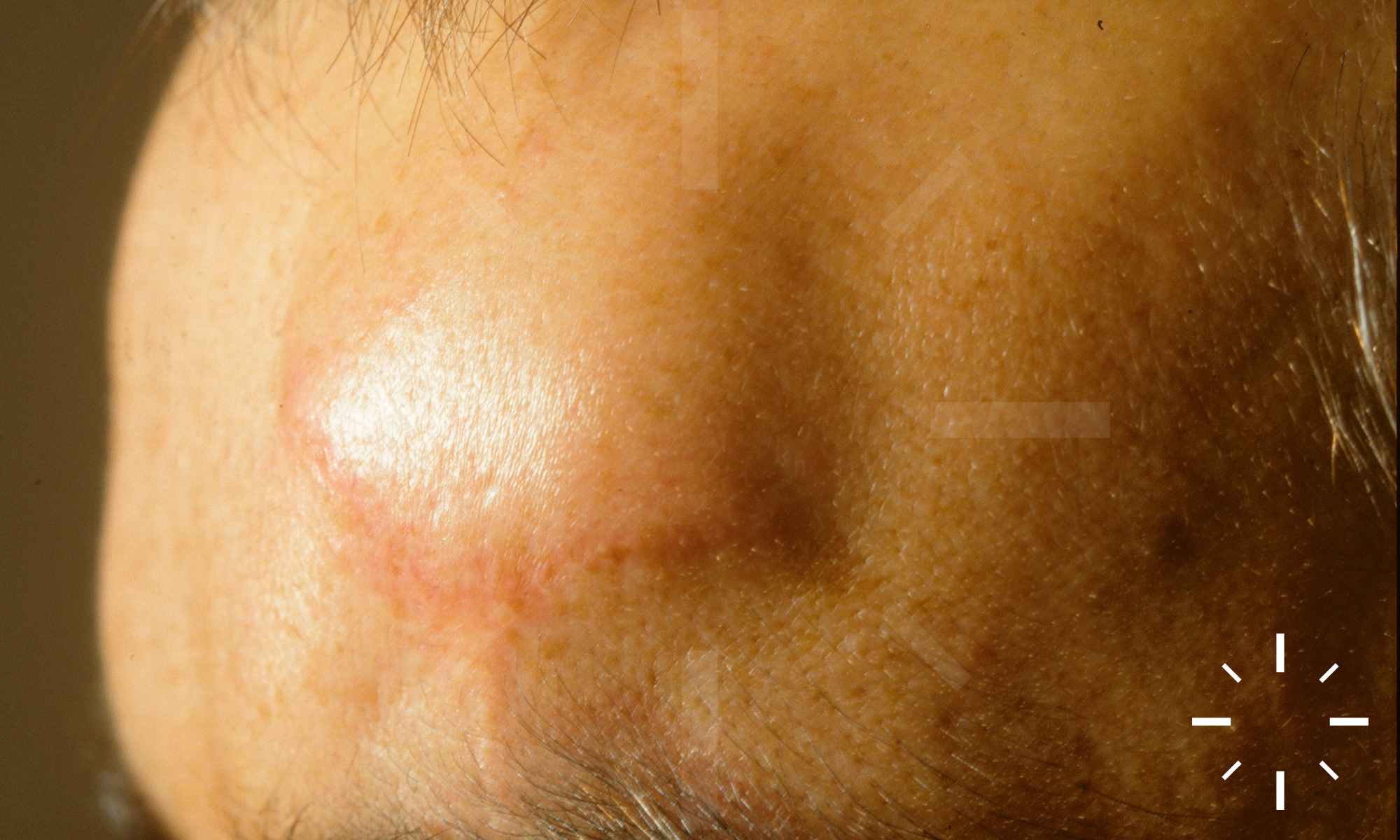
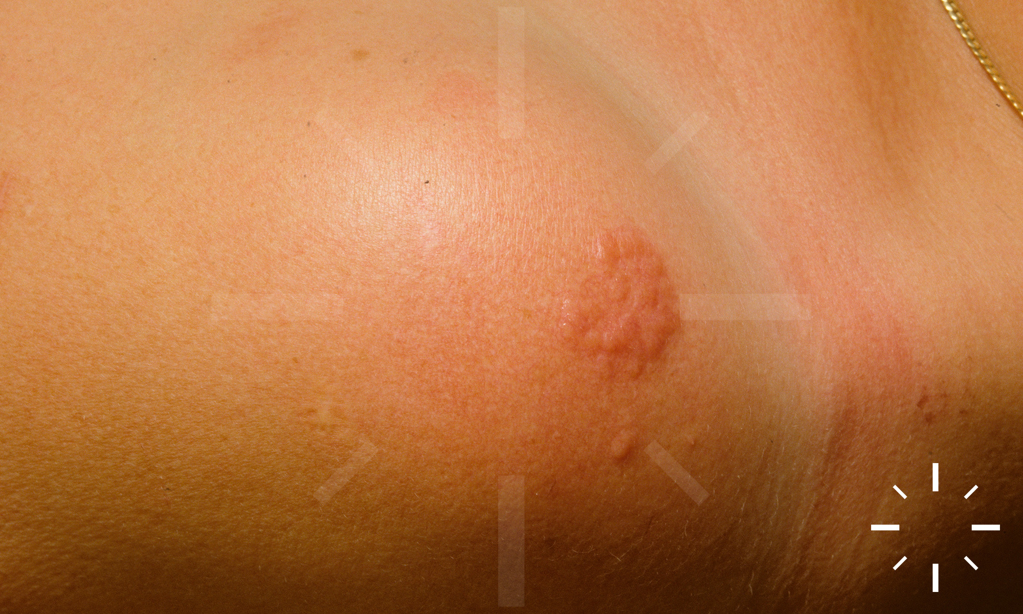
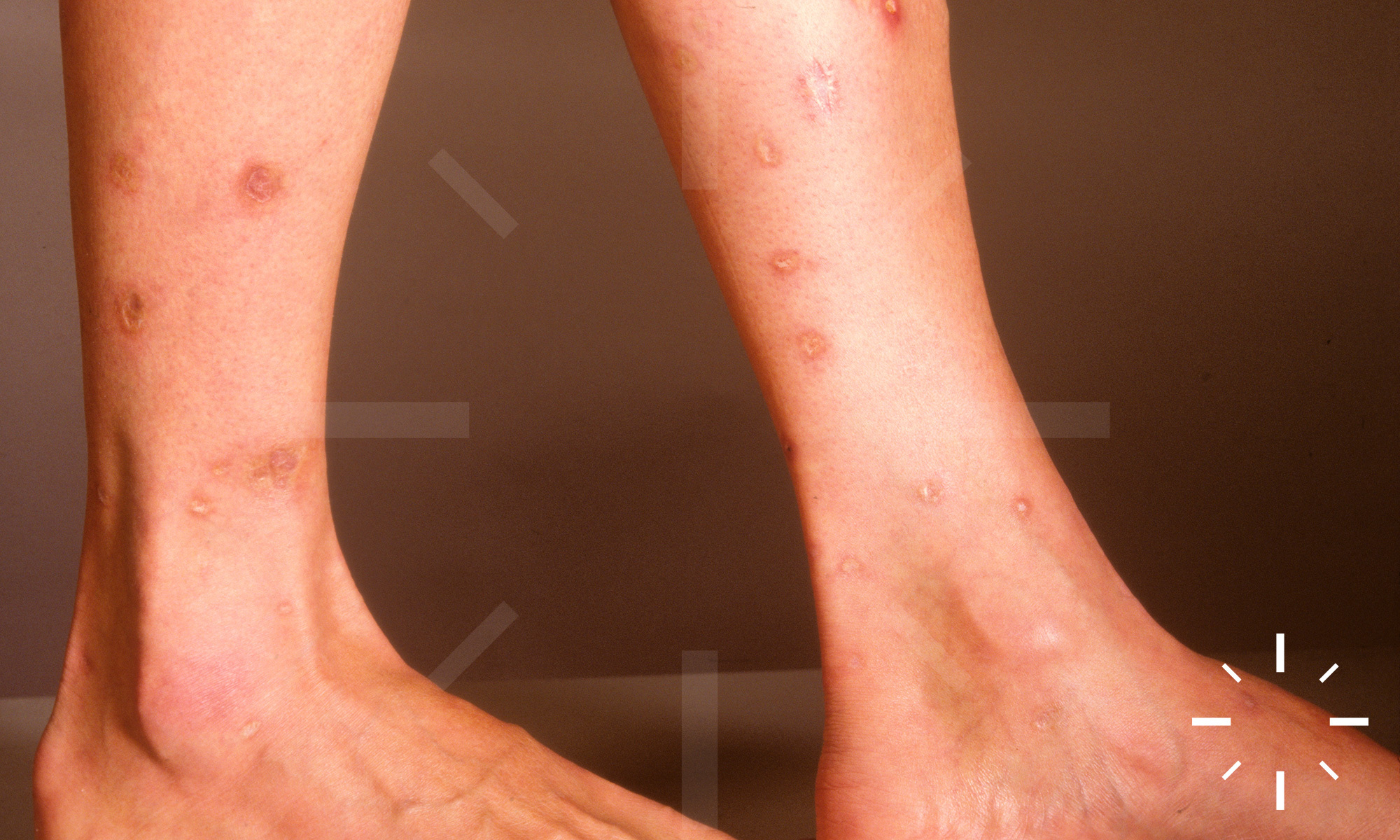
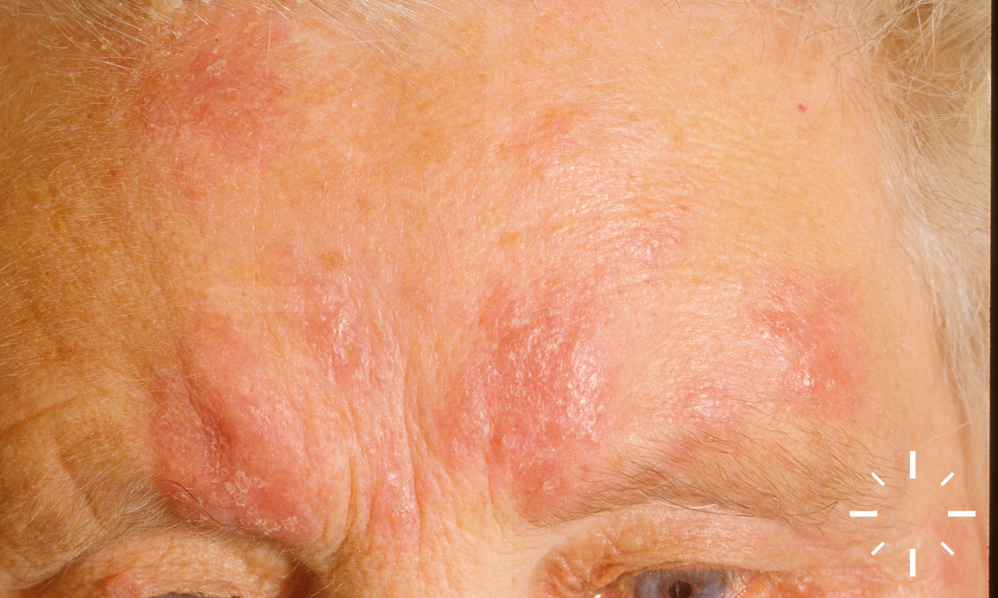
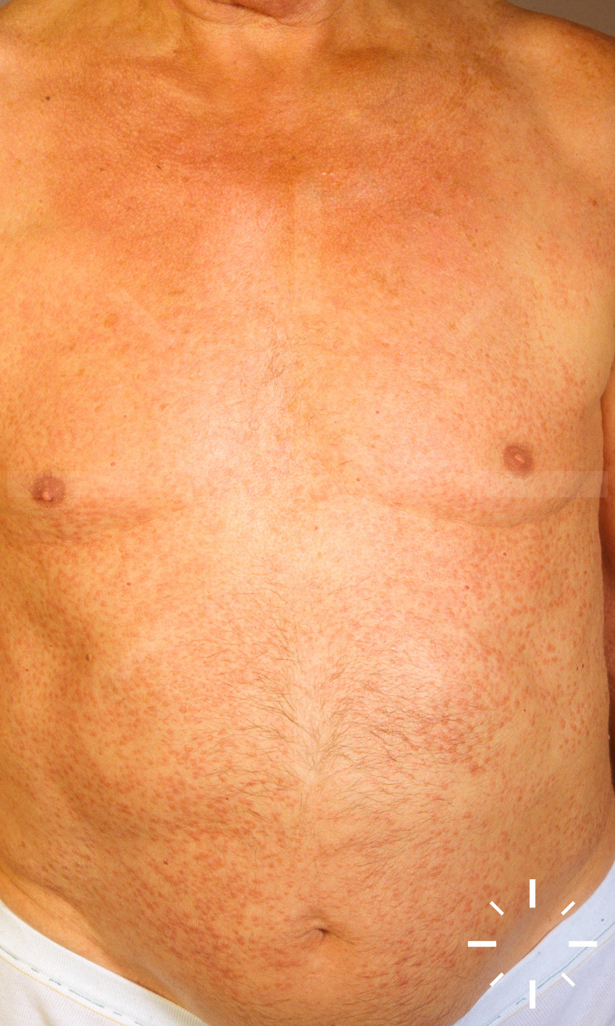
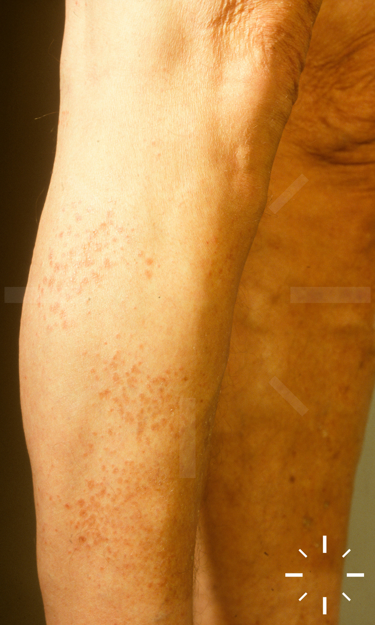
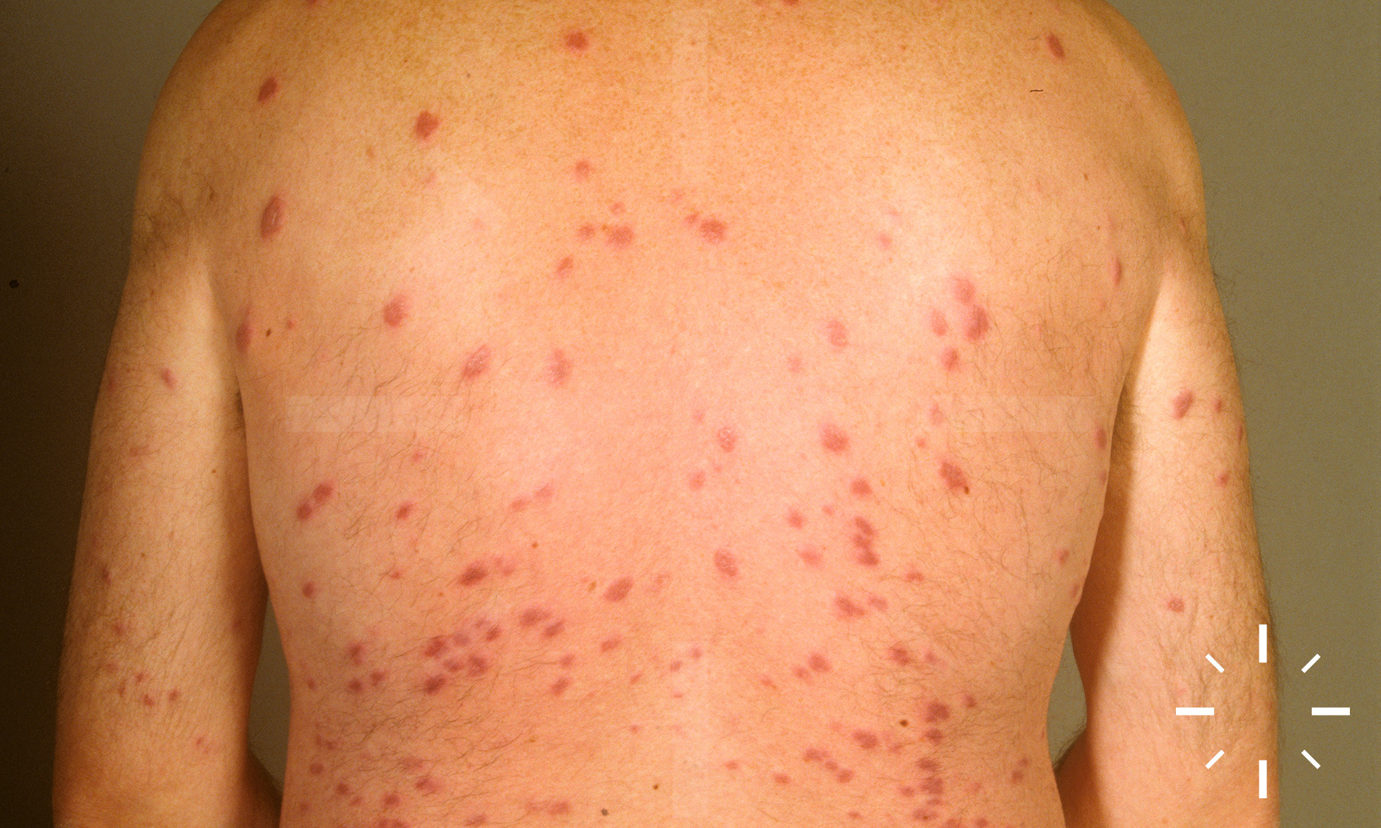
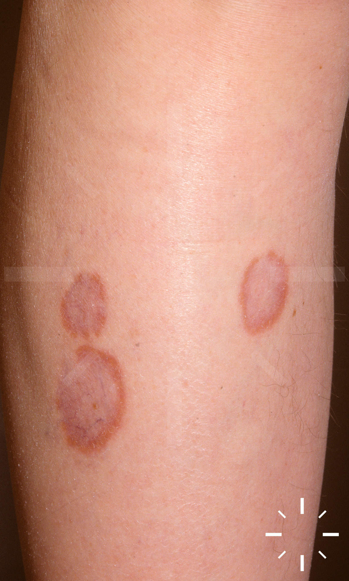
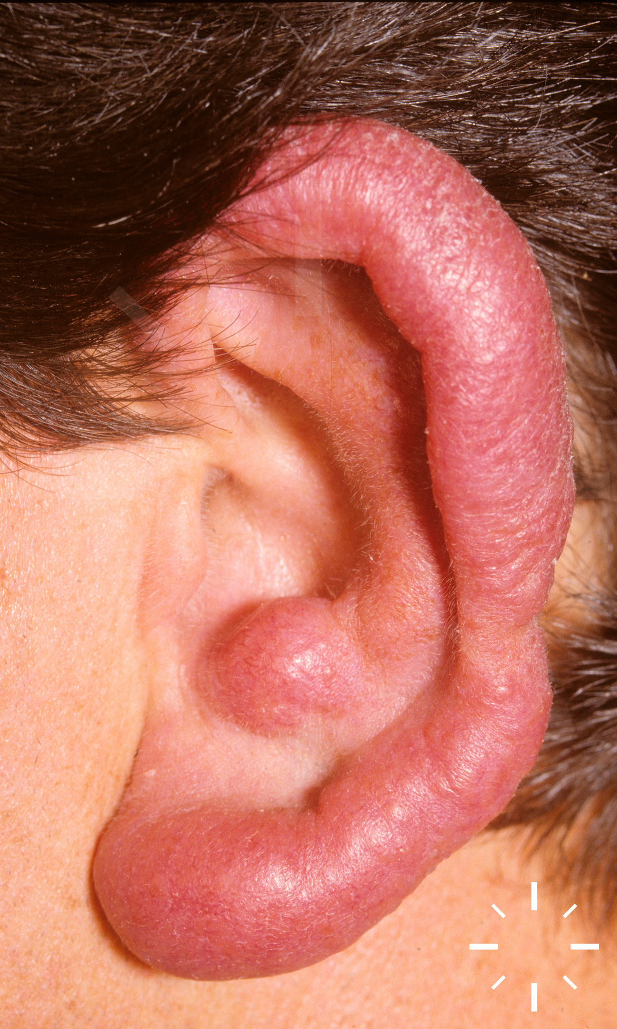
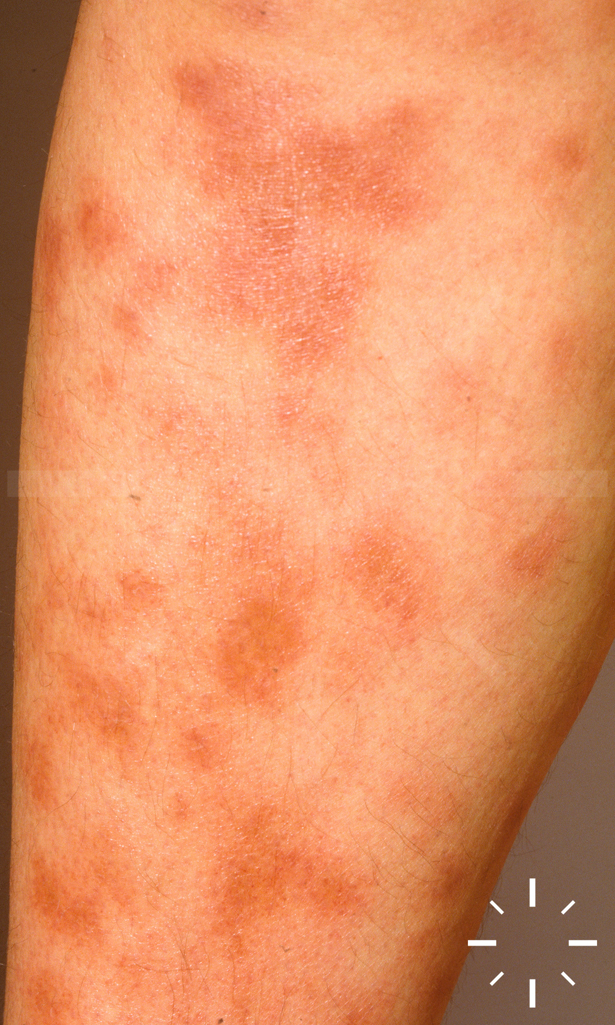
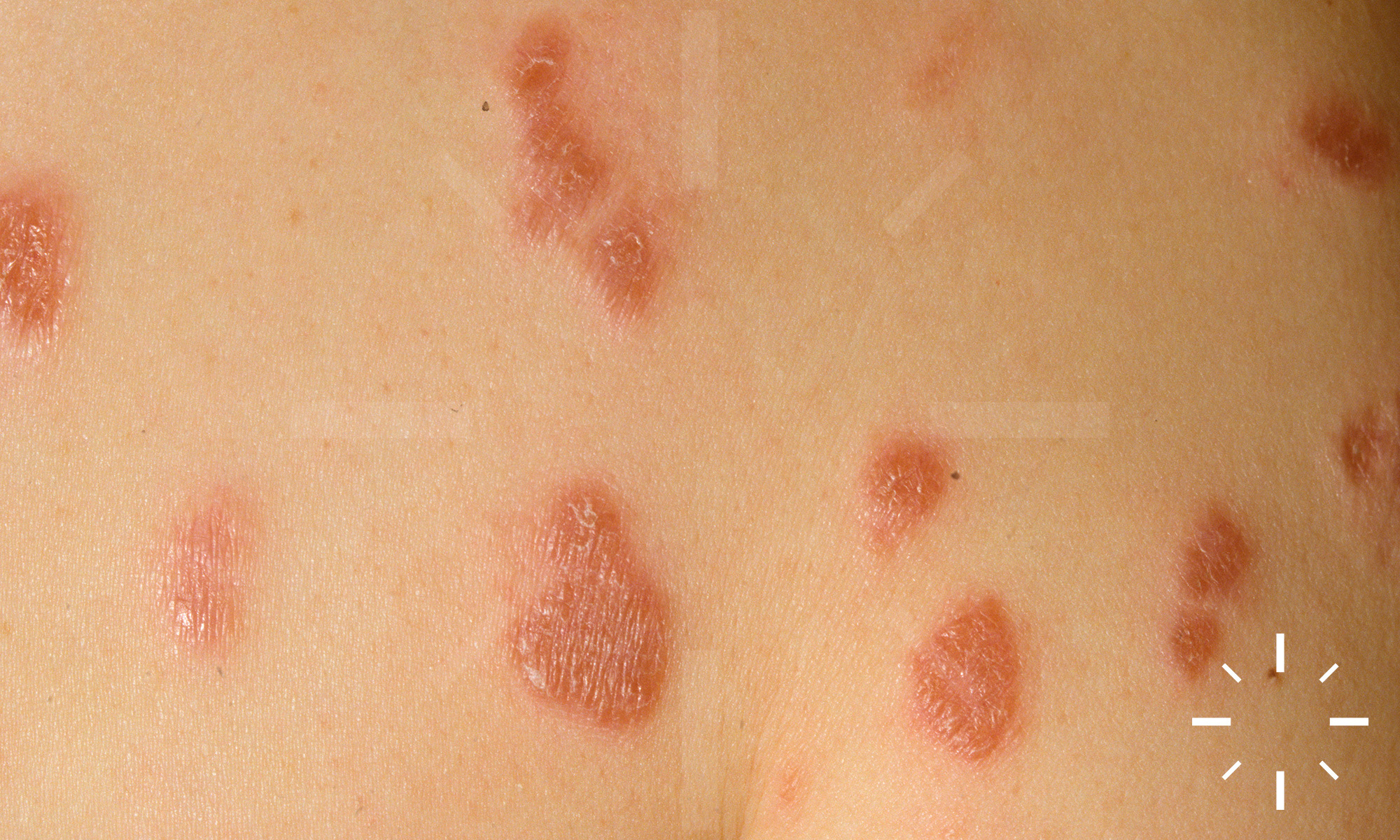
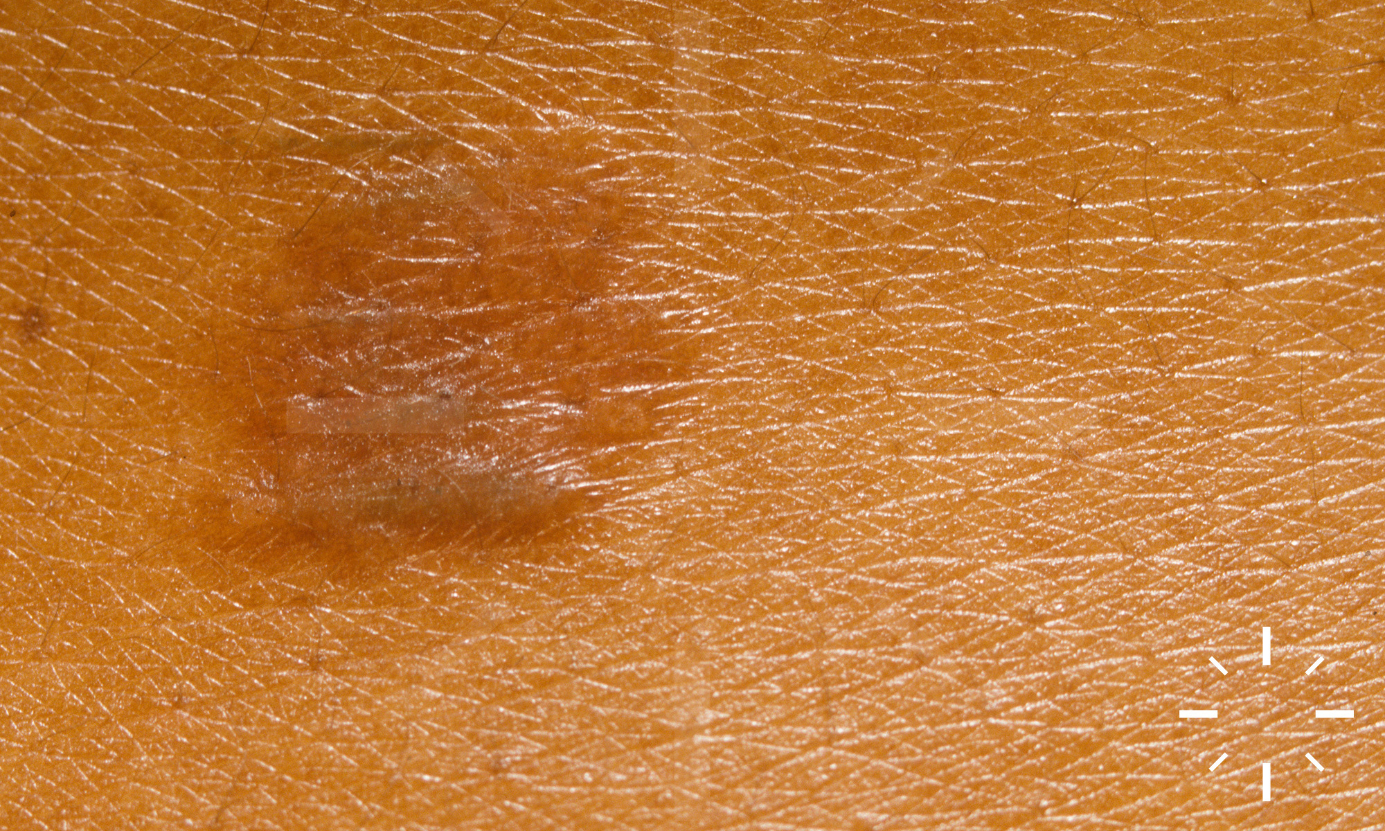
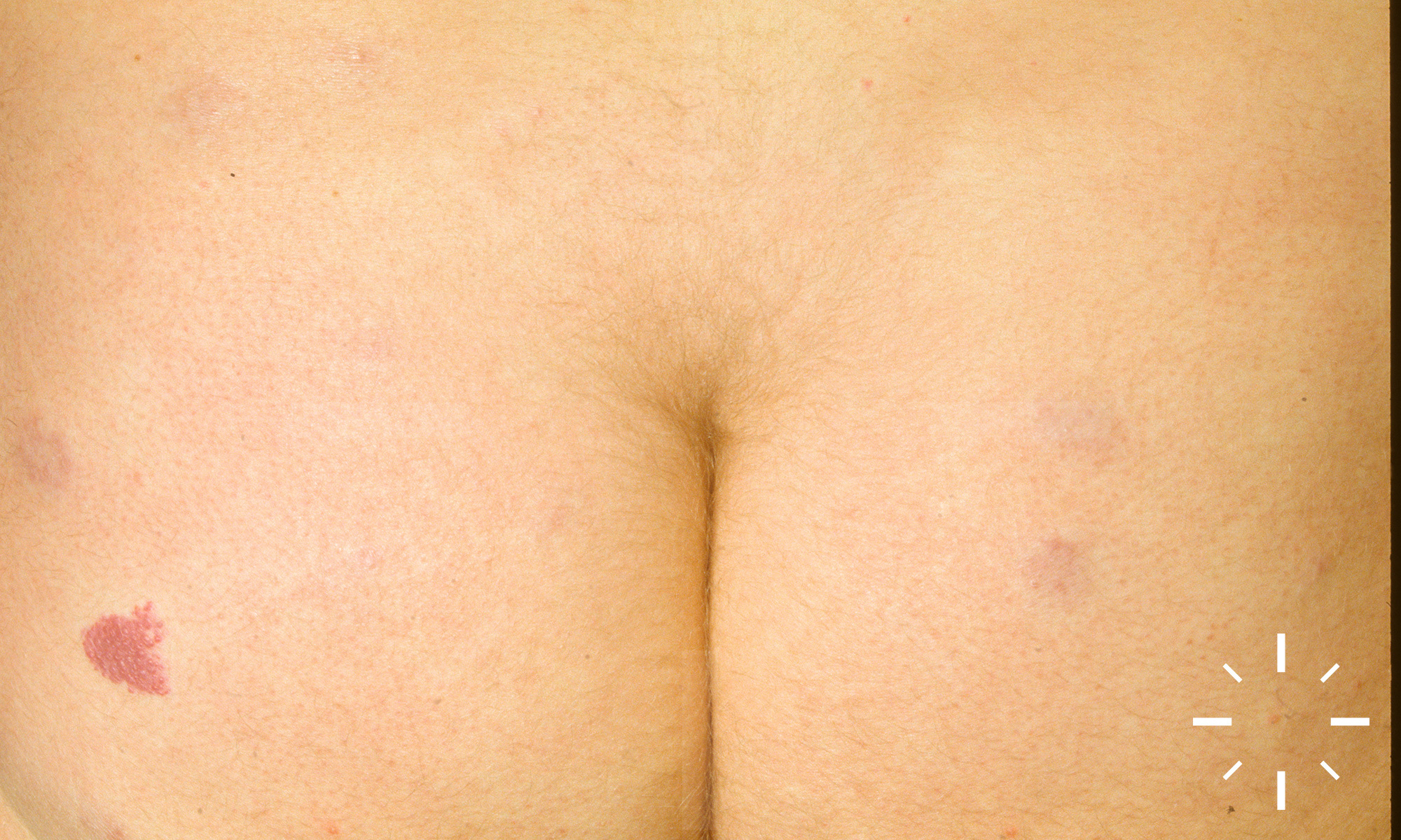
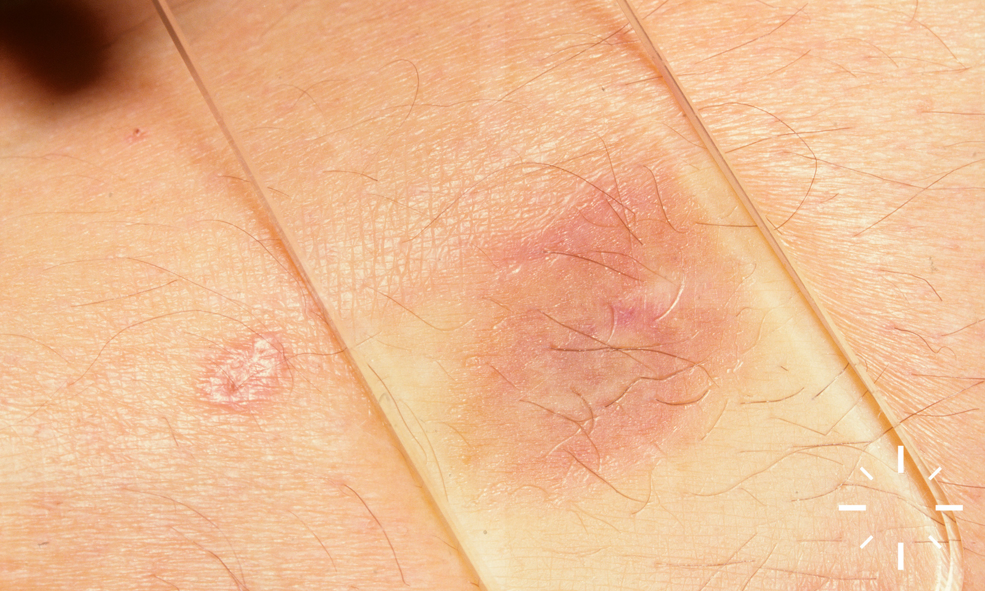
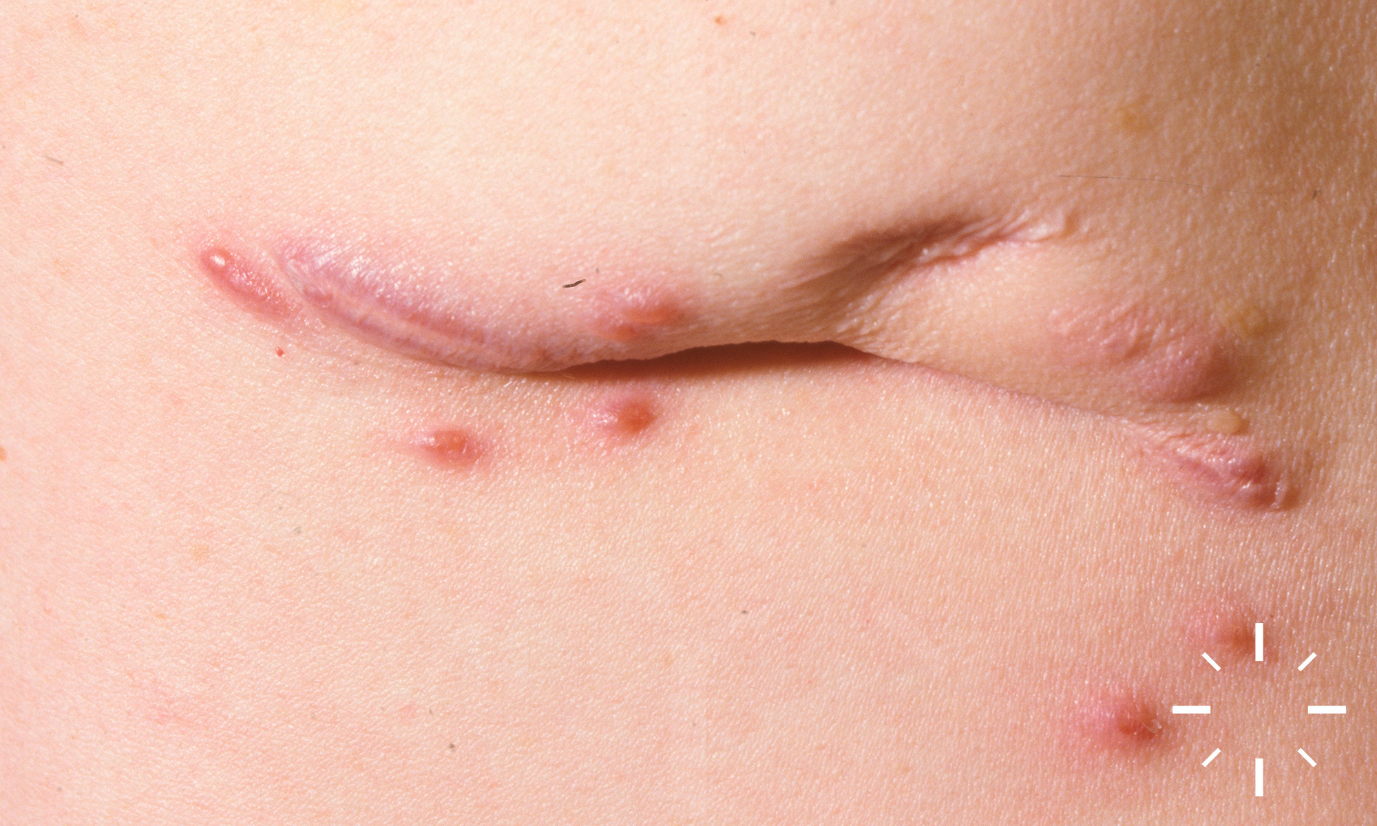
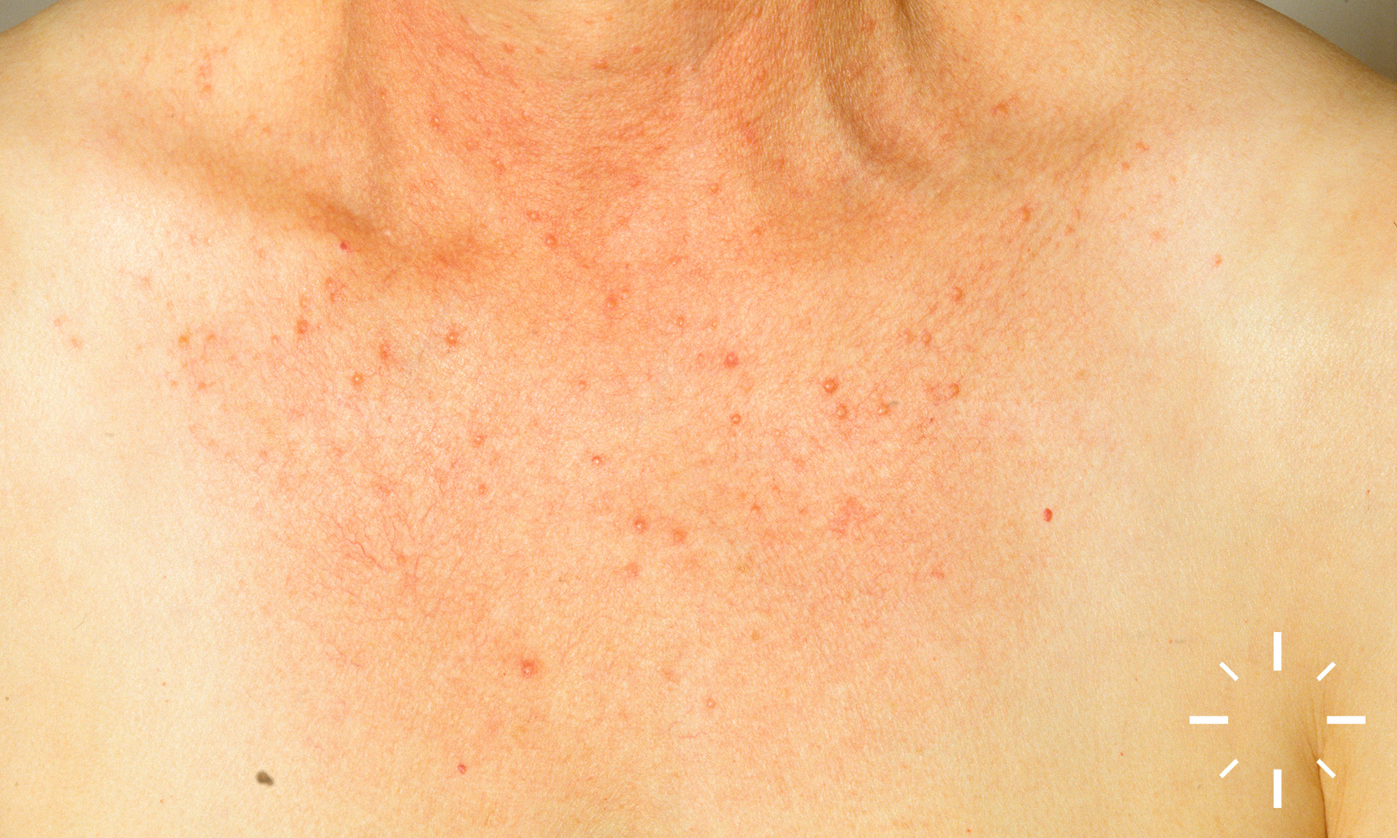
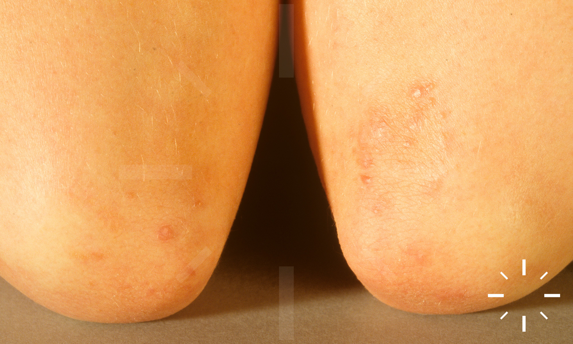
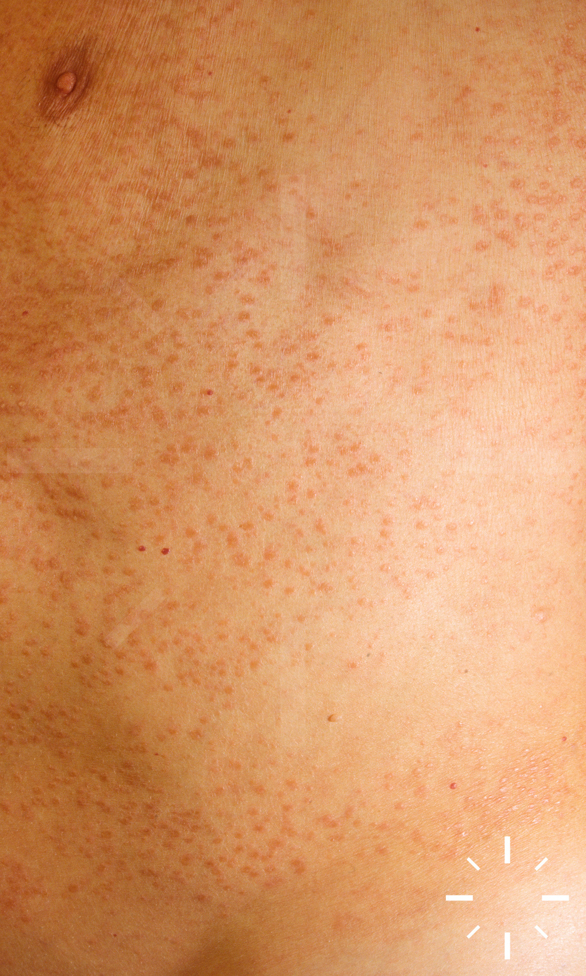
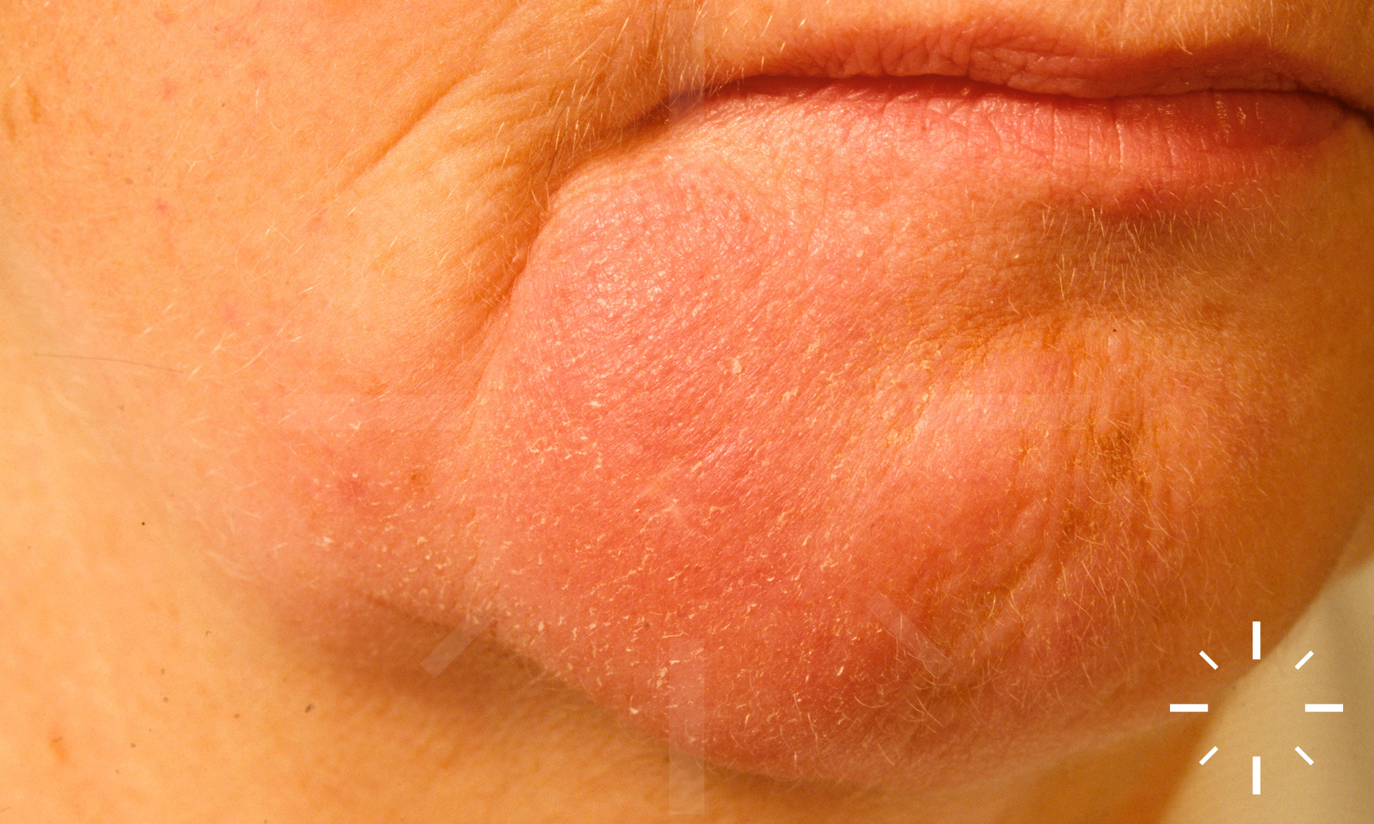
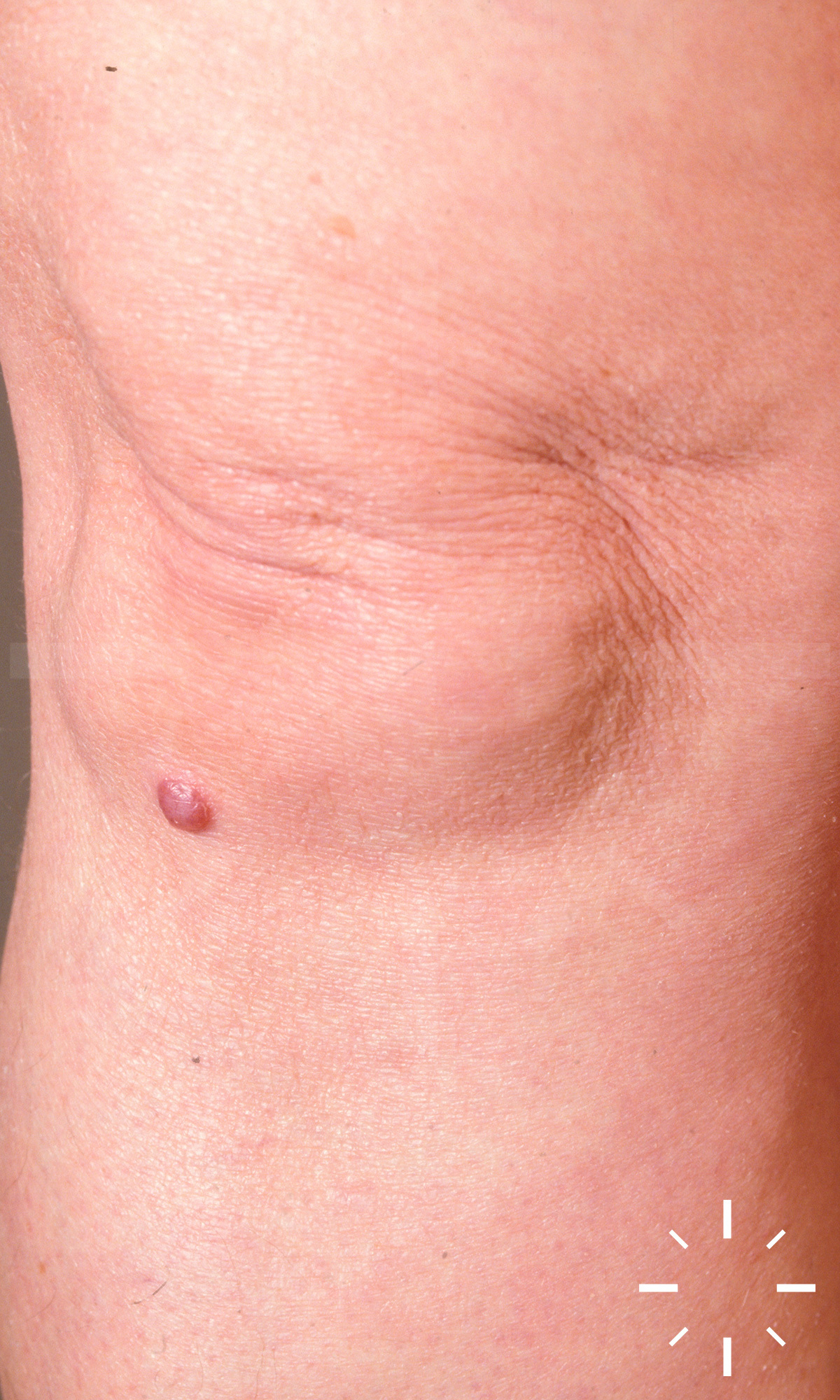
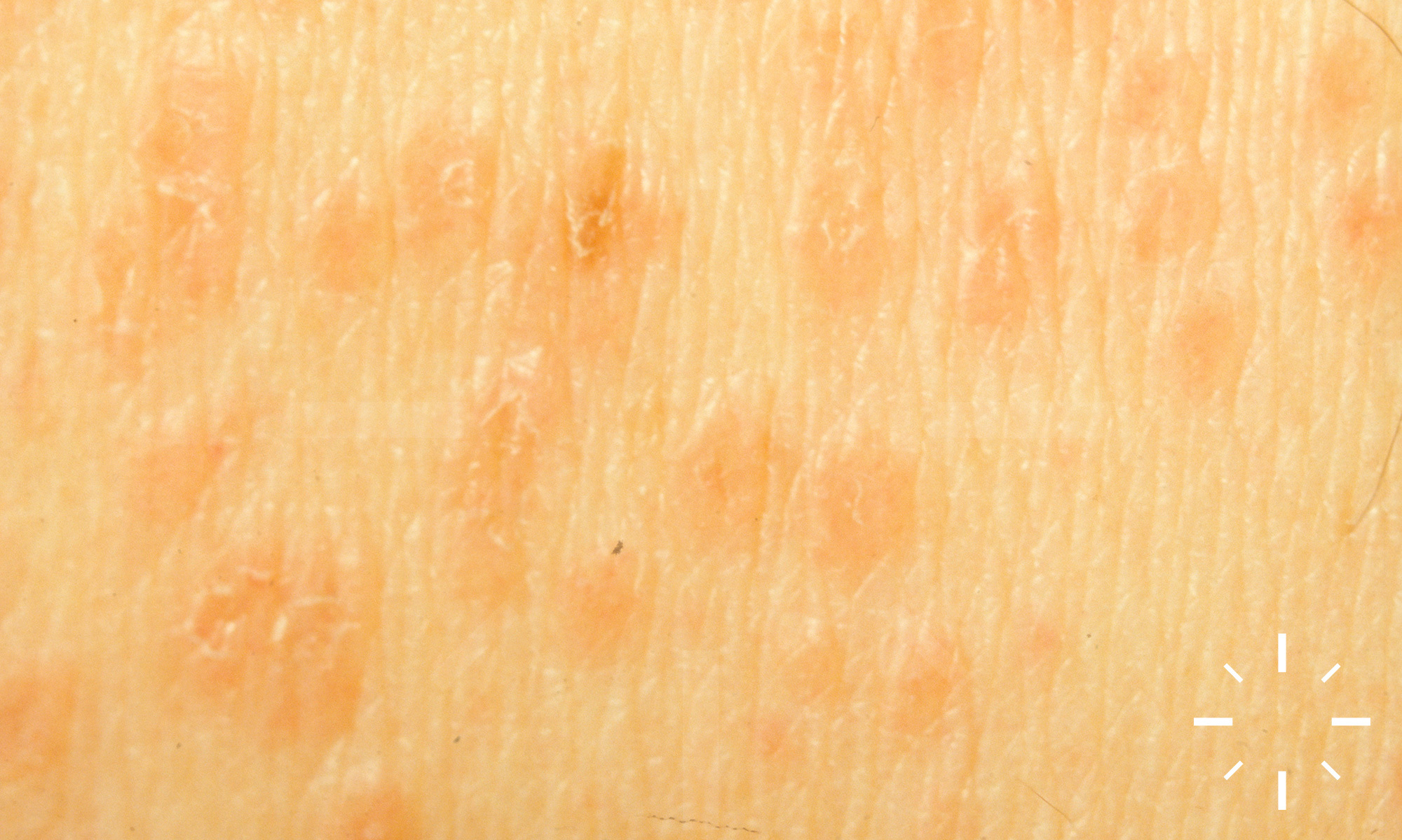
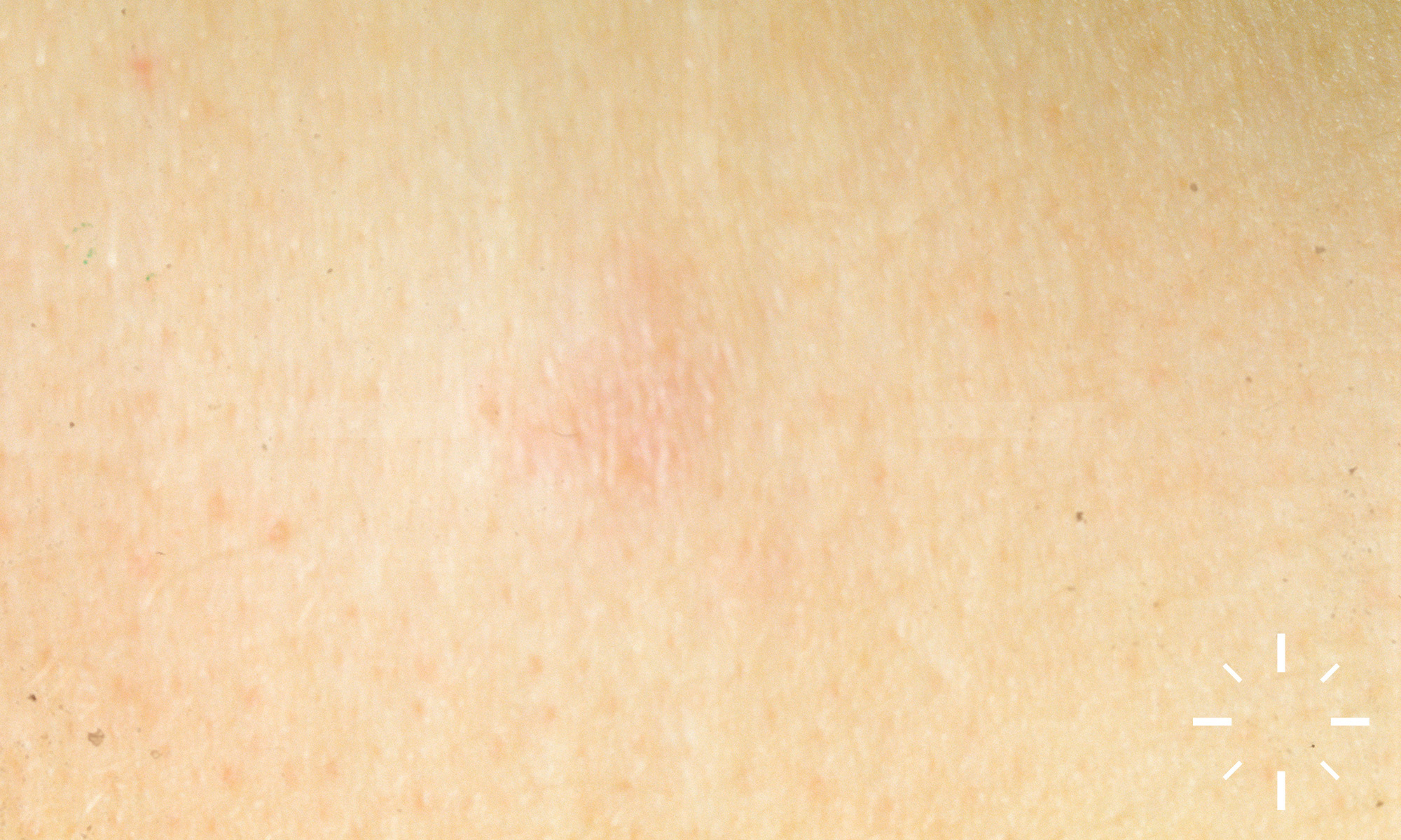
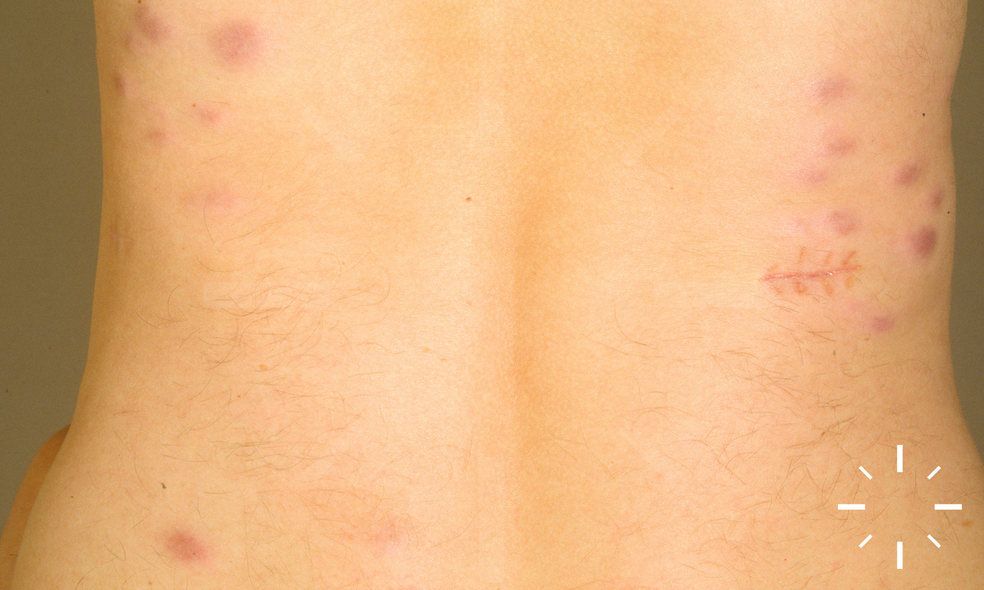
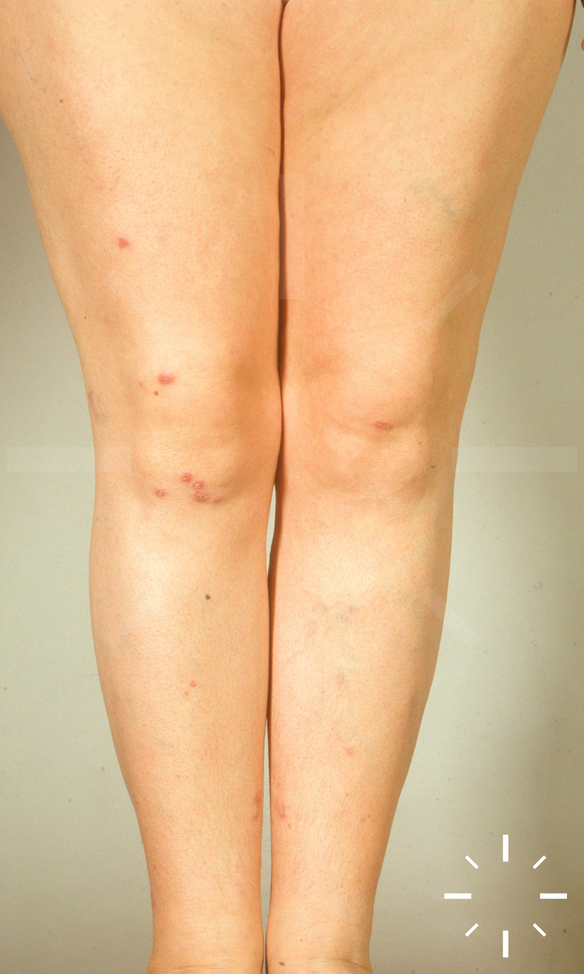
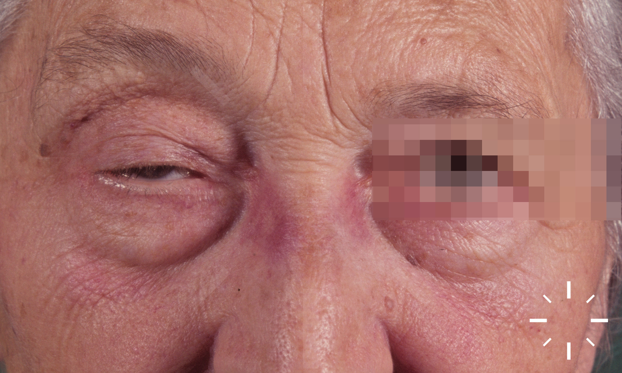
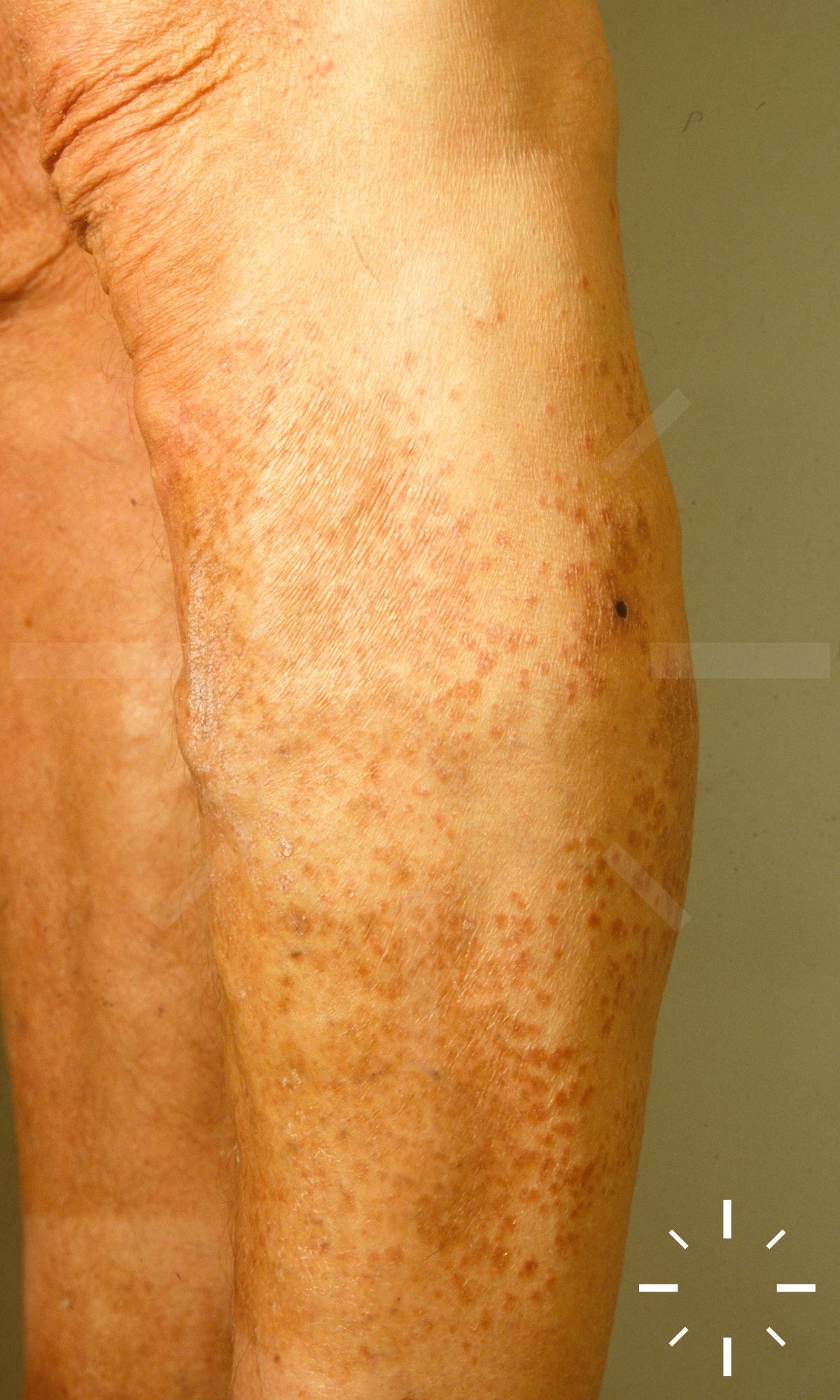
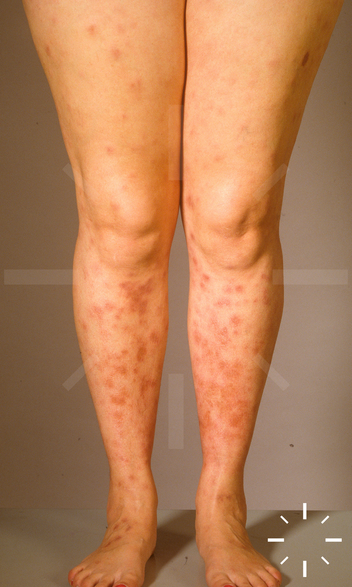
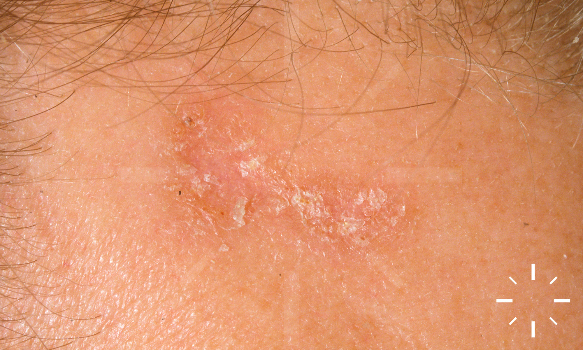
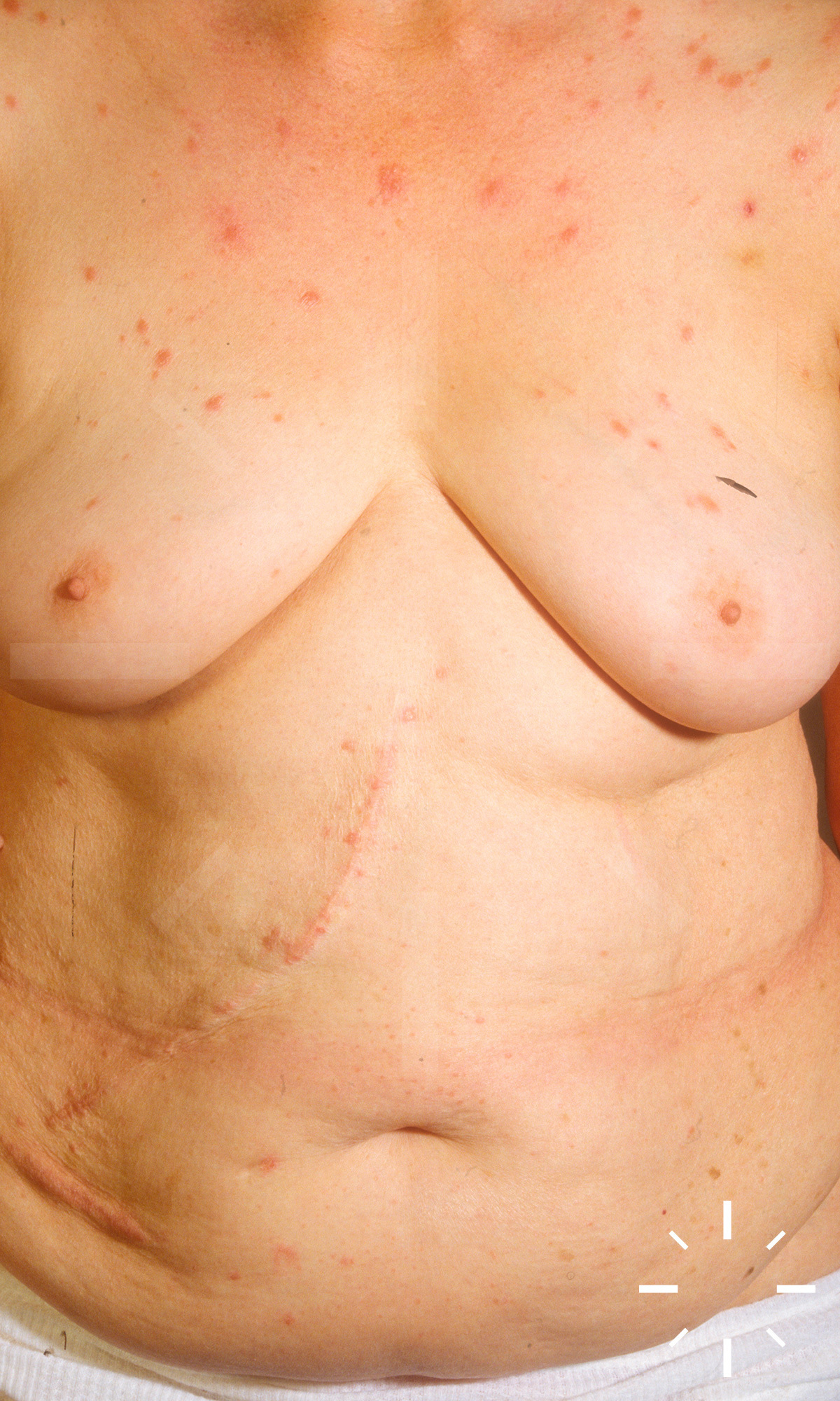
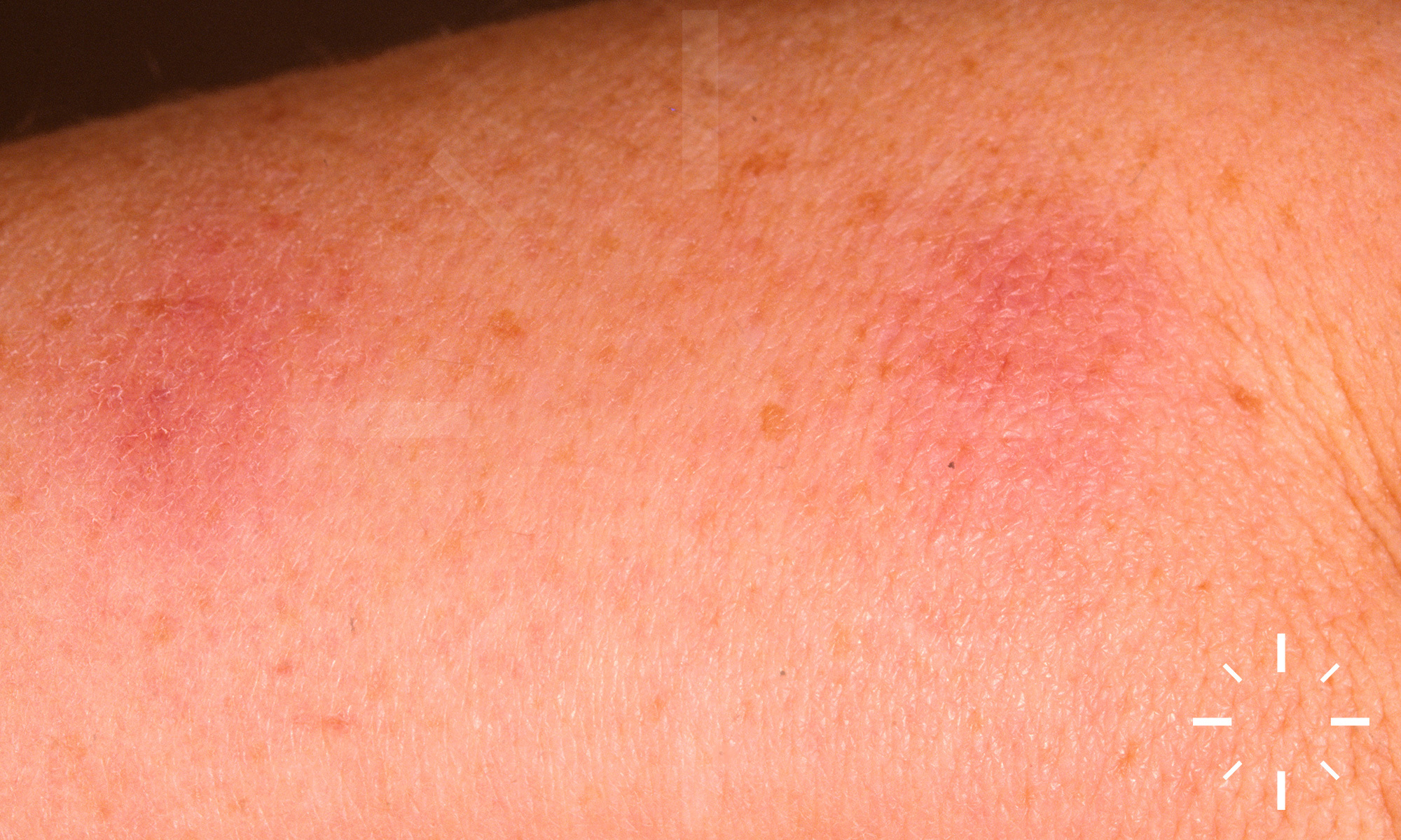
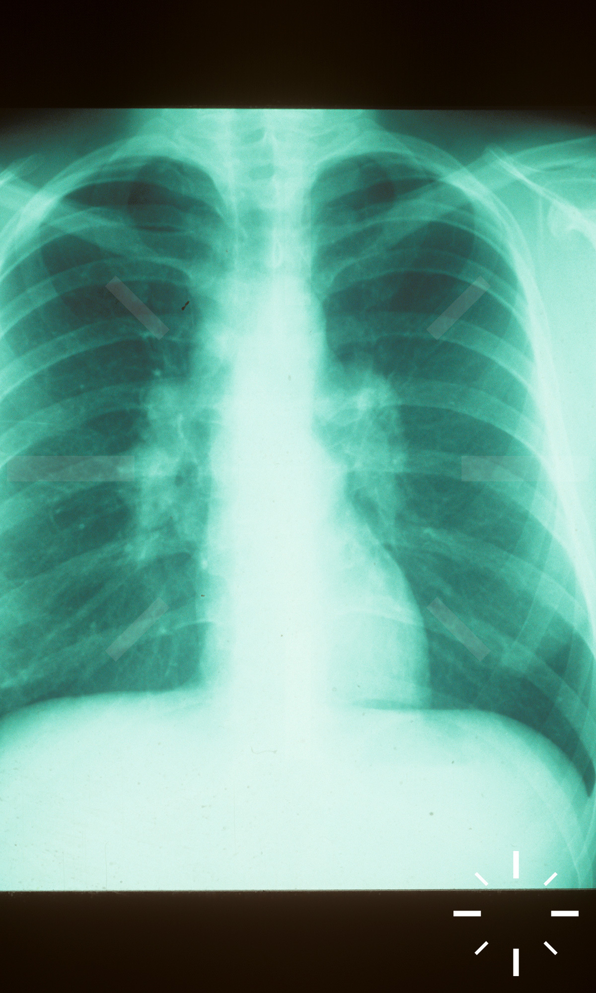
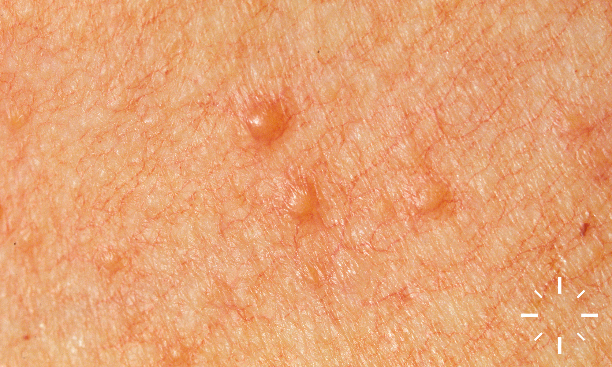
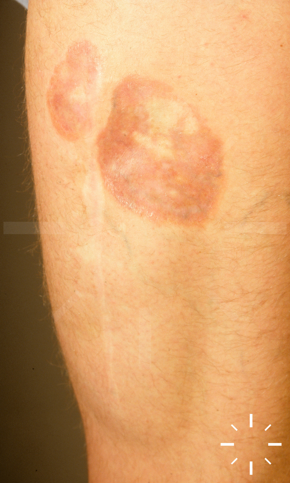
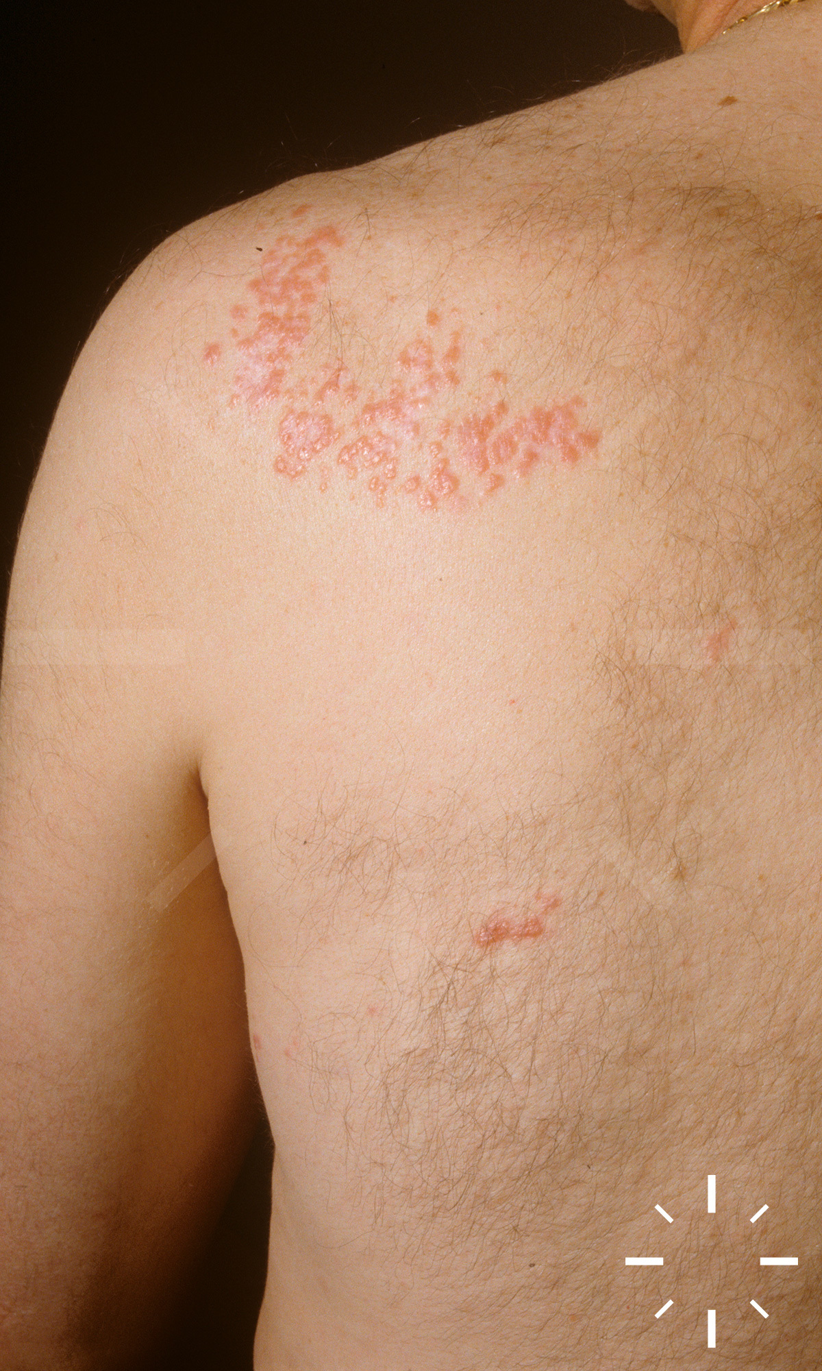
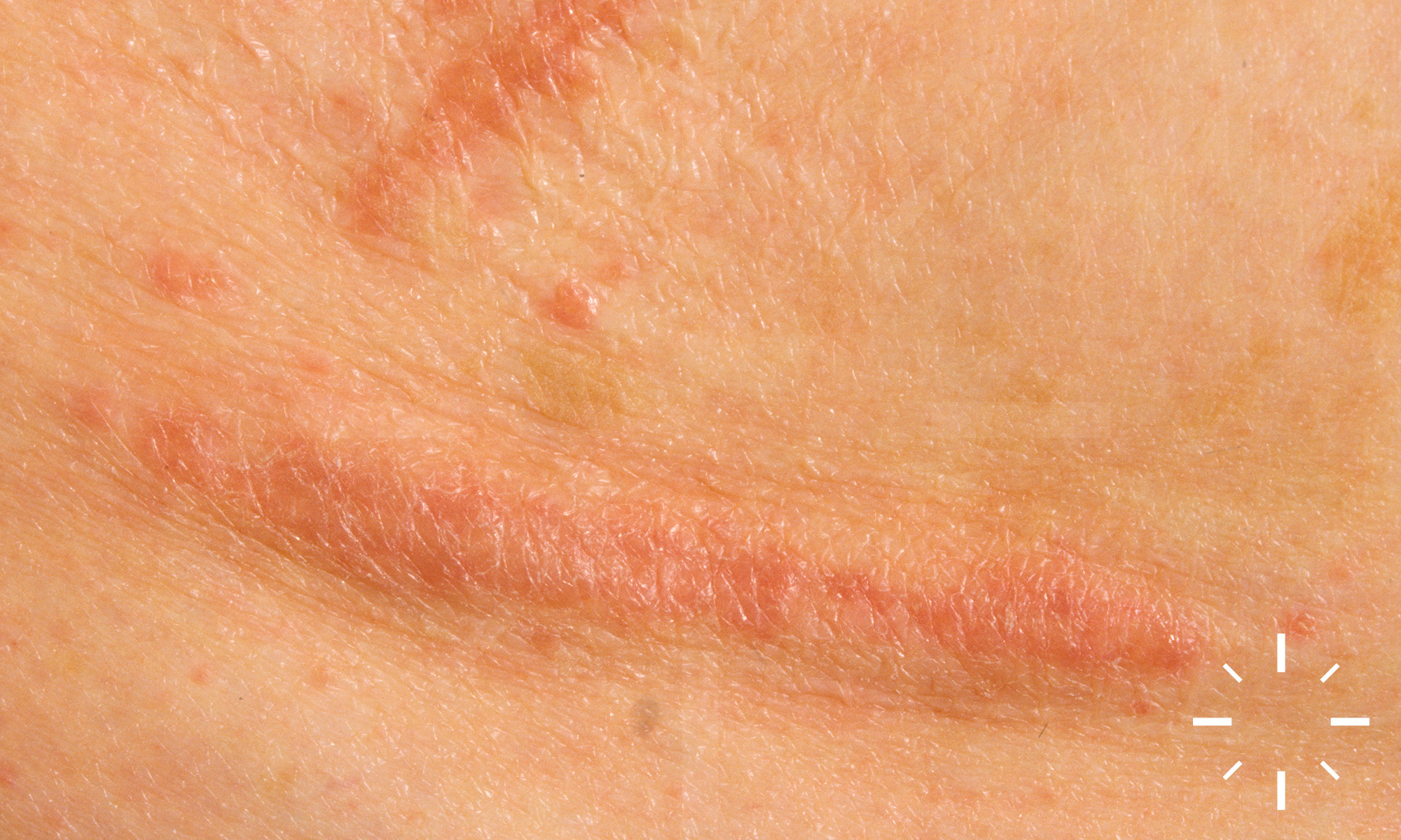
Sarcoidosis

Last Updated: 2023-07-07

Author(s): Anzengruber F., Navarini A.

ICD11: 4B20.Z

  • Jonathan Hutchinson 1875
  • Ernest Besnier 1889
  • Cäsar Boeck 1899
  • Schaumann 1916-1917

Boeck's morbus, Besnier-Boeck-Schaumann's morbus, sarcoidosis.

Granulomatous multisystem disease characterised by non-caseating epithelioid cellular granulomas.

  • Women > Men
  • North-South/gradient
    • 10-14/100'000/year (Caucasians in the USA)
    • 35-64/100'000/Jahr. (African Americans in the USA)
    • 64/100'000/year (Swedes)
    • 20/100'000/year (UK)
    • 1.4/100'000/year (Spain and Japan)

Division into acute, subacute and chronic form. According to organ involvement.

Types of skin sarcoidosis:

  • Large nodular form
    • Special form: lupus pernio
  • Small nodular disseminated form
  • Erythema nodosum
  • Anular, circular form
  • Subcutaneous nodular form
  • Scarring sarcoidosis
  • Angiolupoid sarcoidosis

  • It is an immune disease. CD4+ helper cells (Th1 subtype) are activated, leading to a shift in the CD4/CD8 ratio (↑CD4+ cells over 3.5-fold). There is ↑ production of Th1- cytokines and B-cell stimulation with consequent hypergammaglobulinaemia. This leads to an initiation of a tissue reaction
  • A genetic component is considered certain, as there have been familial clusters reported
  • Association:
    • HLA-B8, -q, -B8, -B13, DR3 and -DR5. HLA DR 11, 12, 14, 15, 17
      • Comment: very different information here depending on the source
  • In addition, polymorphisms of the gene encoding angiotensin-converting enzyme are found in collectives of sarcoidosis patients

  • Cutaneous symptoms show up in about up to 35%

Division

  • Acute form (Löfgren's syndrome)
    • 5% of cases
    • Trias:
      • Erythema nodosum (associated with benign course, often spontaneous regression after years)
      • Arthritis
      • Bihilar adenopathy
        • Stage I: Bihilary lymph node enlargement
        • Stage II: Parenchymal involvement
        • Stage III: Fibrosis
    • Familial blue syndrome --> start <5 years of age. Triad of granulomatous changes in skin, joints and eyes
  • Subacute form
  • Chronic form
    • In 95% of cases
    • Years-long course
    • Organ involvement:
      • Lung (60%)
      • Lymphatic system (20%)
      • Liver (20%)
      • Eyes (18%)
      • Parotis (5%)
      • Tear gland (Heerfordt syndrome)
      • Bones (ostitis multiplex cystoides Jüngling)
      • Joints (oligoarticular arthritis)
    • General symptoms
      • ↓ general condition
      • Fever
      • Fatigue
      • Flu-like symptoms

Skin symptoms

  • Large nodular shape:
    • Mostly on the face and extremities, there are reddish, bluish, sometimes brownish, derb palpable nodules and plaques with telangectasias. In some cases, central regression is possible
  • Small nodular form:
    • Erythematous, sometimes bluish papules and nodules are visible, especially on the face and extensor sides of the extremities
  • Anular or circular form:
    • Polymorphous, partly roundish/ring-shaped/annular or circine erythematous to brownish plaques and nodules with central atrophic regression
  • Subcutaneous nodular form:
    • Subcutaneously localised, indolent, nodular space-occupying lesions under mostly inconspicuous, in some cases slightly livid discoloured skin

  • Clinical image
  • Diascopy ("apple jelly" colour of infiltrates on pressure)
  • Biopsy
  • Chest X-ray
  • Pulmonary function
  • X-ray of the hands
  • ECG
  • Laboratory
    • BSG ↑
    • Angiotensin converting enzyme (ACE) ↑ in about 75% of non-treated patients. but poorly specific and sensitive, role as a progression parameter unclear.
    • Soluble interleukin-2 receptor (sIL2R) ↑ in up to 80%
      • Lab parameter with the highest sensitivity. Possible marker for extrapulmonary involvement.
    • ↑ Levels may also occur in other T-cell mediated diseasesAdenosine deaminase and serum amyloid A ↑ (low specificity/sensitivity)
    • Procollagen III peptide (marker for pulmonary fibrosis)
    • Calcium ↑ in urine (30-60%), hypercalcaemia (15%)
    • Eosinophilia (3%)
    • Leukopenia (5-10%), but hypergammaglobulinaemia (30-80%)
    • Lymphocytopenia
    • Antinuclear antibodies are elevated in 1/3 of patients - do not be confused, does not argue against sarcoidosis.
  • Upper abdominal sonography
  • If necessary, liver biopsy
  • Neck sonography
  • Ophthalmological consultation
    • Ruling out involvement with visual field impairment
  • When pulmonary involvement is suspected
  • Bronchoscopy with bronchoalveolar lavage (T4/T8 quotient ↑)
  • Biopsy from mediastinal or pulmonary lymph node
  • Mediastinoscopy with lymph node extirpation
  • Tbc exclusion
  • When cardiac involvement is suspected
    • Echocardiography
    • Long-term ECG
    • Cardiac MRI
    • Cardiac scintigraphy
  • In cases of suspected neurosarcoidosis
    • Liquor puncture & cranial MRI

Mostly where ↓ blood flow e.g.:

  • Front, nose, cheeks, earlobes, as well as the extensor sides of the extremities (flexor sides are not affected)
  • Scars

  • Spontaneous healing frequent in over 50%
  • Lung involvement prognostic

  1. Judson M. Faculty of 1000 evaluation for Methotrexate is steroid sparing in acute sarcoidosis: results of a double blind, randomized trial.  F1000 - Post-publication peer review of the biomedical literature: Faculty of 1000, Ltd.
  2. Martinetti M, Tinelli C, Kolek V, et al. "The sarcoidosis map": a joint survey of clinical and immunogenetic findings in two European countries. Am J Respir Crit Care Med 1995;152:557-64.
  3. Newman LS, Rose CS, Maier LA. Sarcoidosis. New England Journal of Medicine 1997;336:1224-34.
  4. Georgiou AMEPS. Cutaneous Sarcoidosis: Complete Remission after Oral Isotretinoin Therapy. Acta Dermato-Venereologica 1998;78:457-9.
  5. Voelter-Mahlknecht S. Treatment of Subcutaneous Sarcoidosis With Allopurinol. Archives of Dermatology 1999;135:1560-1.
  6. English JC, Patel PJ, Greer KE. Sarcoidosis. Journal of the American Academy of Dermatology 2001;44:725-46.
  7. Yanardag H, Pamuk mN, Pamuk GlmE. Lupus Pernio in Sarcoidosis. JCR: Journal of Clinical Rheumatology 2003;9:72-6.
  8. Marcoval J, Moreno A, Mana J. Foreign bodies in cutaneous sarcoidosis. J Cutan Pathol 2004;31:516-.
  9. Heffernan MP, Smith DI. Adalimumab for Treatment of Cutaneous Sarcoidosis. Arch Dermatol 2006;142.
  10. Iannuzzi MC, Rybicki BA, Teirstein AS. Sarcoidosis. New England Journal of Medicine 2007;357:2153-65.
  11. Marcoval J, Mañá J, Rubio M. Specific cutaneous lesions in patients with systemic sarcoidosis: relationship to severity and chronicity of disease. Clinical and Experimental Dermatology 2011;36:739-44.
  12. Rkiouak A, Zinebi A, Rabhi M, et al. [Pulmonary nocardiosis and sarcoidosis]. Rev Pneumol Clin 2013;69:139-43.
  13. Zuk J, Bazan-Socha S, Zarychta J, et al. Disseminated nocardiosis mimicking exacerbation of pulmonary sarcoidosis. Sarcoidosis Vasc Diffuse Lung Dis 2013;30:65-9.
  14. Rajagopala S, Dangeti G. Disseminated nocardiosis in a patient with sarcoidosis. Indian J Med Res 2016;143:118-9.
  15. Lebwohl, Mark. Treatment of Skin Disease: Comprehensive Therapeutic Strategies. Elsevier, 2014. Print.