Scarlet fever

Last Updated: 2023-07-07

Author(s): Anzengruber F., Navarini A.

ICD11: 1B50

Sydenham, 1676.

Scarlet fever, Scarlatina, Streptococcal sore throat with rash, Canker rash.

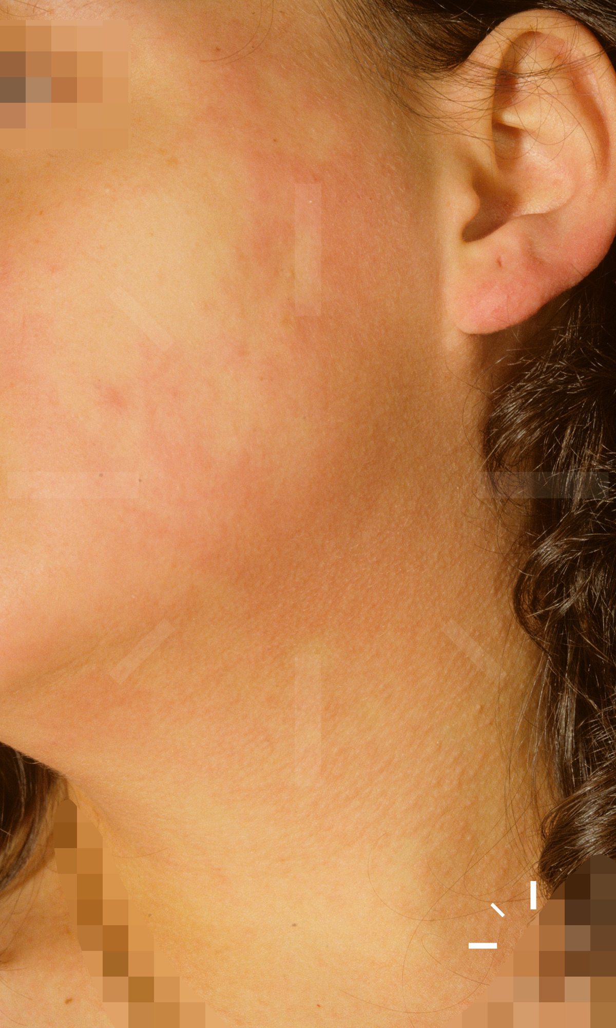
Notifiable acute infection with group A β-haemolytic streptococci leading to angina, general symptoms and macular exanthema

The age peak is between 3-15 years of age

.

  • The disease is caused by group A β-haemolytic streptococci (over 60 serotypes are counted in total). Scarlet fever usually occurs in connection with angina. However, other diseases with group A streptococci can also be causative
  • Transmission occurs through droplet infection, contact or food and is favoured by cold weather. Ports of entry include the nasopharynx and wounds
  • The streptococci produce erythrotoxin (A, B, C), which has superantigenic activity and stimulates proinflammatory cytokines
  • Incubation period: 2-5 days

  • Reduced general condition with febrile temperature, headache, sore throat, vomiting and reddened throat ring (pharyngotonsillitis) with locoregional lymphadenopathy
  • Exanthema with pin-sized follicularly bound papules and small vesicles (Miliaria scarlatinosa) may occur, possibly with a time delay. In some cases, however, there is no cutaneous manifestation. Mostly initially inguinal, on the triangle of the thighs or the crooks of the arms. In most cases, there is generalisation (including the palms and feet), usually with exclusion of the face
  • Dot-like erythematous patches are seen on the dorsum of the hands and feet. If the eruption is severe, both cheeks are reddened. Facies scarlatinosa is the name given to the characteristic perioral area free of skin findings.
  • The "raspberry tongue" is typical. A macular enanthema occurs in the area of the palatinum molle
  • Subicterus is observed in some cases. The capillaries show increased tearability during the disease (see Rumpel-Leede test)

  • Clinic
  • BB (leukocytosis, in the course eosinophilia)
  • BSG, CRP elevated
  • Bact. smear of the pharynx for pathogen detection
  • ASL titre (elevation after approx. 1-2 weeks after infection)
  • A white dermographism occurring after 20 seconds is also typical
  • In the past, the Dick test was performed by injecting 0.1 ml Dick toxin (erythrogenic streptococcal toxin) i.c.. If there is no reddening of the skin >1 cm after up to 20 hours, the existence of antibodies against erythrogenic toxins can be assumed. In this case, the patient is immune to scarlet fever
  • The effacement phenomenon occurs up to 20 hours after intracutaneous injection of 0.2 ml scarlet convalescent serum or 0.3 ml antistreptococcal serum as pallor. Provided that a recess of the exanthema is visible, scarlet fever can be assumed. If the test is negative, other triggers of the exanthema must be considered
  • In the Rumpel-Leede test, a blood pressure cuff is inflated to 50 mmHg and left for at least 5 minutes. Provided that more than 5 petechiae are found within 5 cm of the cuff, it is considered positive. In scarlet fever sufferers, this test is often positive.

  • Hyperpyrexia
  • Disturbance of consciousness
  • Convulsions
  • Purpura
  • Sepsis
  • Otitis media
  • Sinusitis
  • Myocarditis
  • Acute glomerulonephritis
  • Polyarthritis
  • Pneumonia
  • Pericarditis
  • Meningitis
  • Hepatitis
  • Glomerulonephritis
  • Rheumatic fever
  • PANDAS-Syndrome
  • Necrotising fasciitis

  • Lethality: 0.5%
  • After experiencing scarlet fever, immunity is lifelong. However, there are 3 different variants of the erythrotoxin, so immunity after scarlet fever only exists against one variant

  1. Schlievert, P.M., Staphylococcal scarlet fever: role of pyrogenic exotoxins. Infect Immun, 1981. 31(2): p. 732-6.
  2. Silva-Costa, C., et al., Scarlet fever is caused by a limited number of Streptococcus pyogenes lineages and is associated with the exotoxin genes ssa, speA and speC. Pediatr Infect Dis J, 2014. 33(3): p. 306-10.
  3. Wang, L.Y. and T.H. Young, Hepatitis, gallbladder hydrops, splenomegaly, and ascites in a child with scarlet fever. Pediatr Emerg Care, 2012. 28(11): p. 1215-7.
  4. Paul, S.P. and P.A. Heaton, At a glance: scarlet fever in children. J Fam Health Care, 2014. 24(3): p. 25-7.
  5. Wessels, M.R., Pharyngitis and Scarlet Fever, in Streptococcus pyogenes: Basic Biology to Clinical Manifestations, J.J. Ferretti, D.L. Stevens, and V.A. Fischetti, Editors. 2016: Oklahoma City (OK).
  6. [Scarlet fever and other infections from Streptococcus pyogenes]. Med Monatsschr Pharm, 2001. 24(3): p. 83-6.