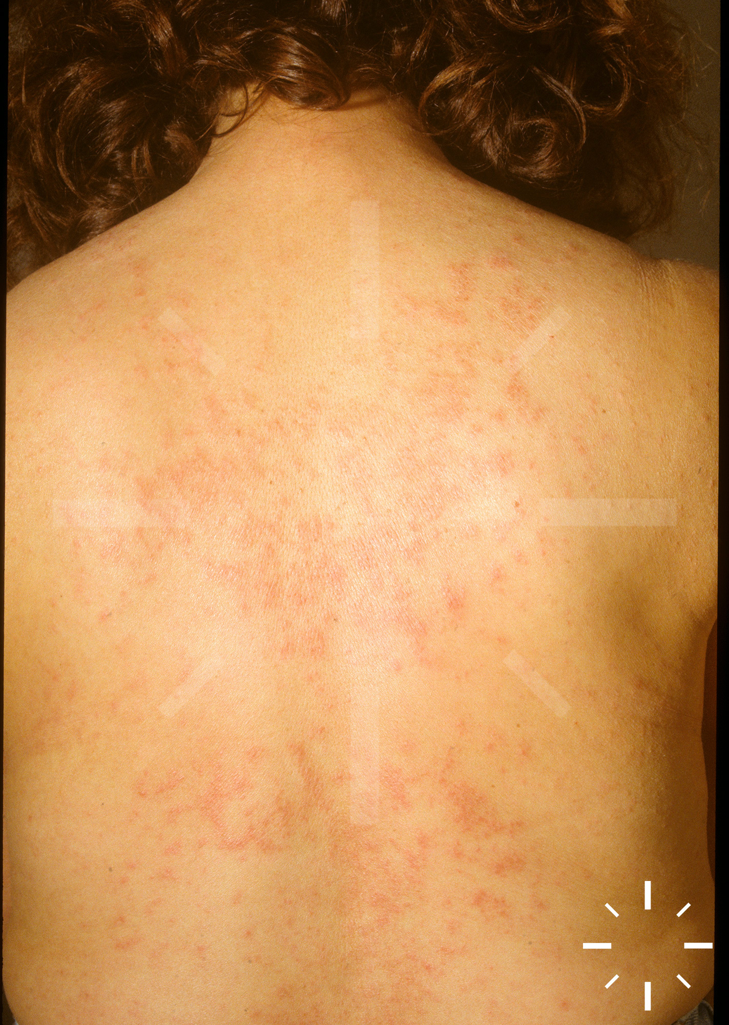
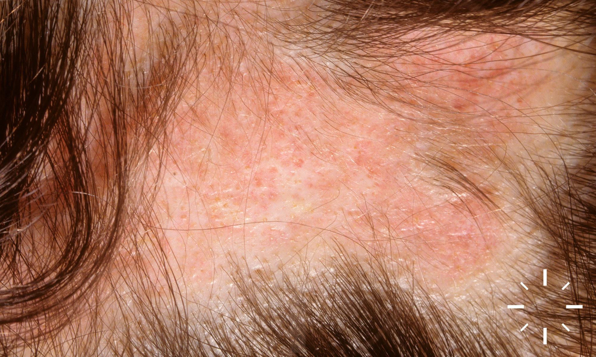
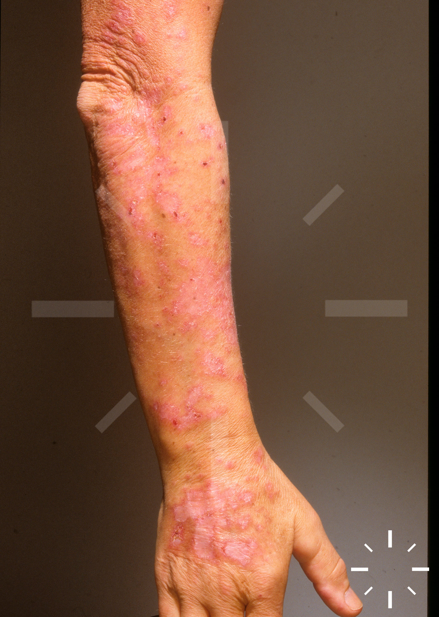
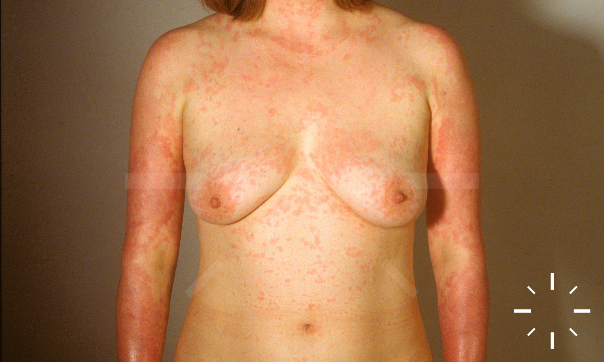
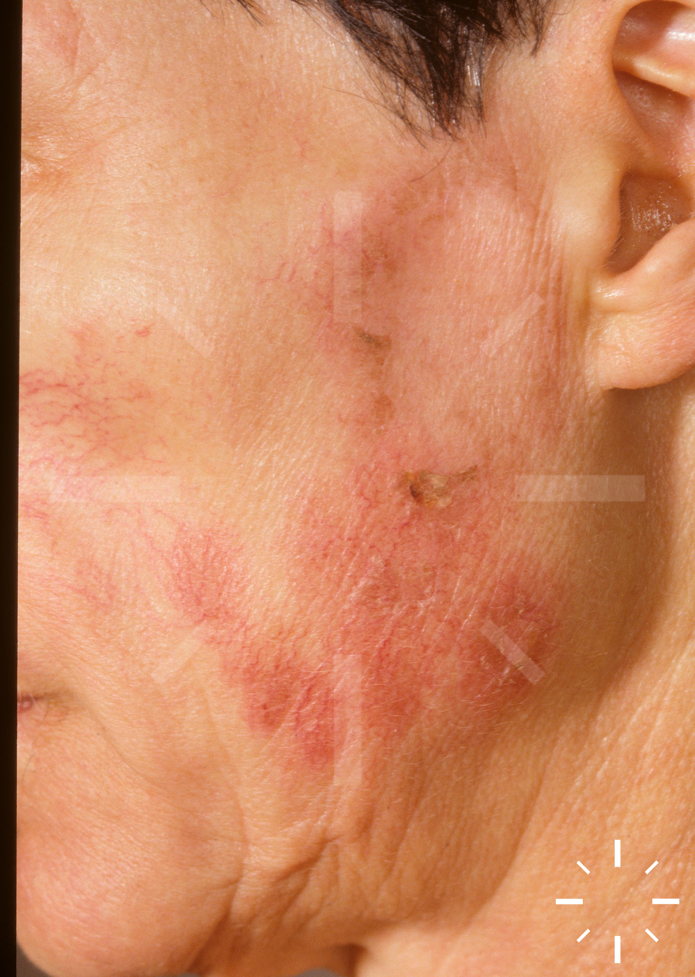
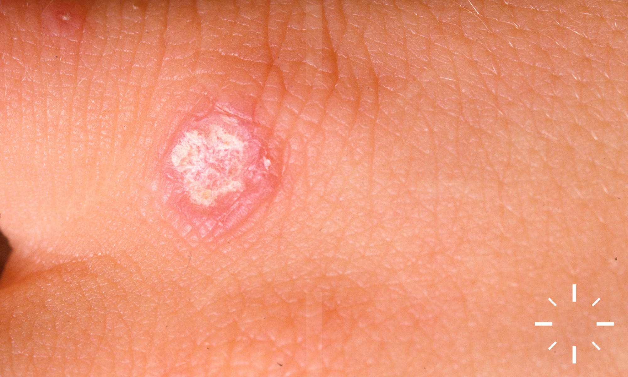
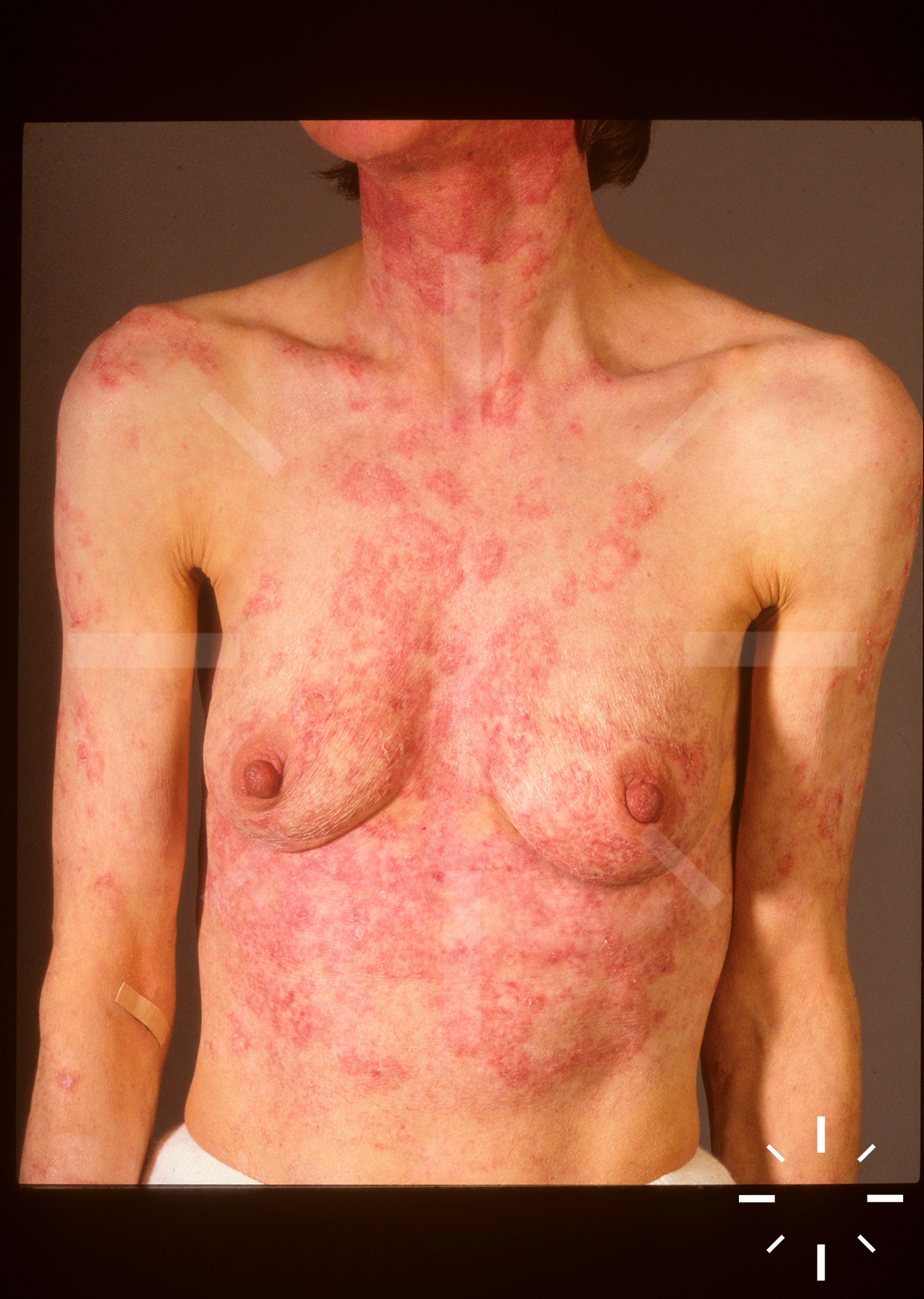
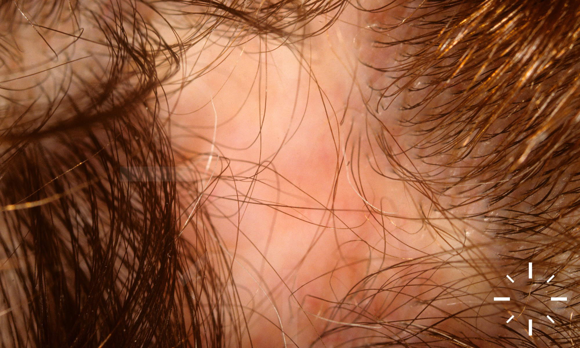
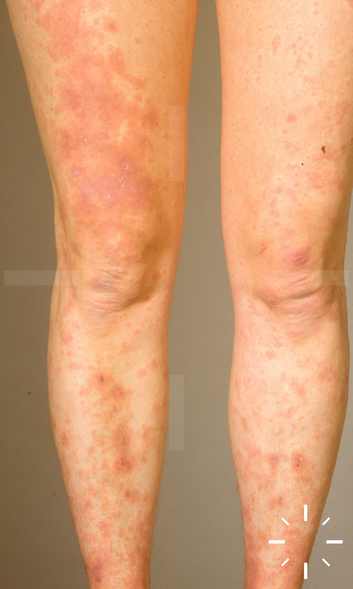
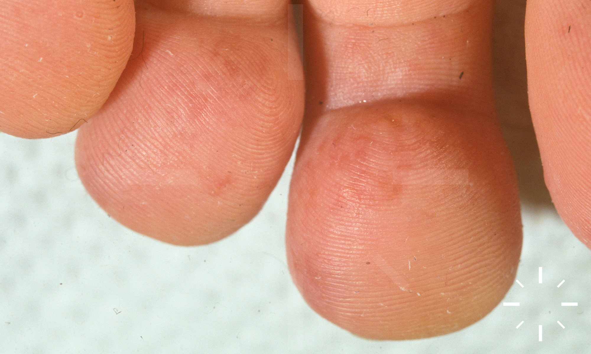
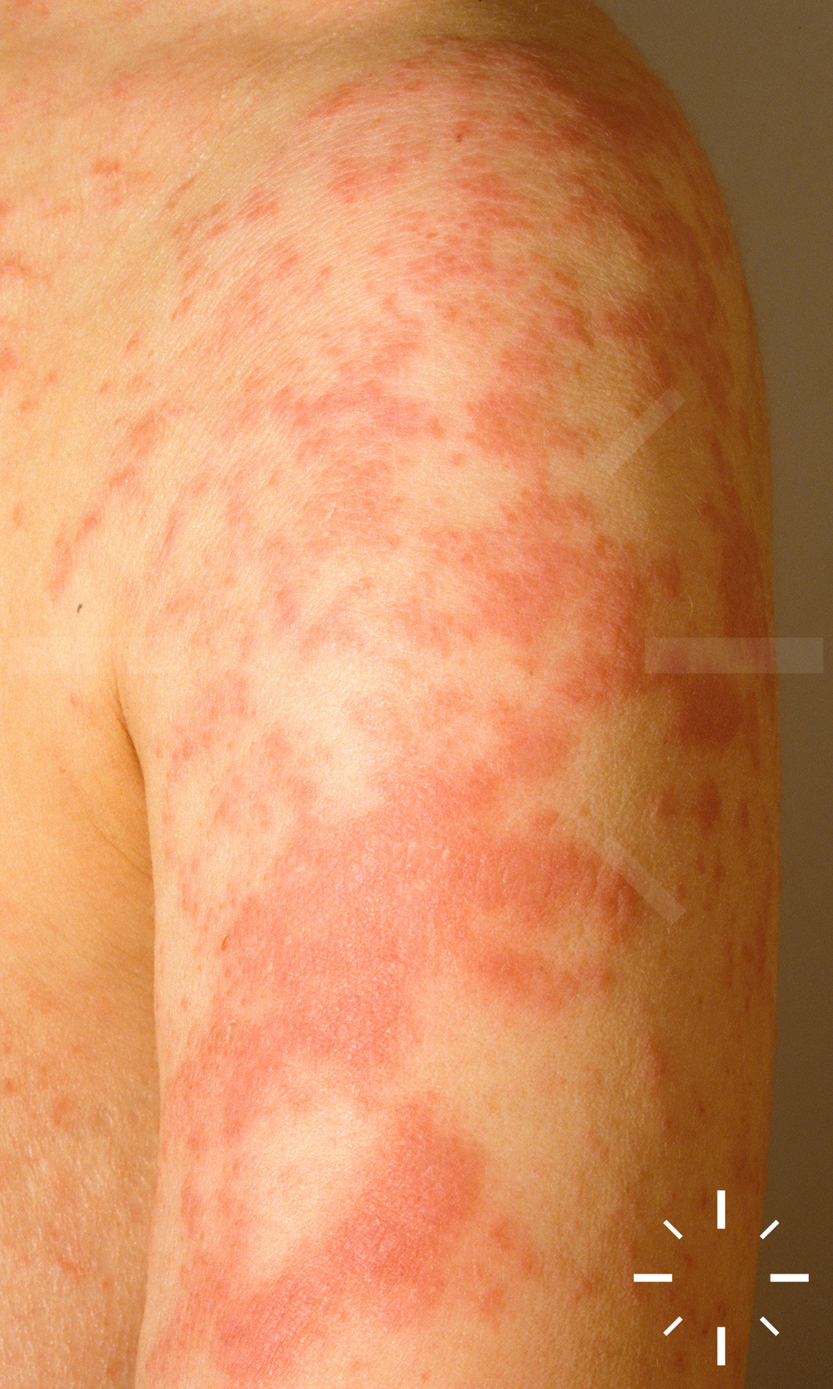
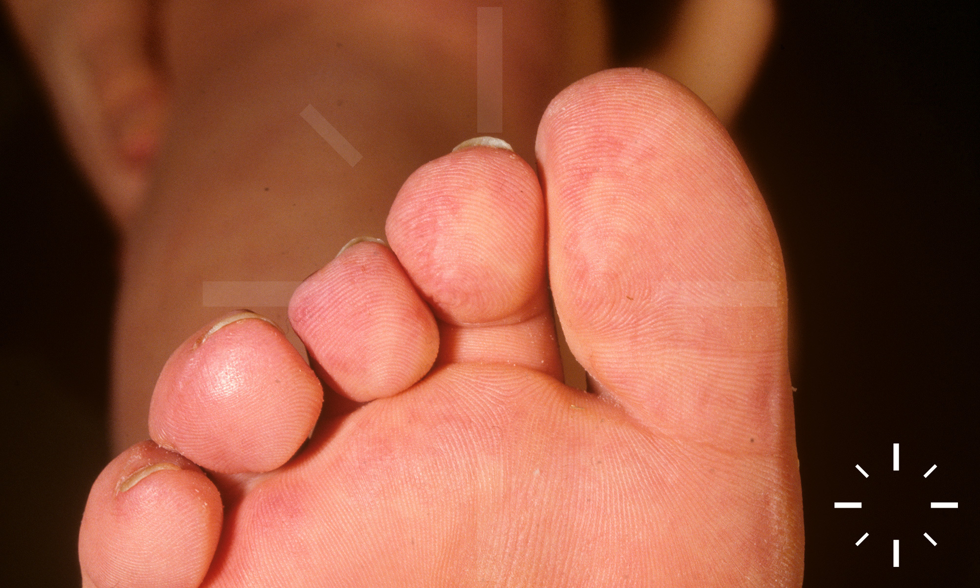
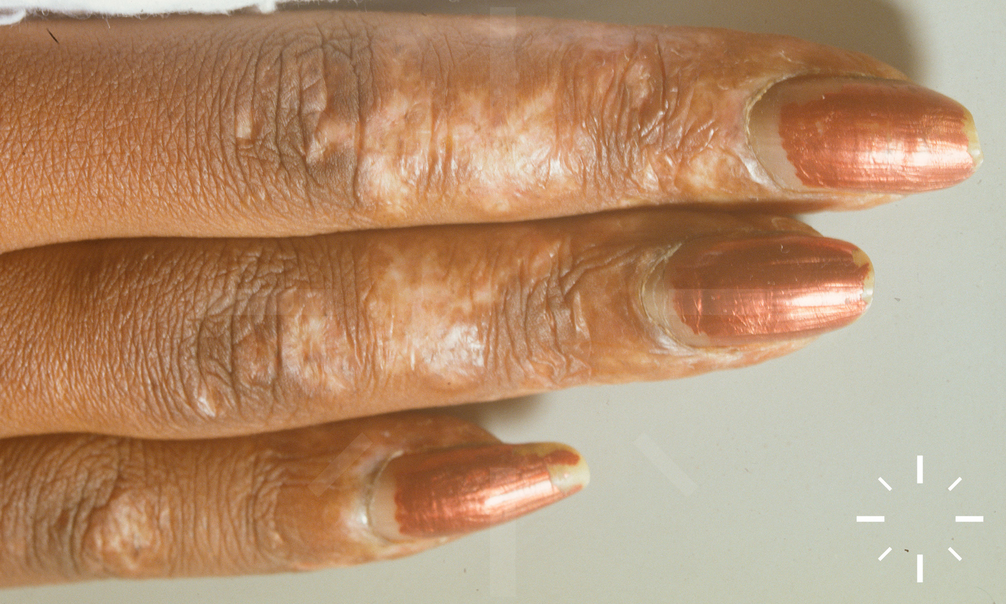
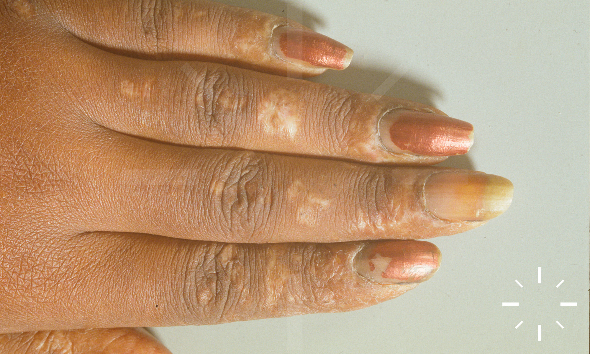
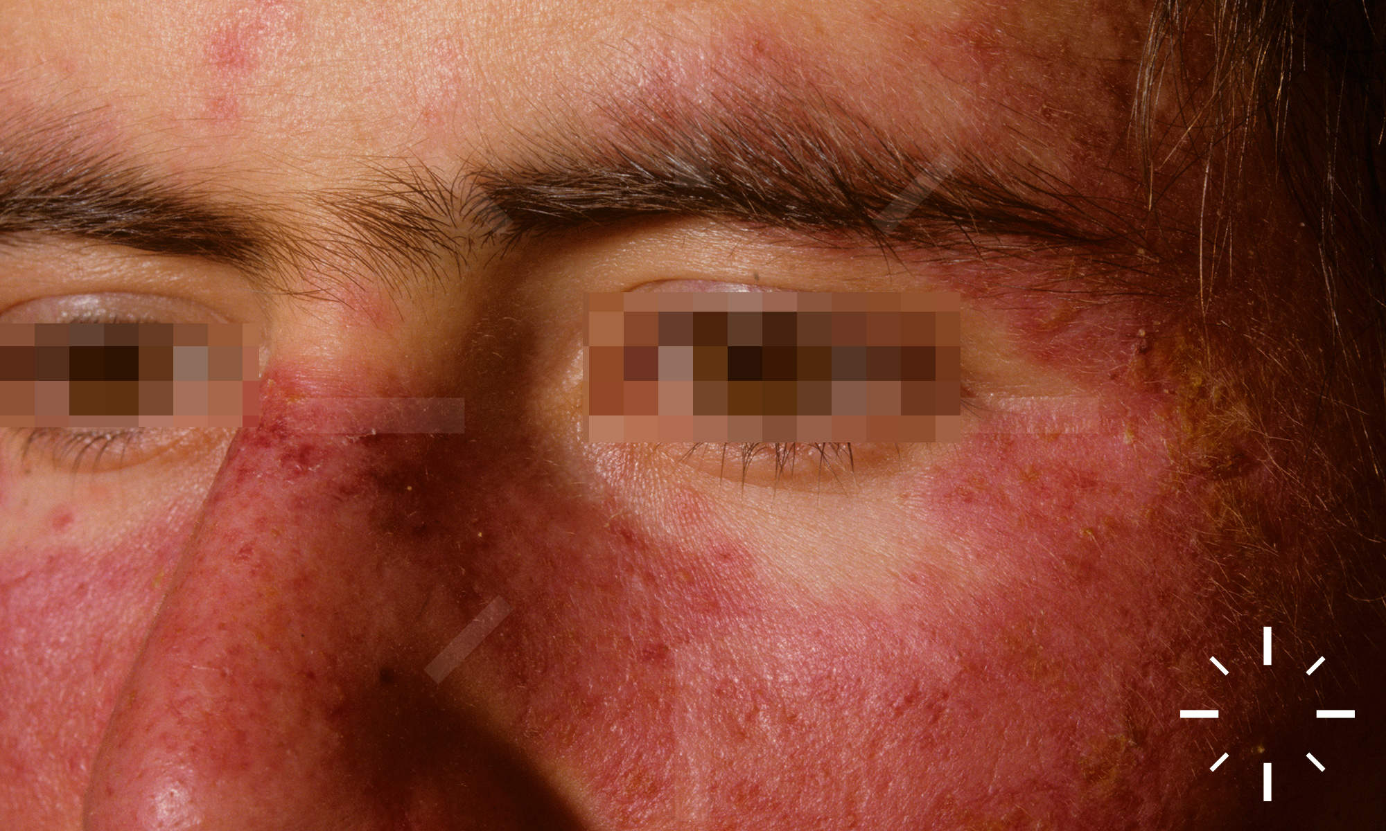
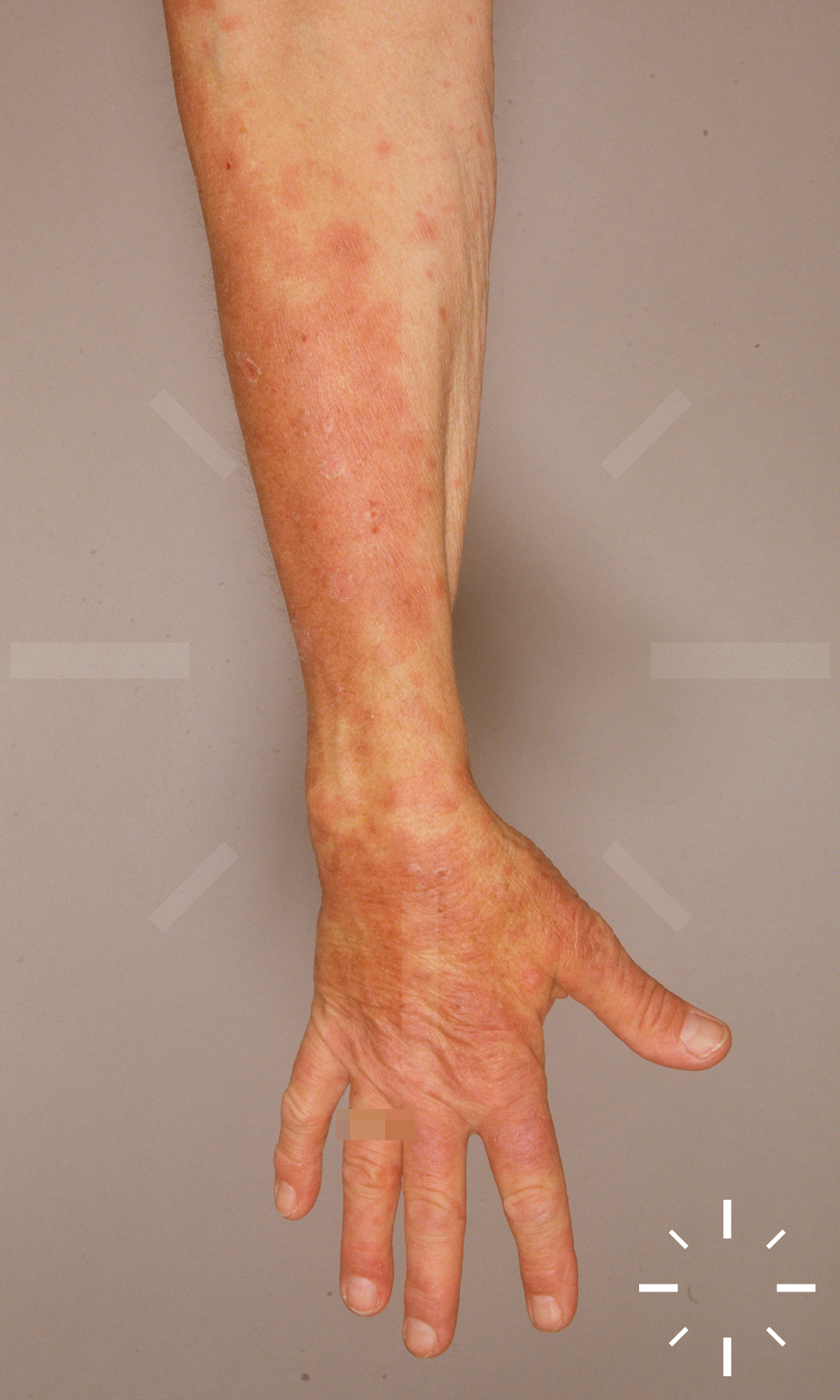
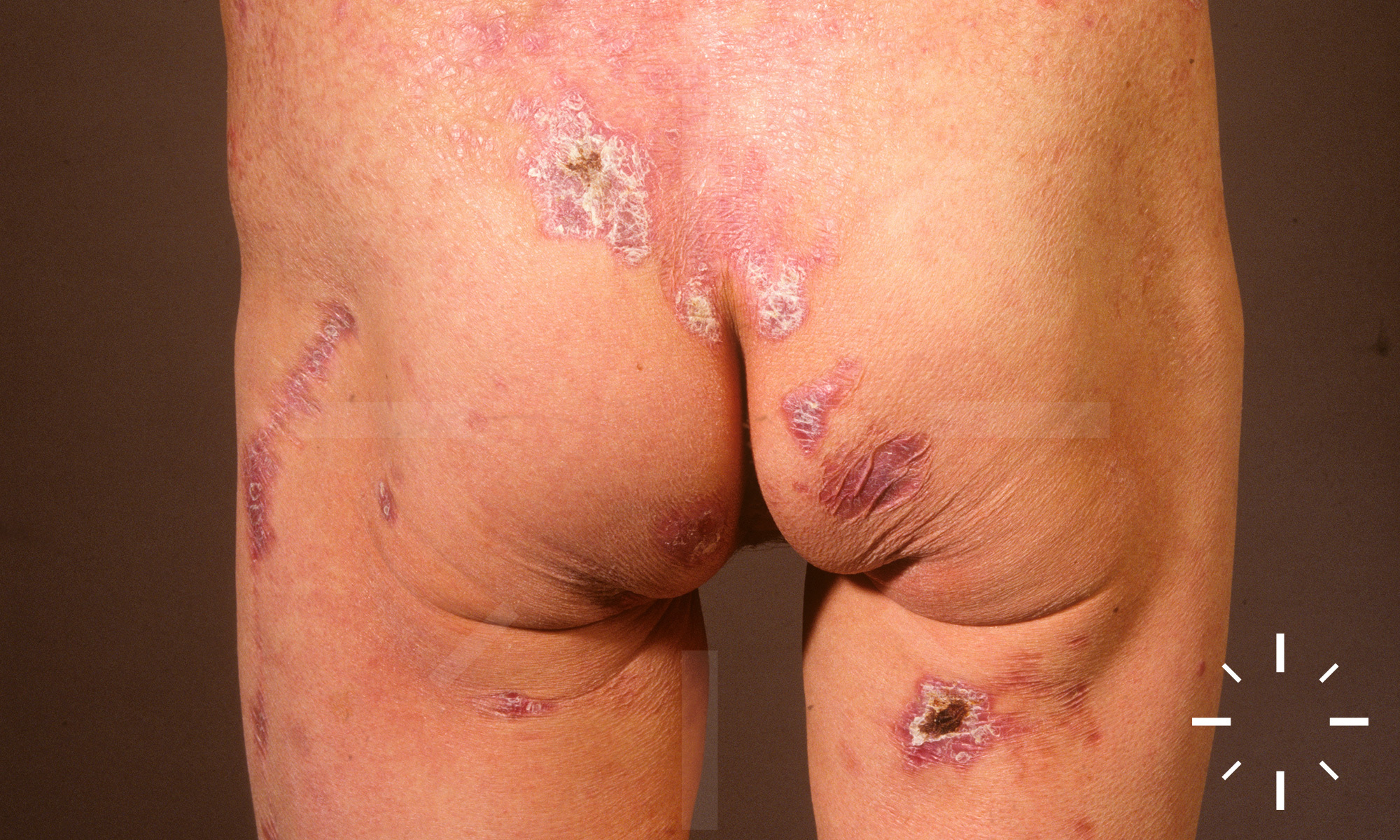
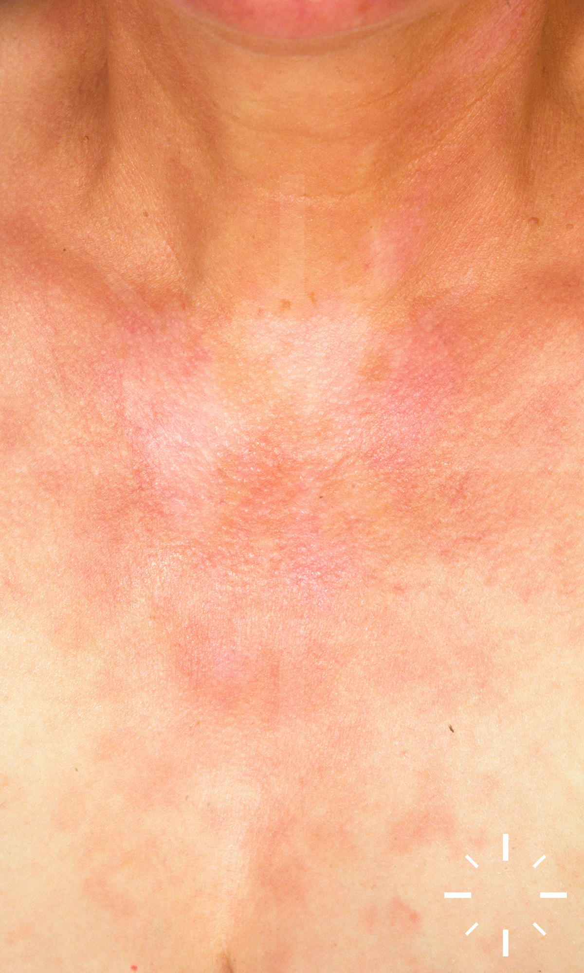
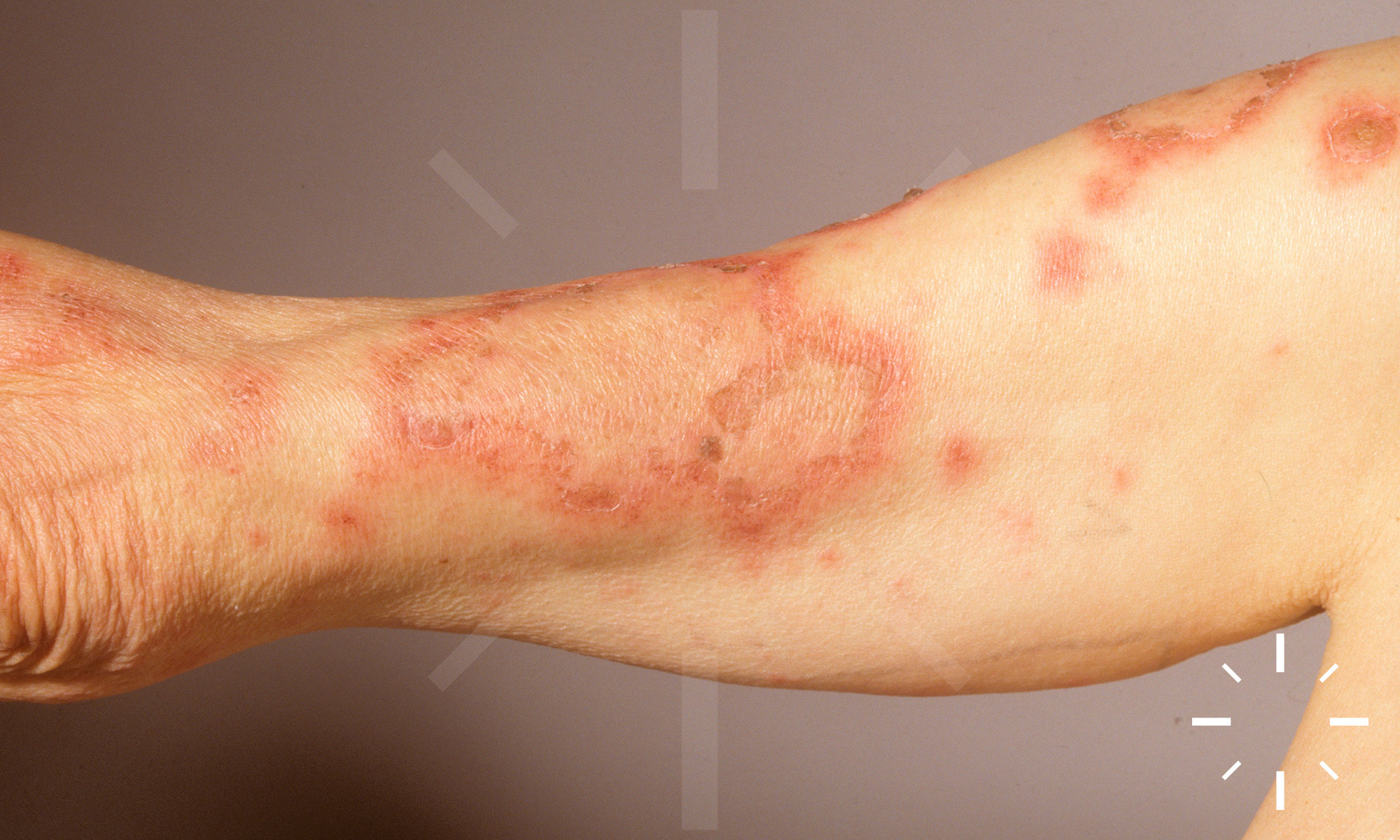
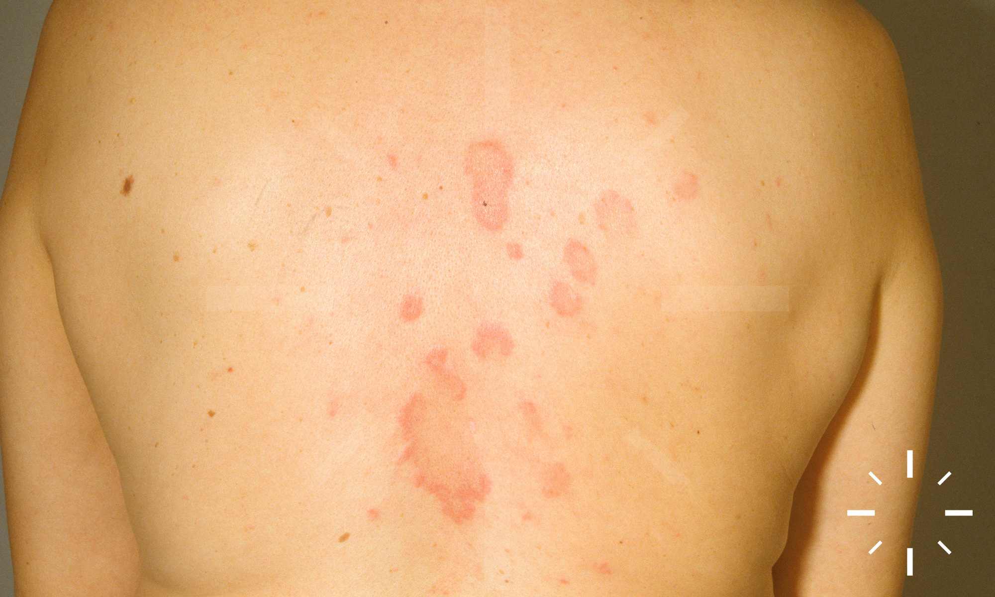
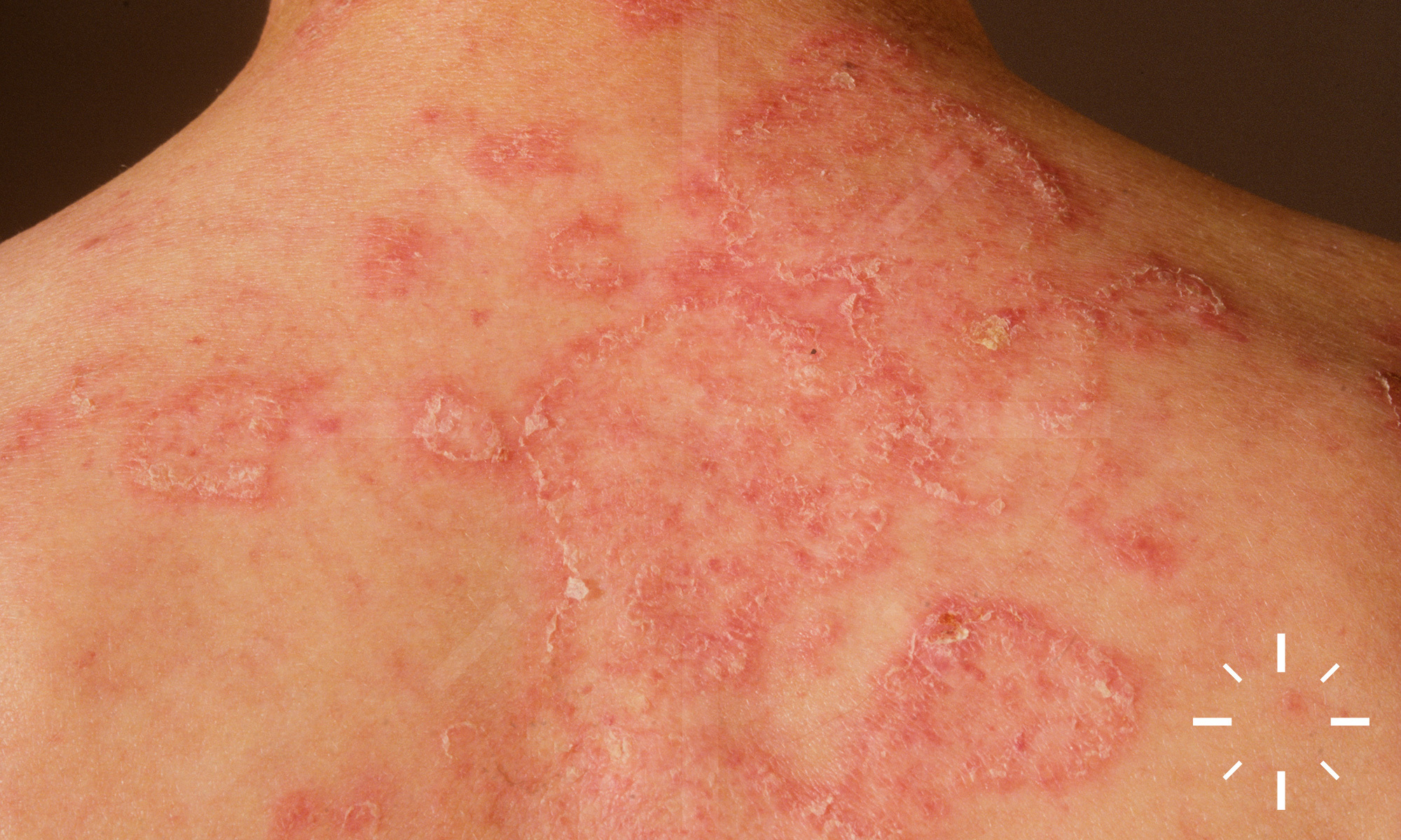
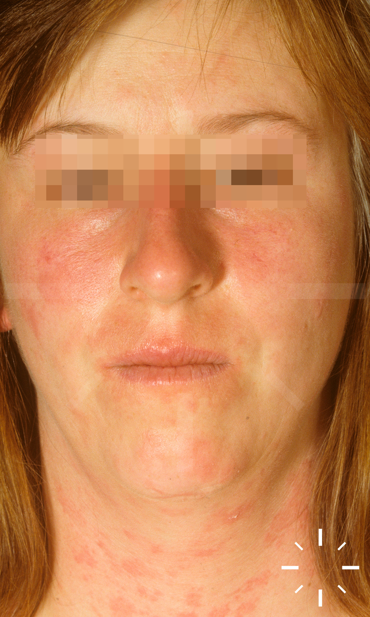
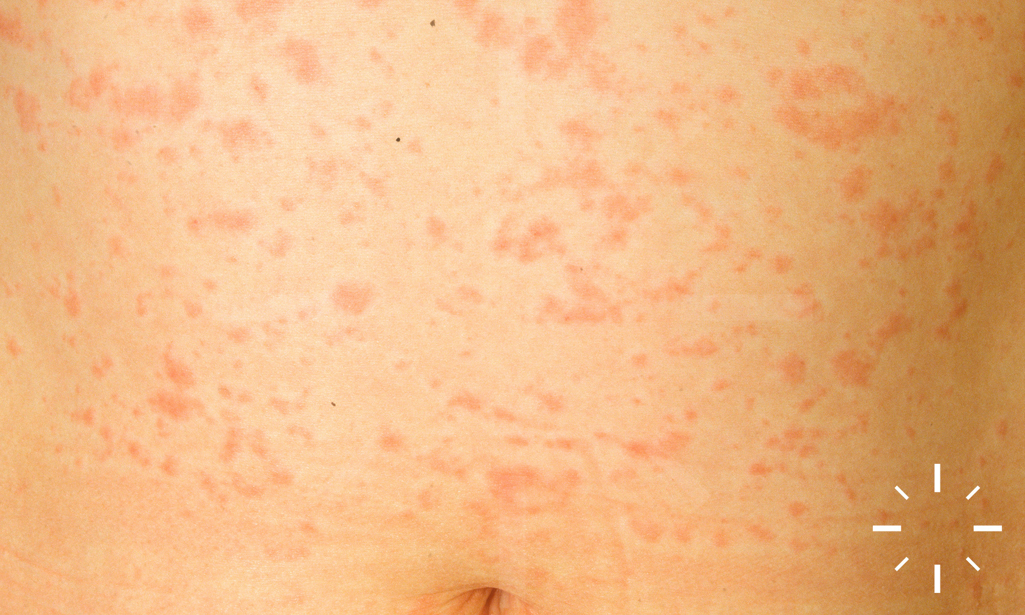
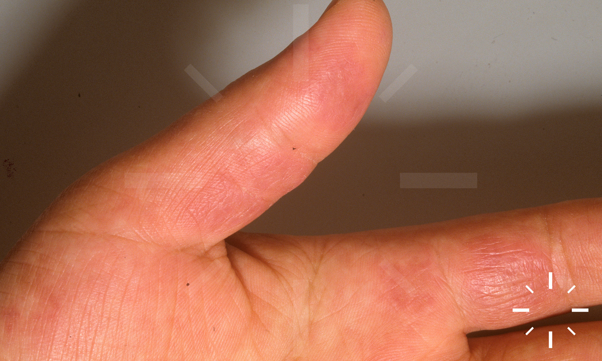
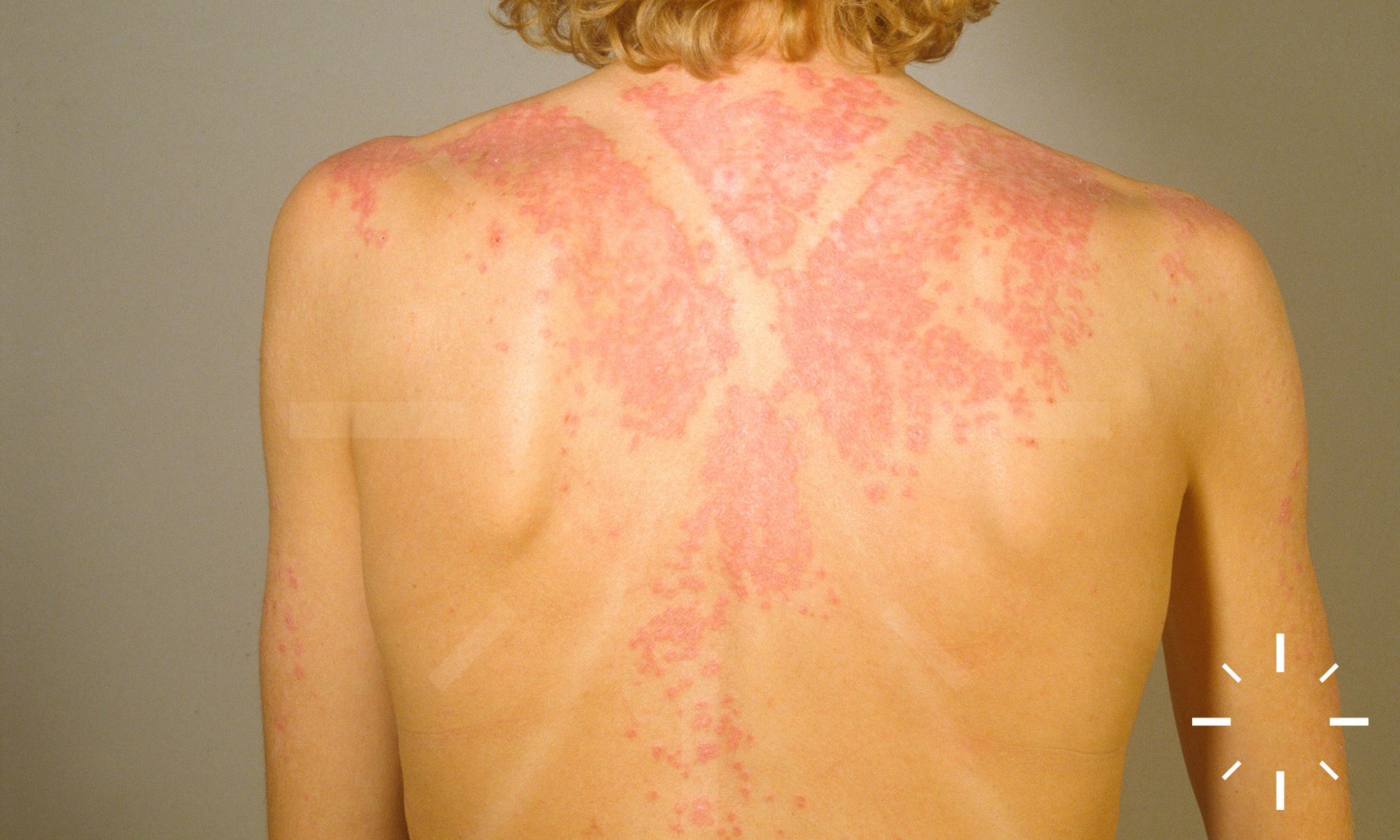
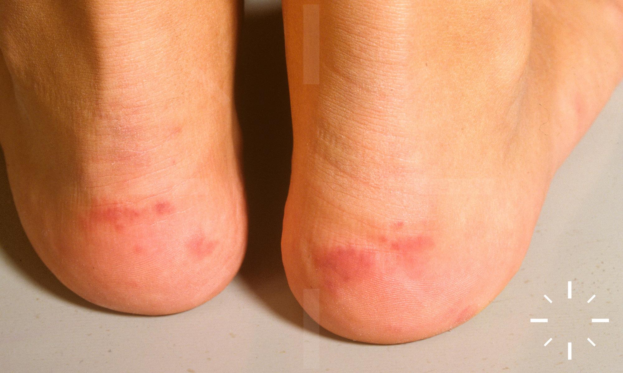
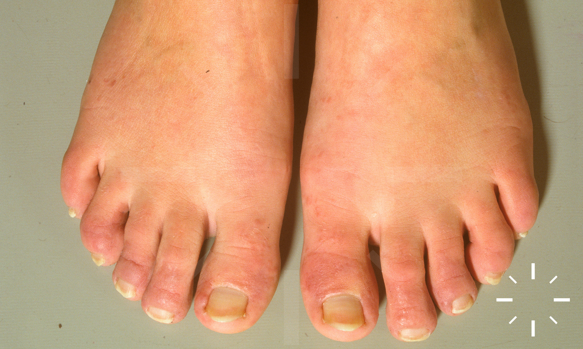
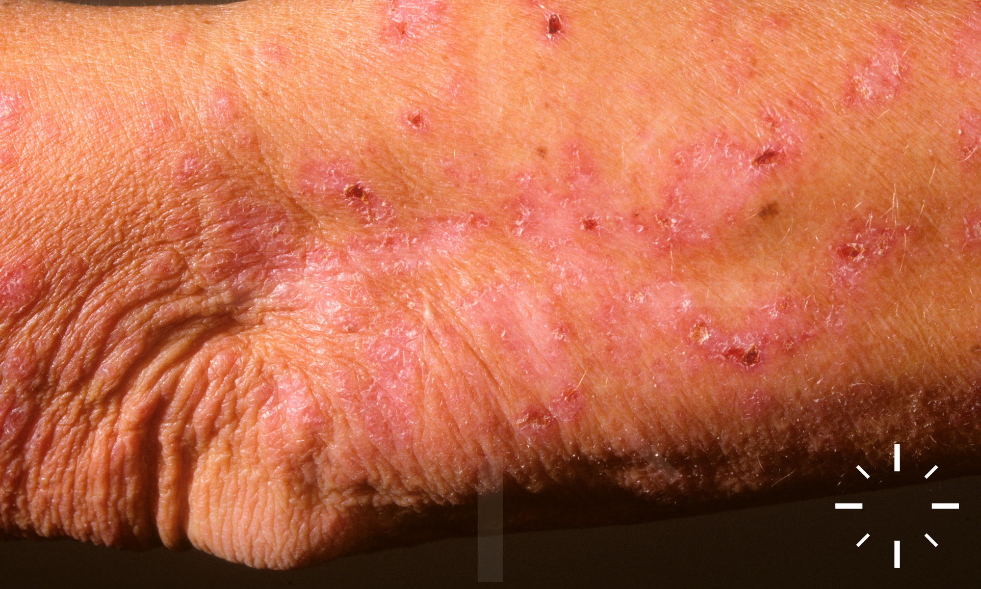
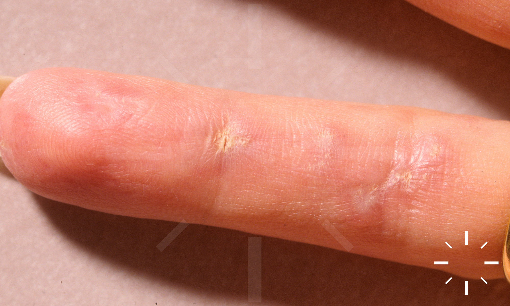
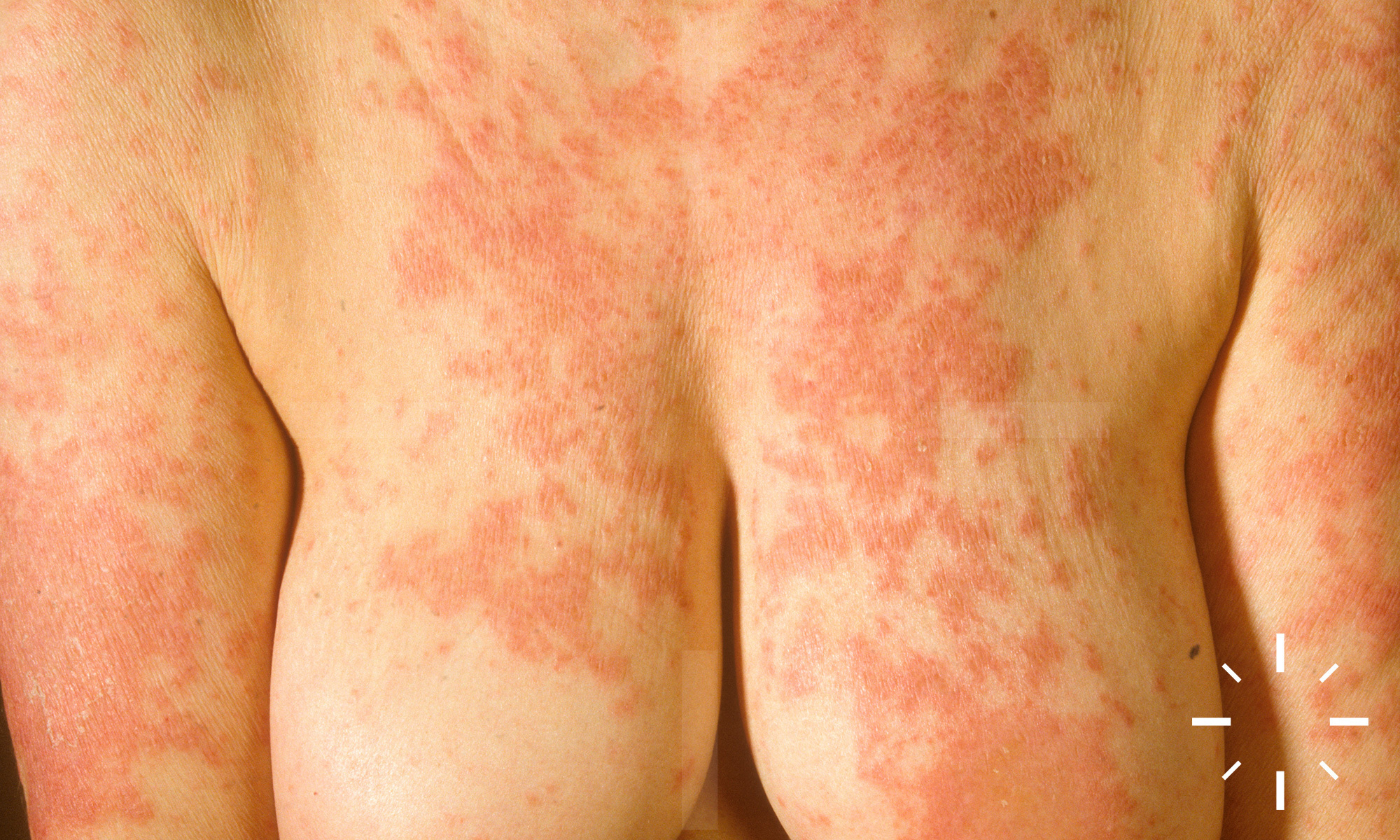
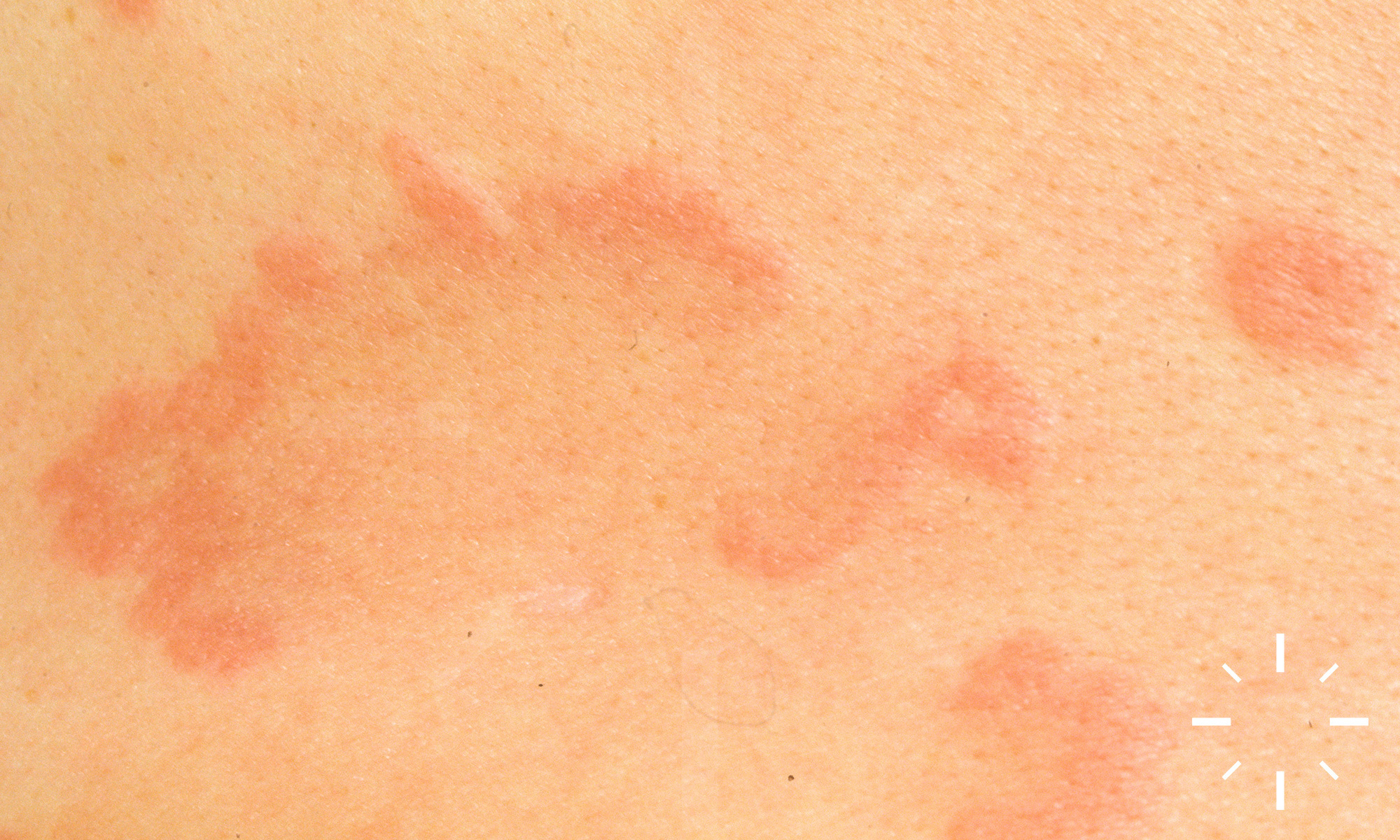
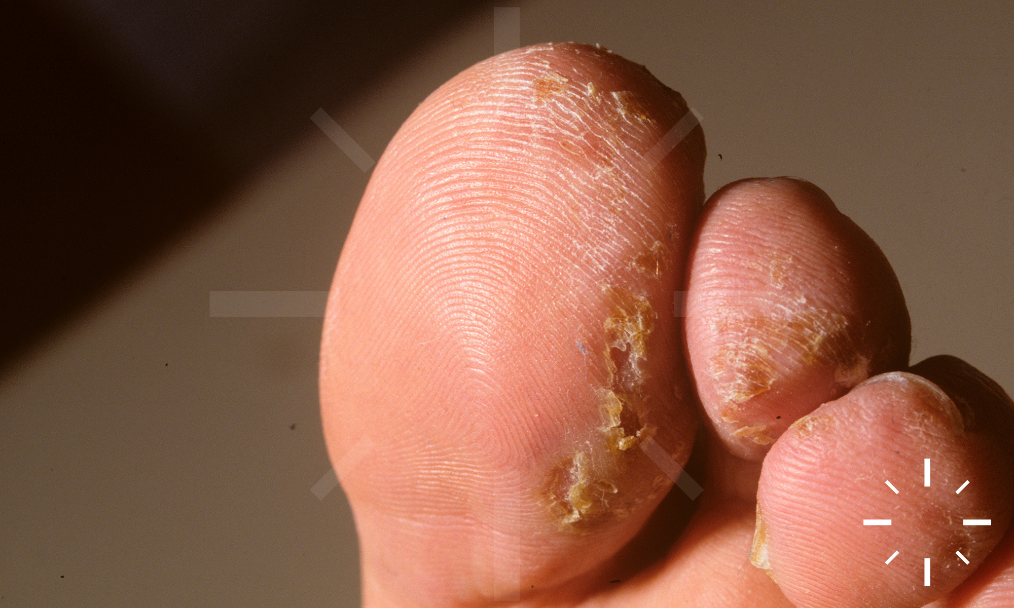
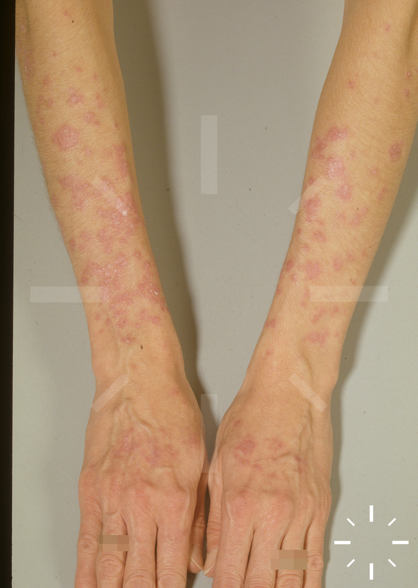
Systemic lupus erythematosus
Last Updated: 2023-10-12
Author(s): Anzengruber F., Navarini A.
ICD11: 4A40.0Z
1/49