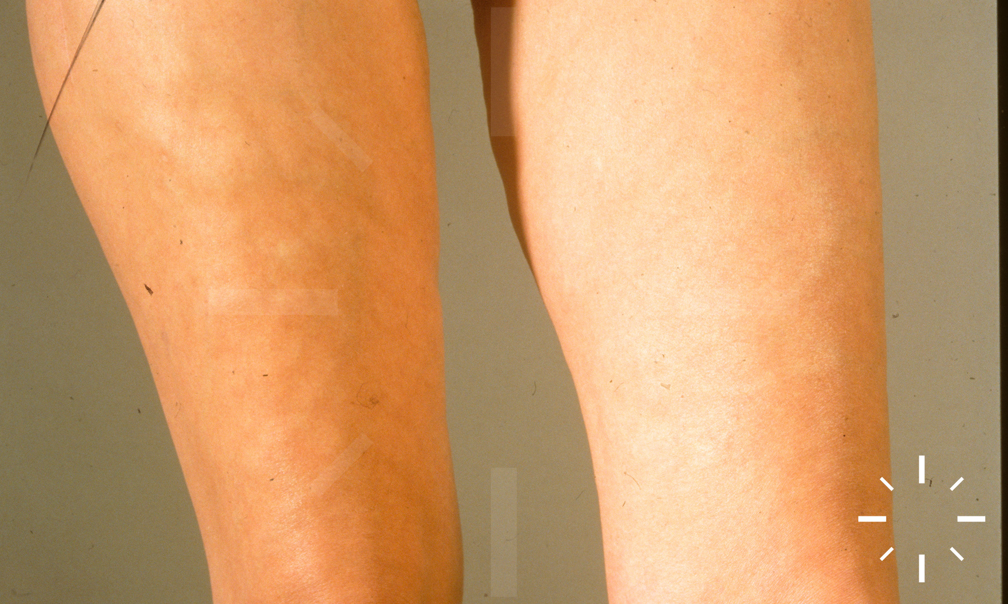
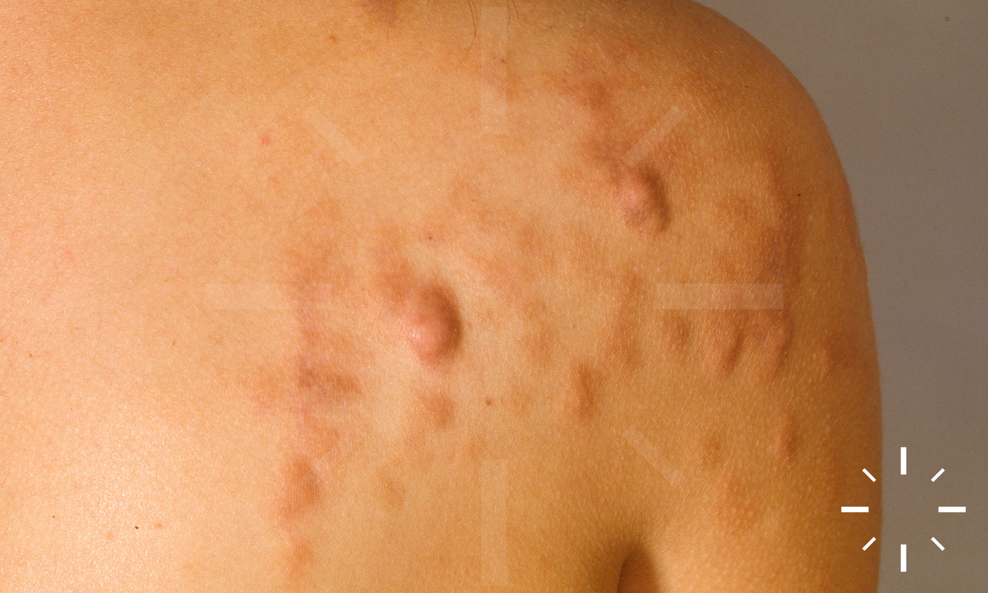
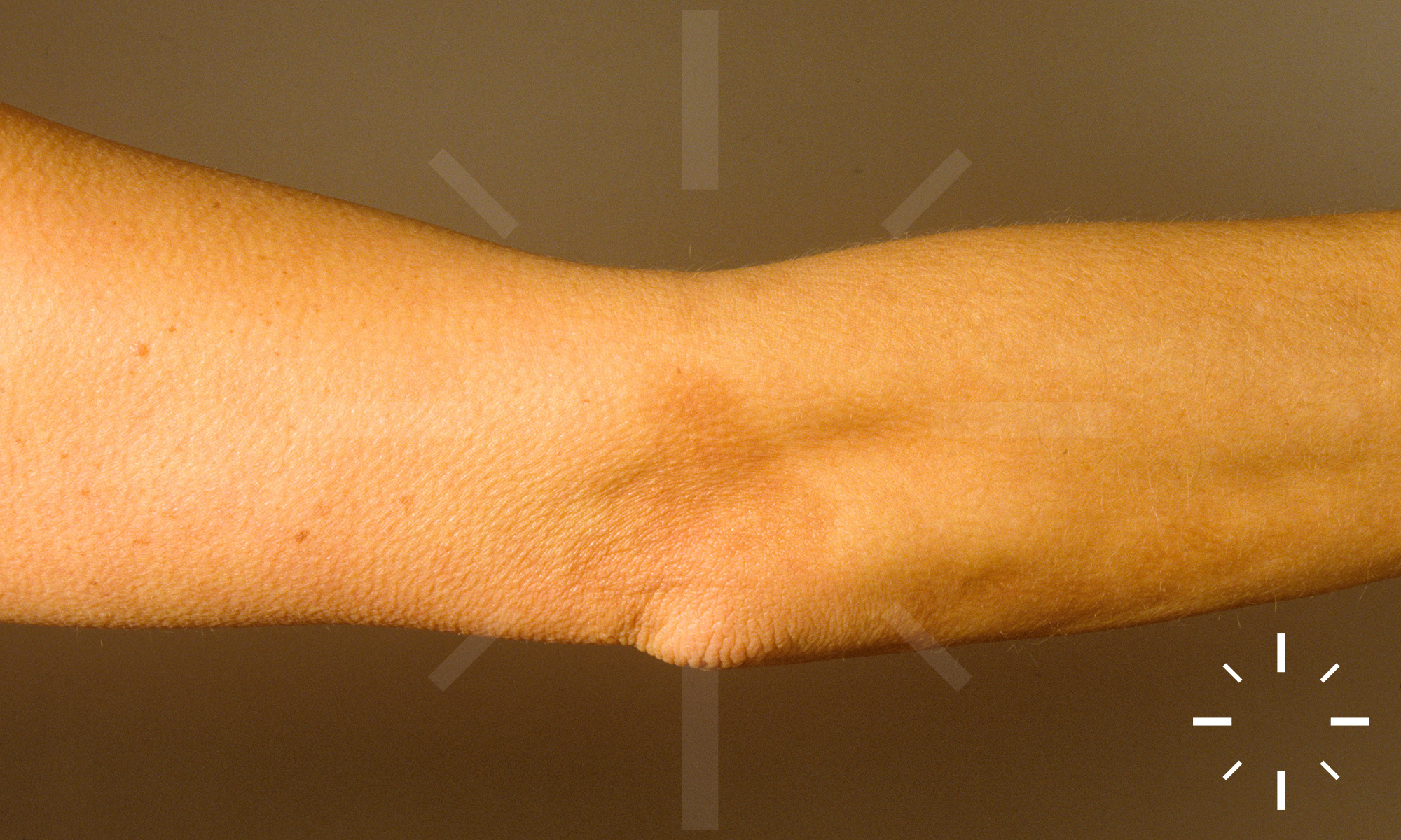
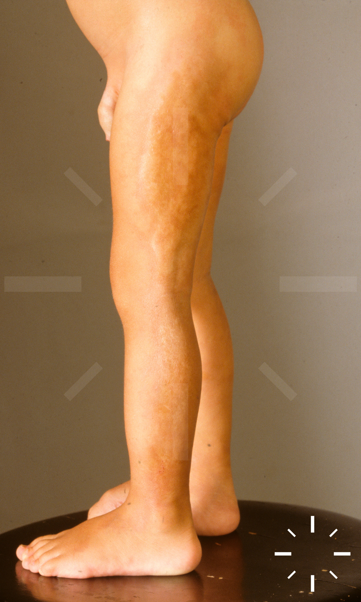
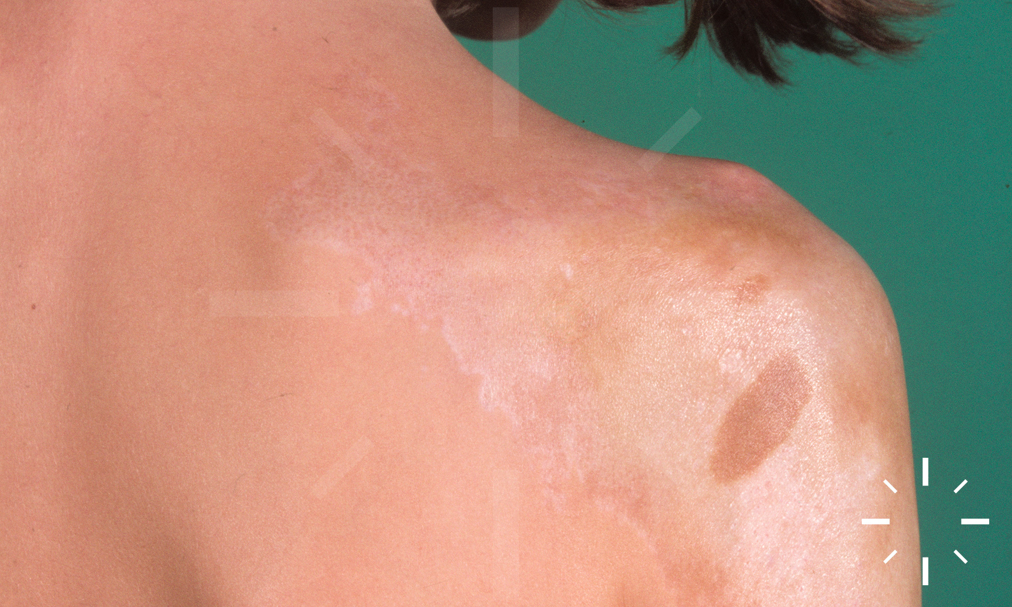
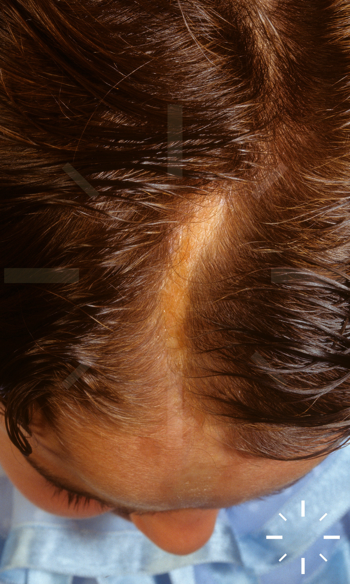
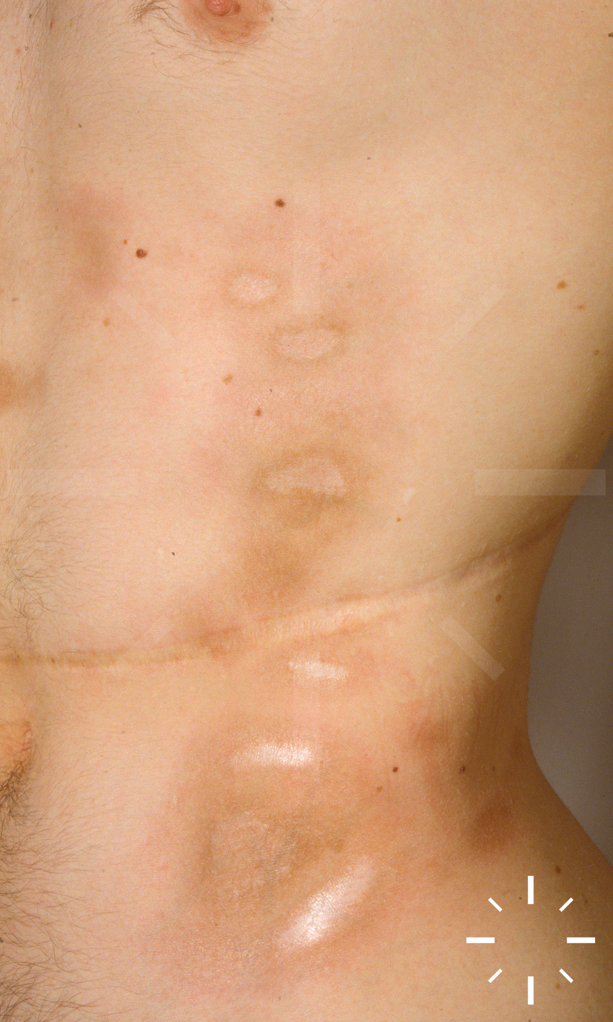
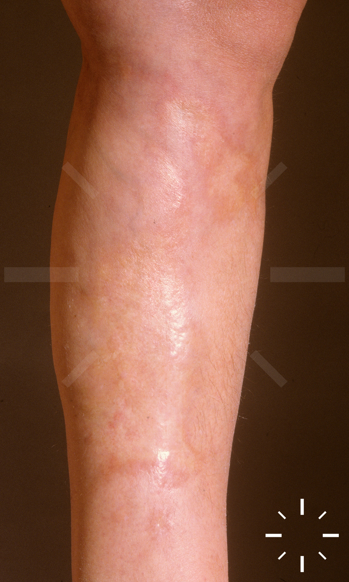
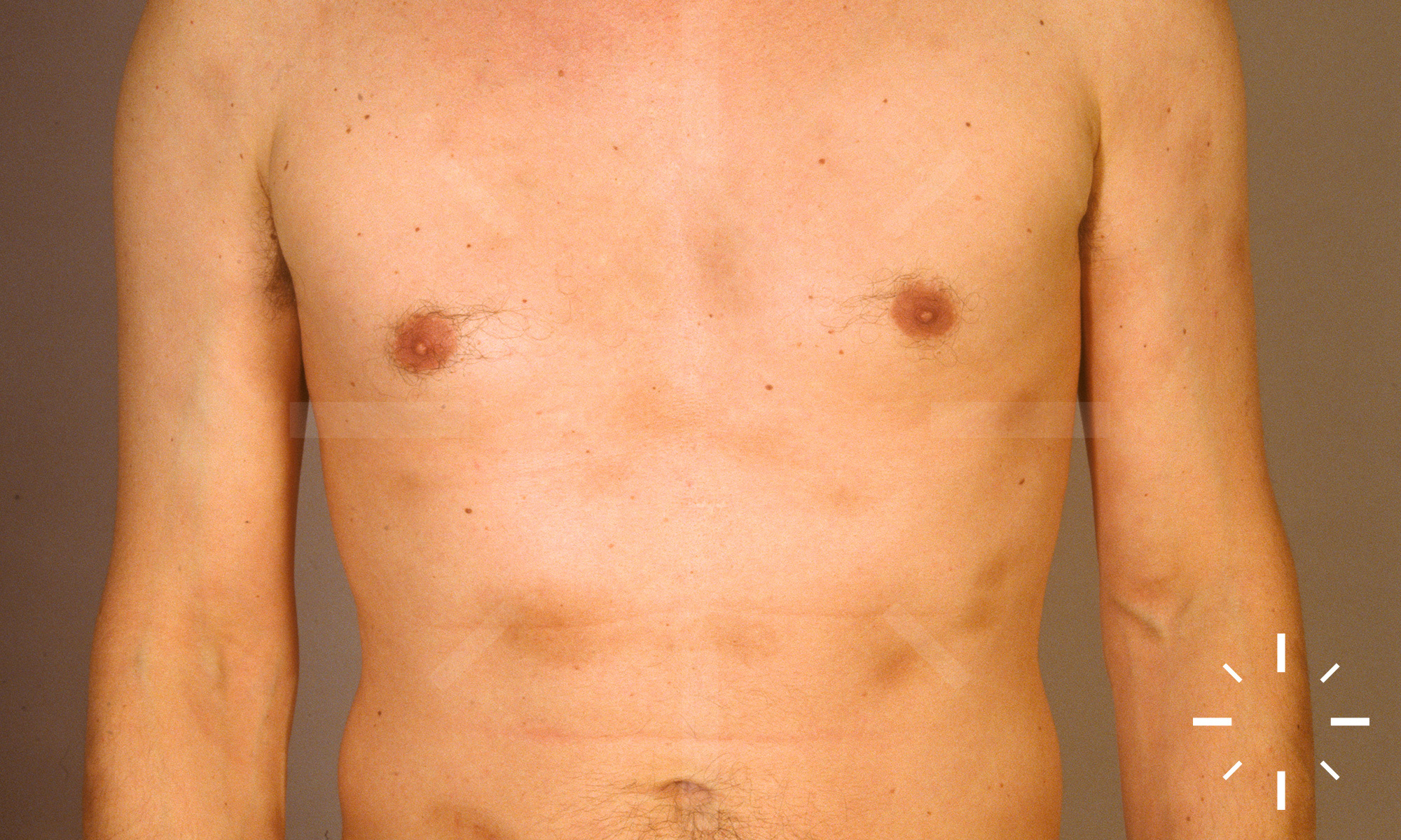
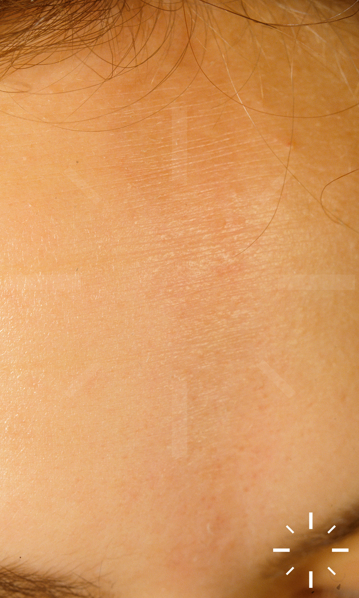
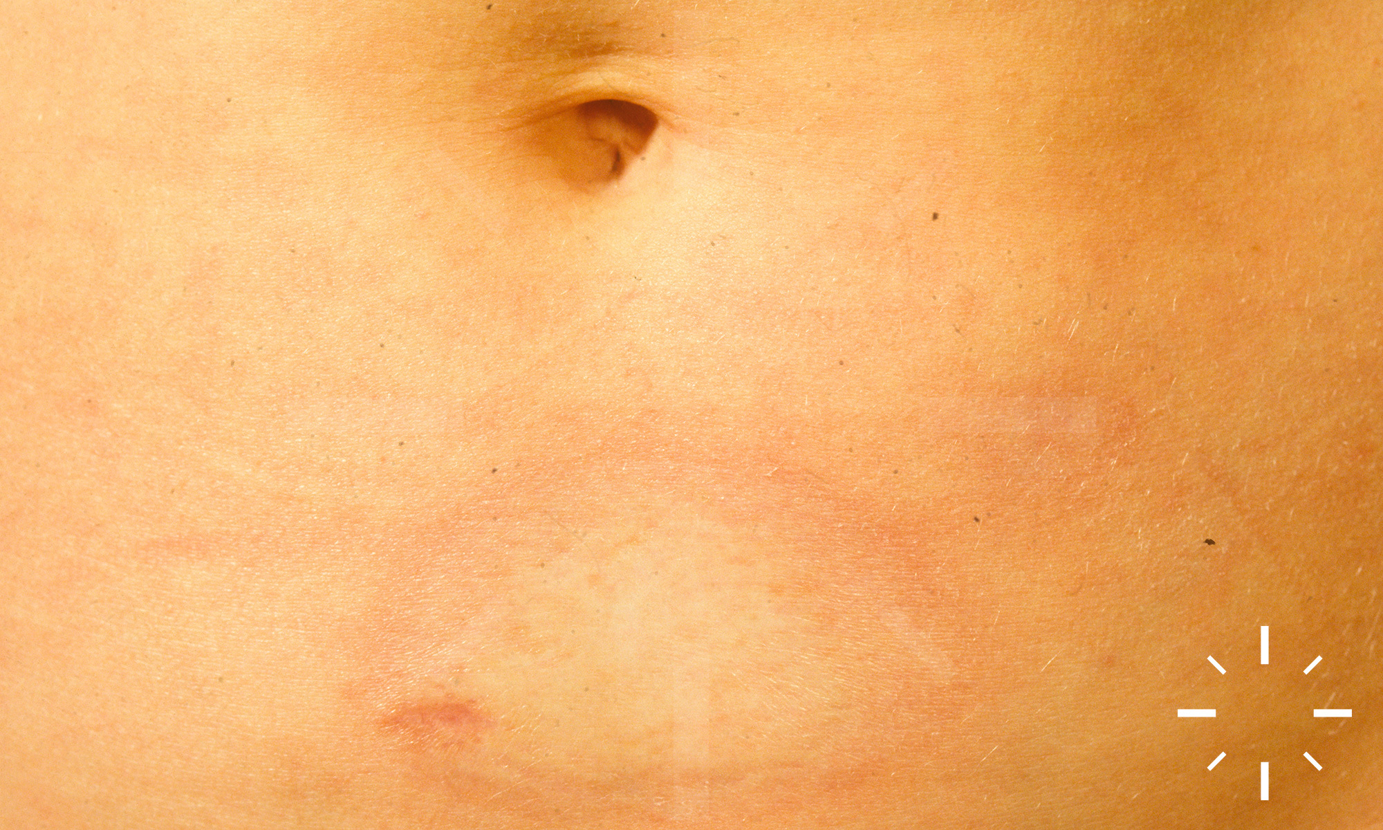
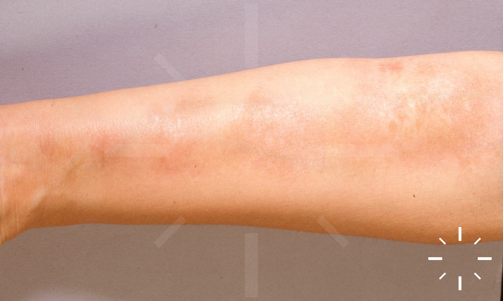
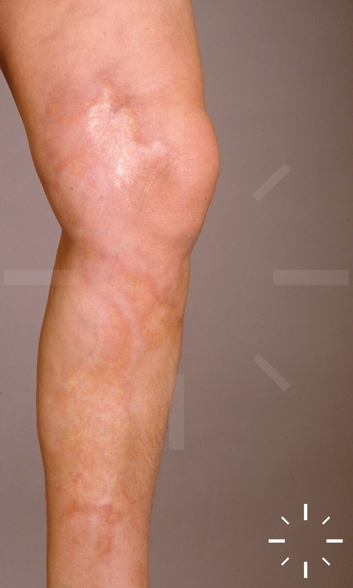
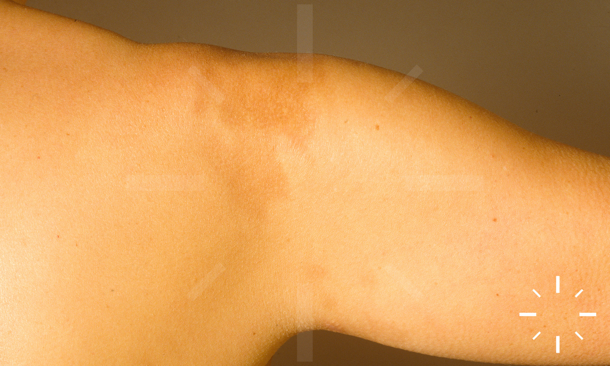
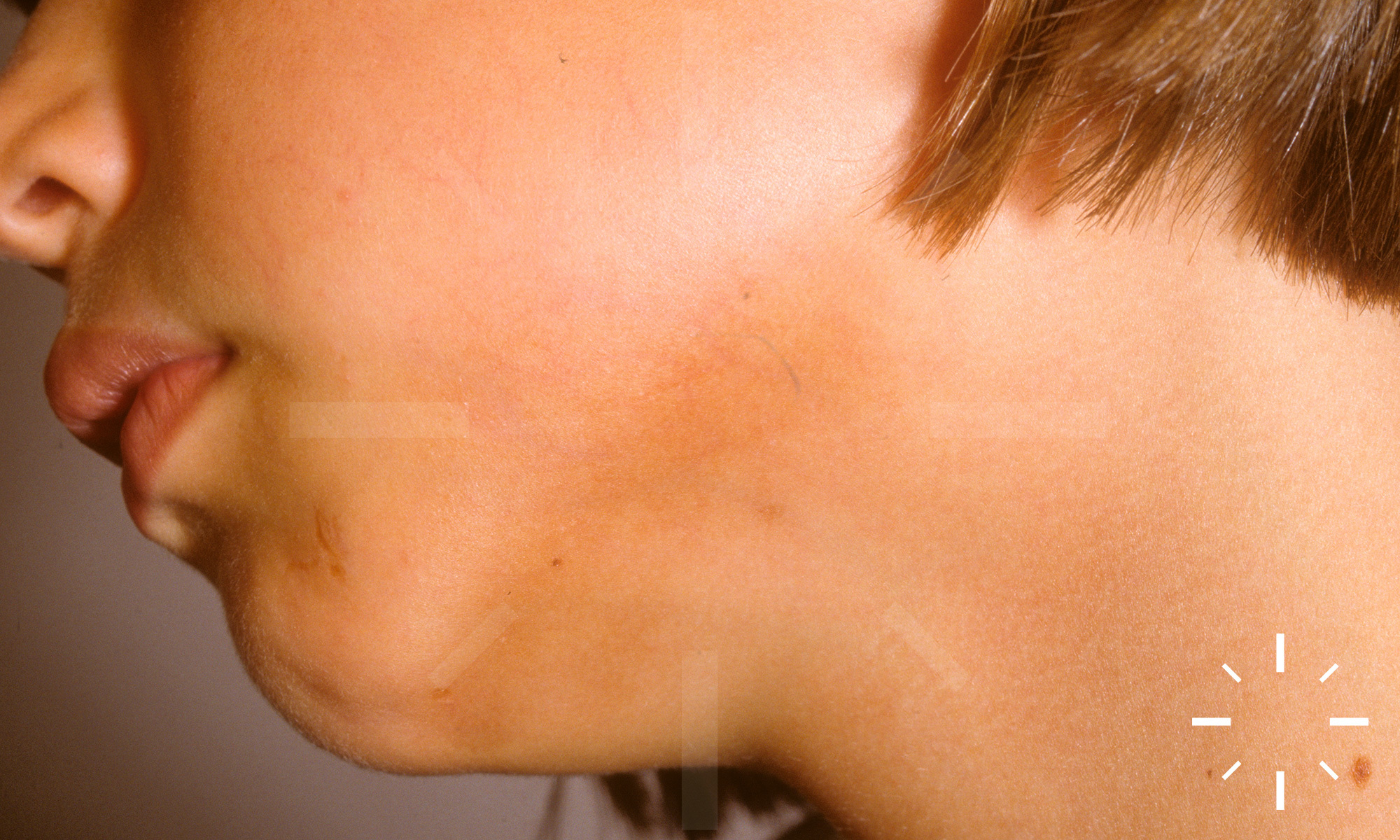
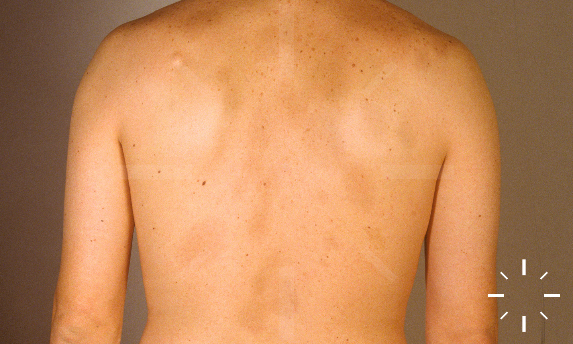
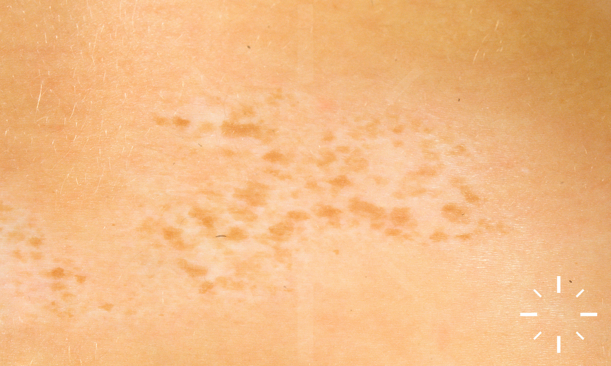
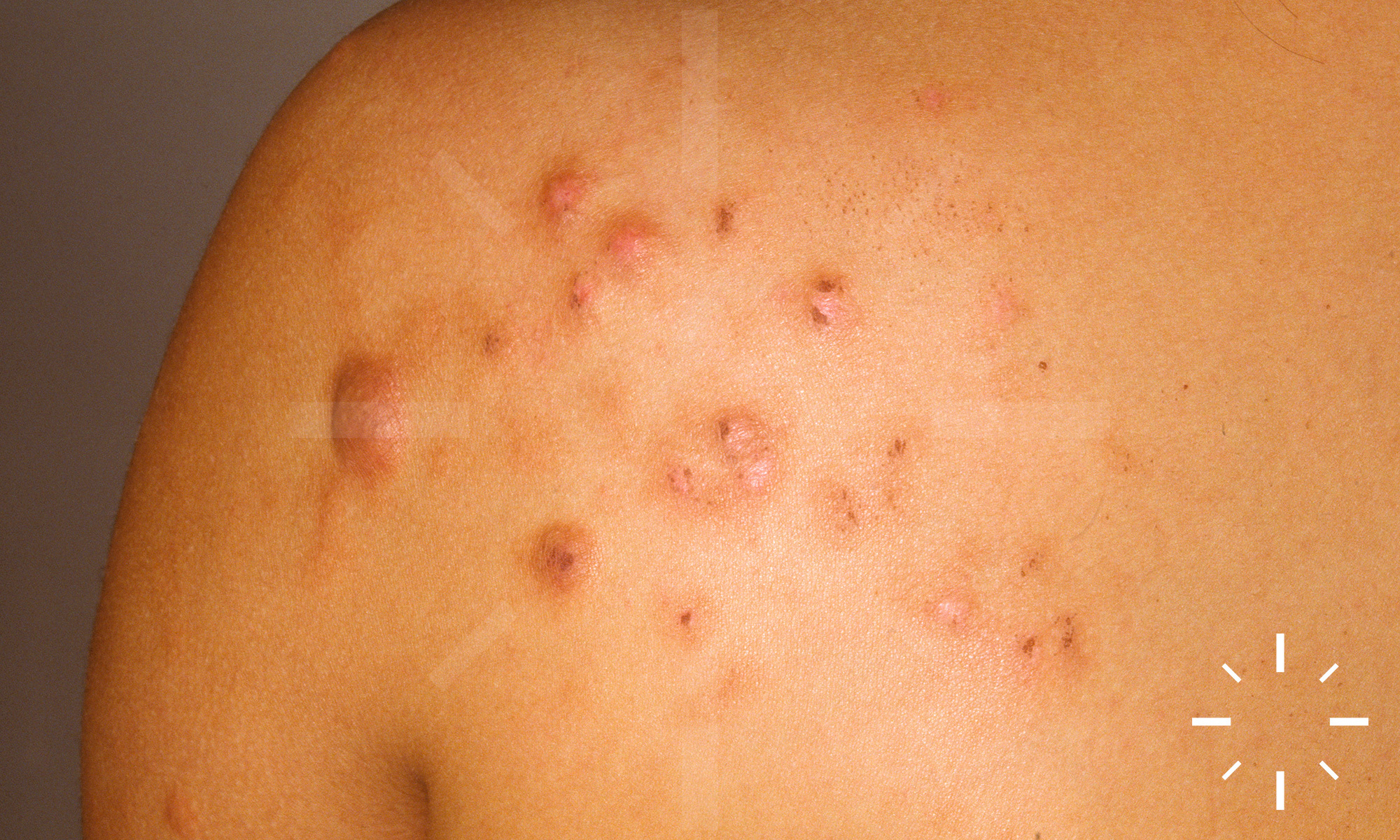
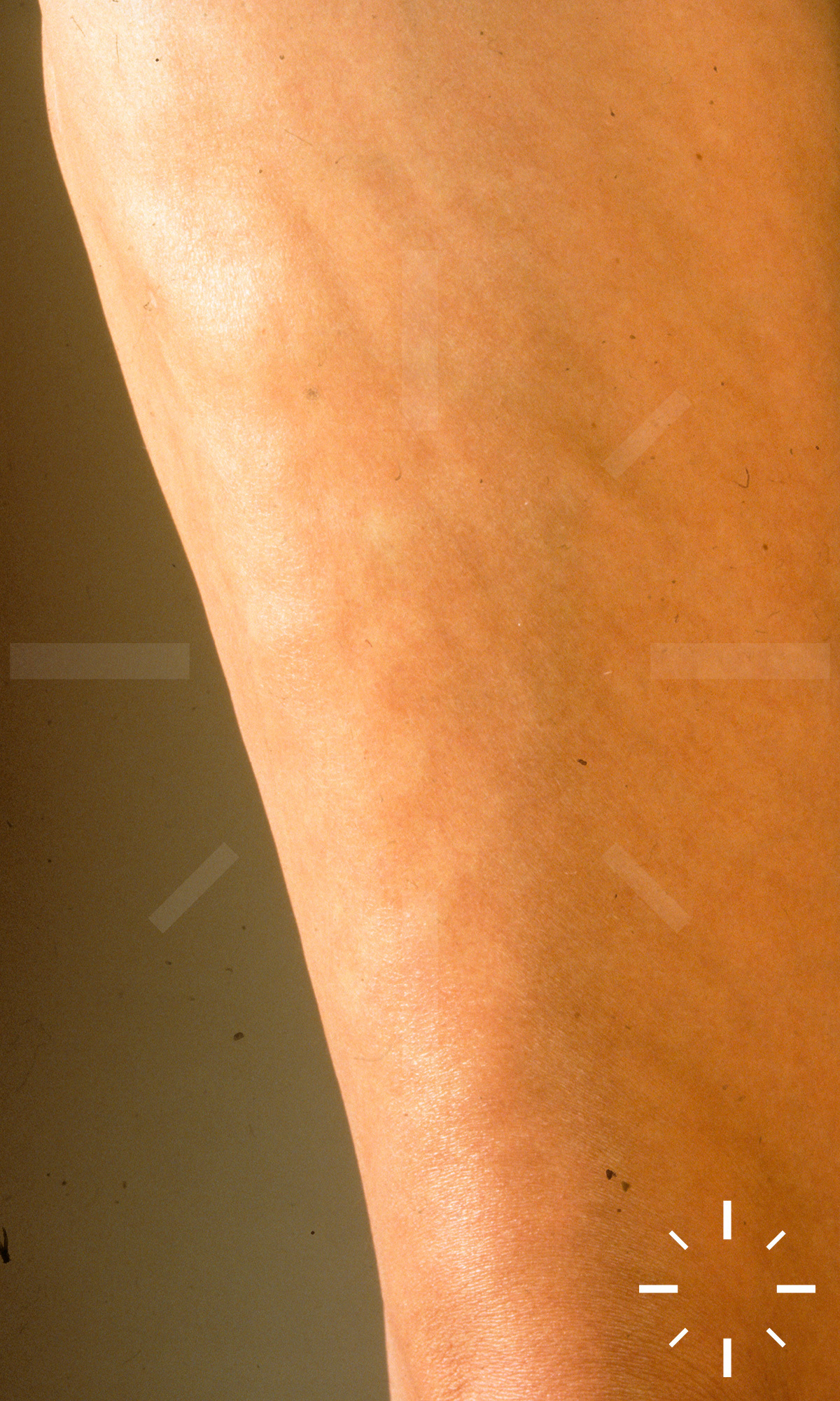
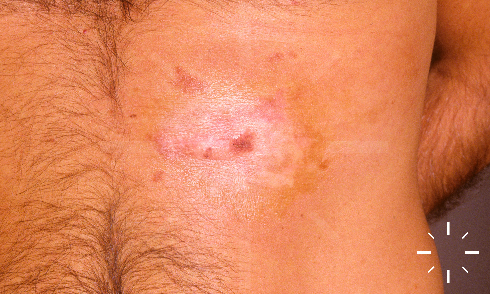
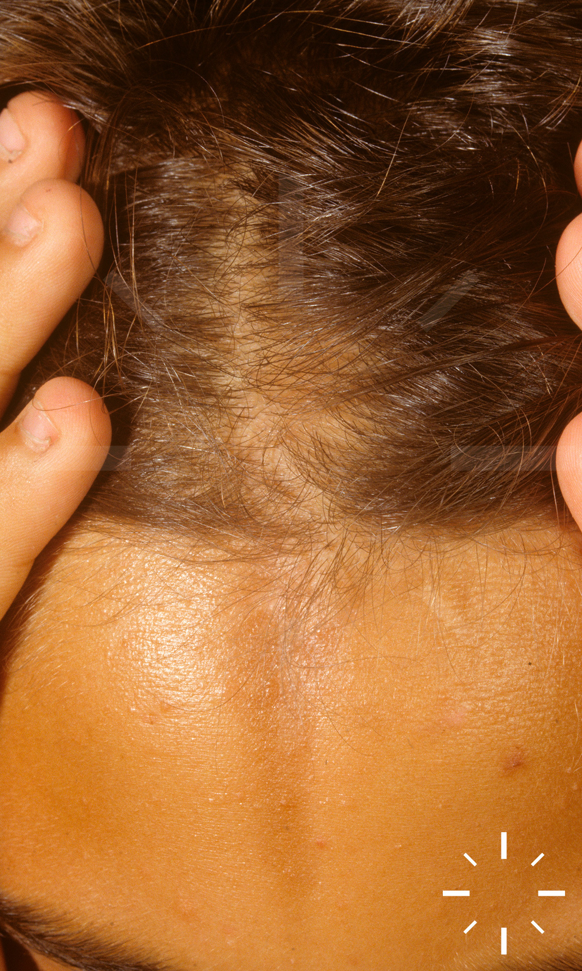
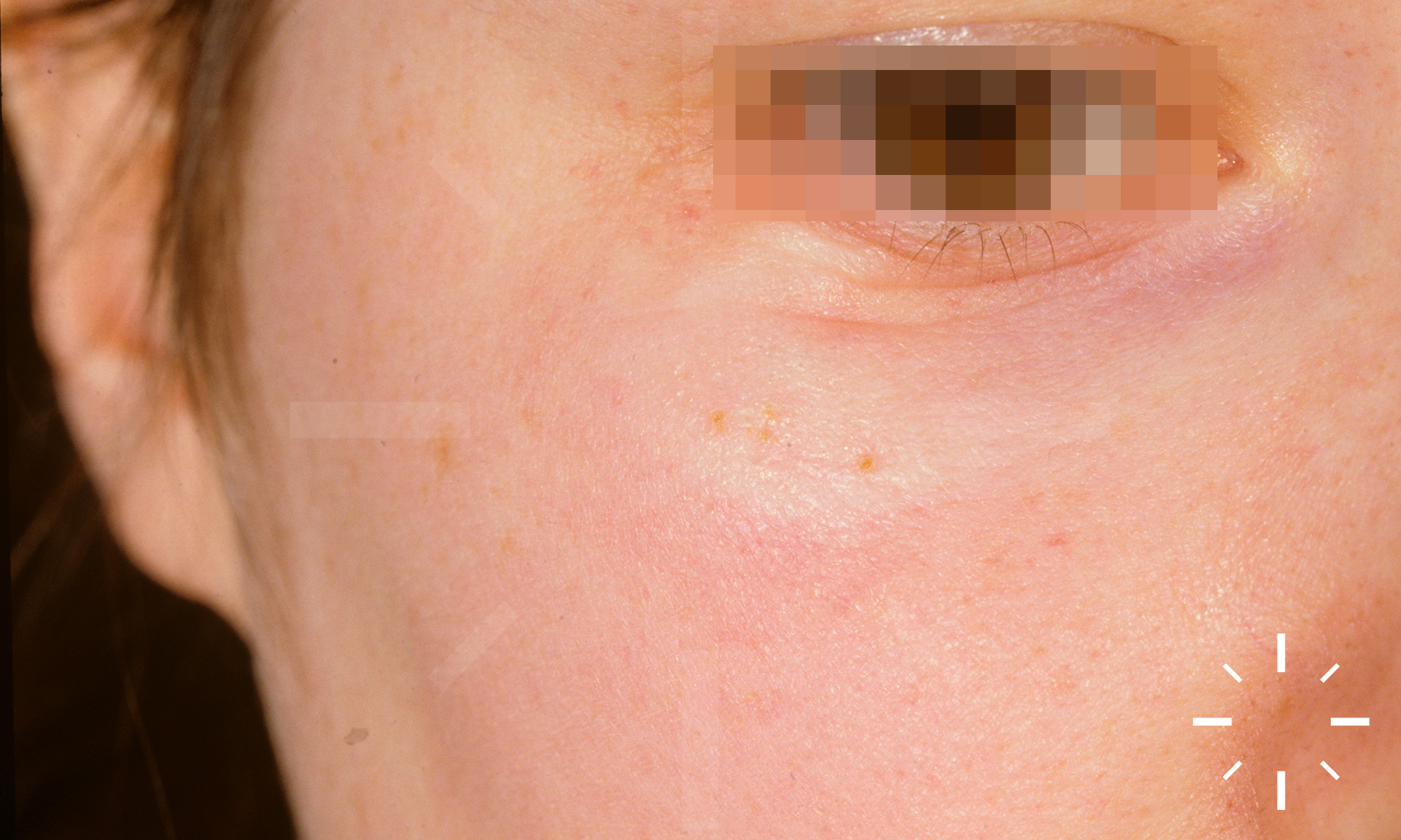
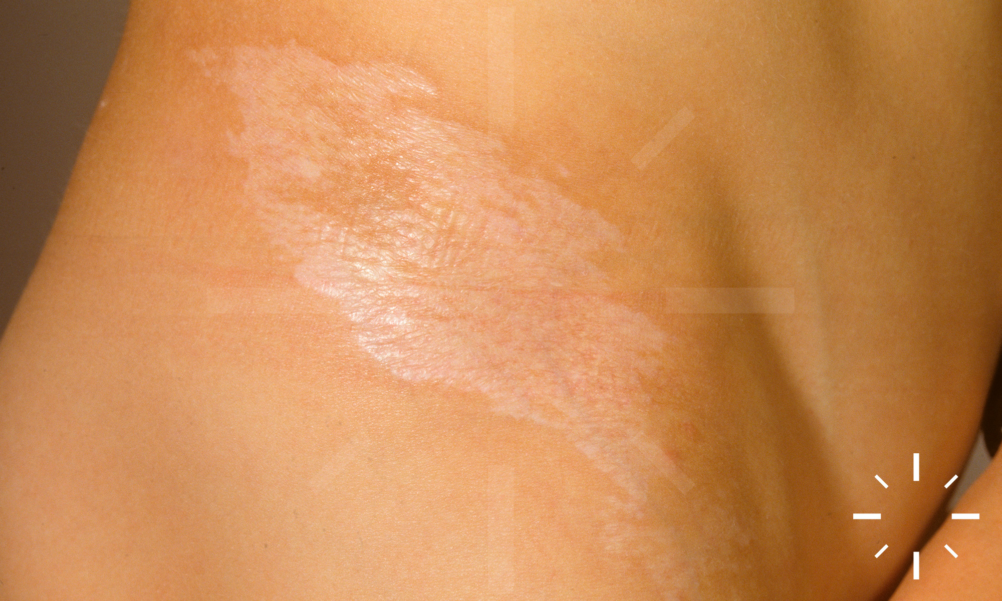
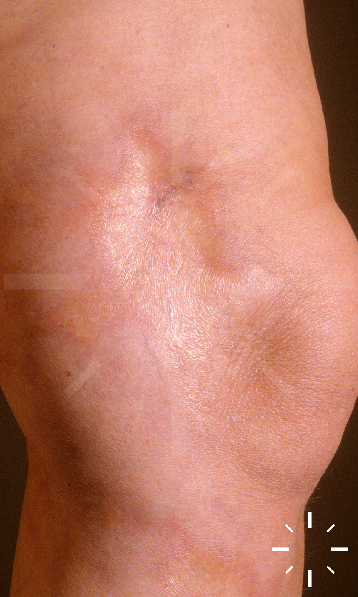
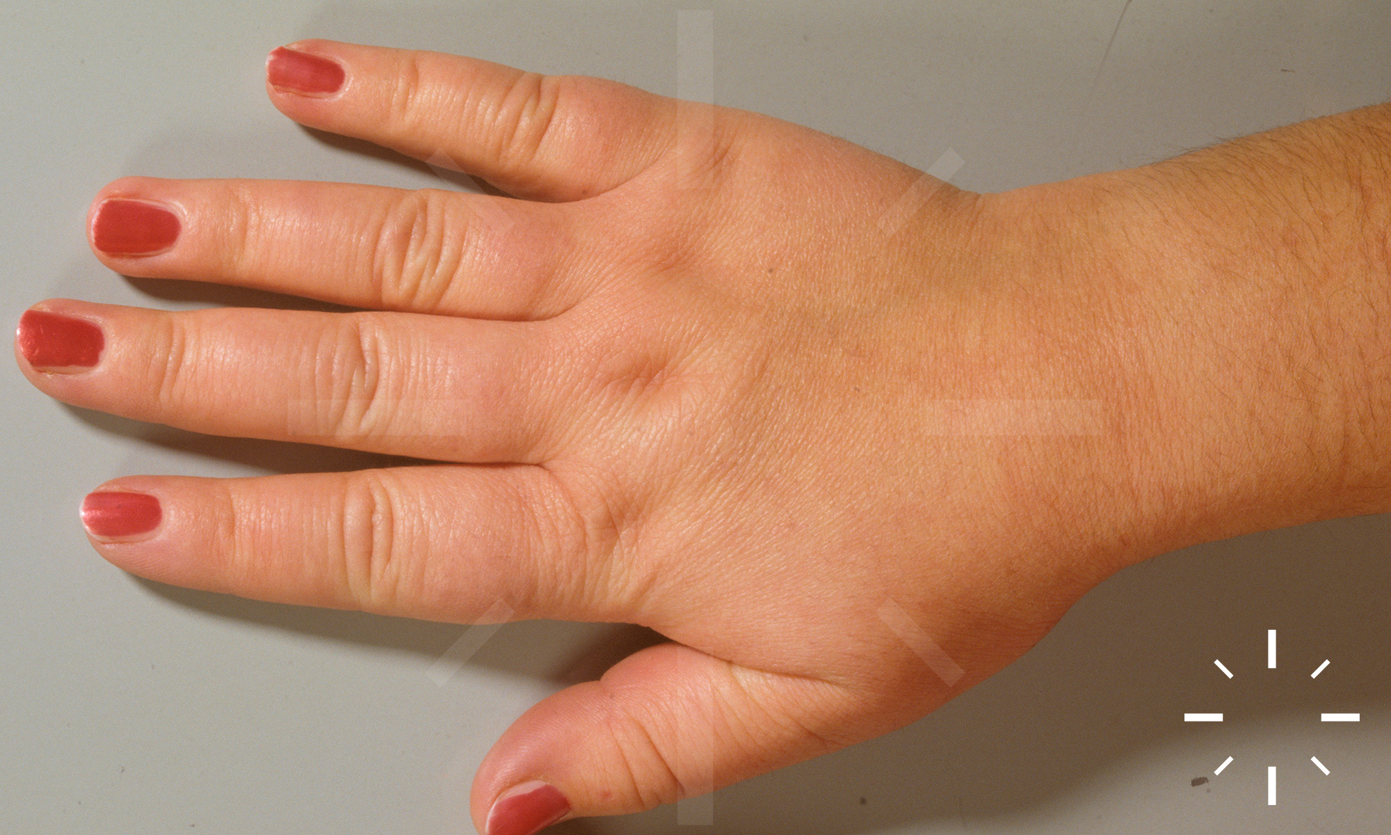
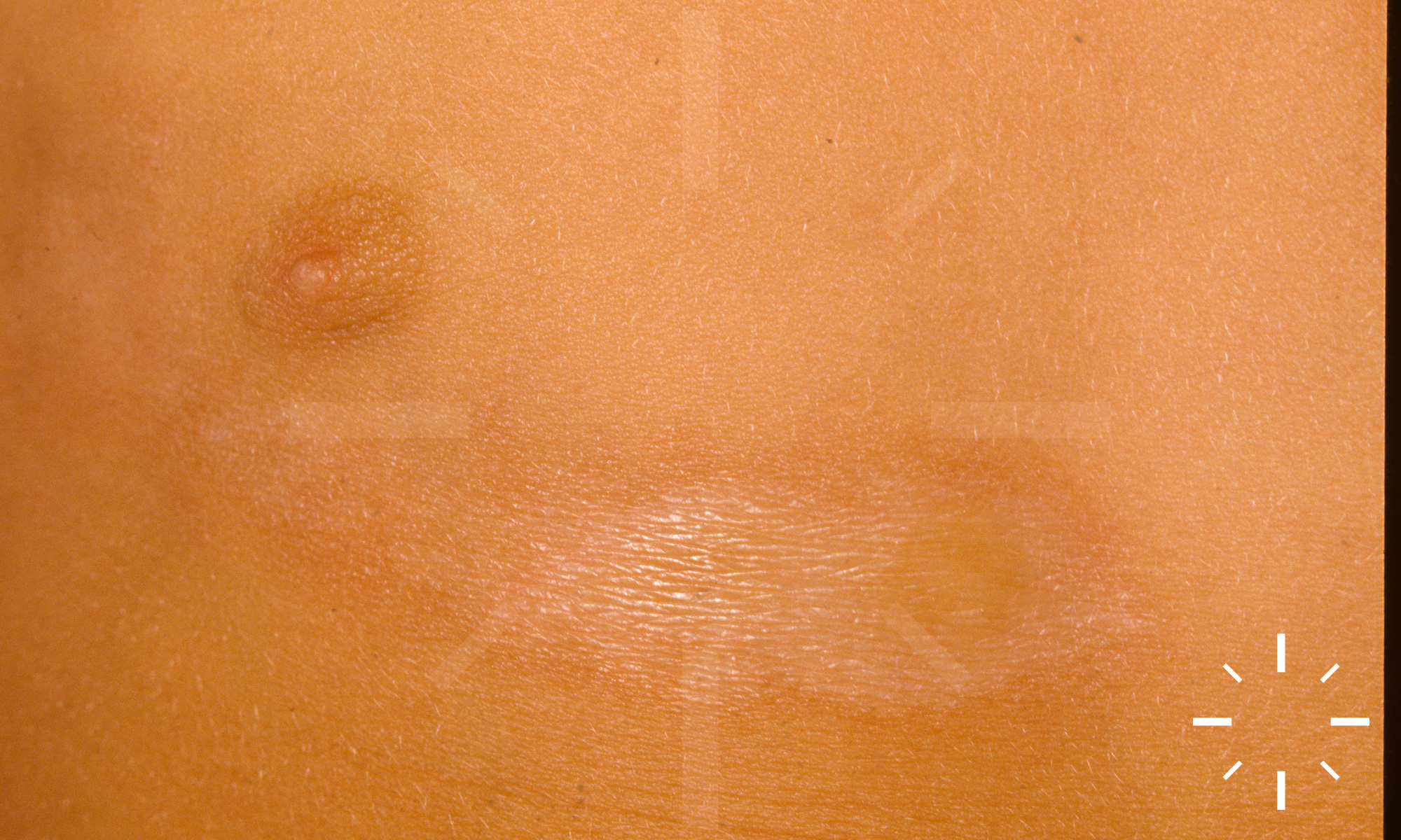
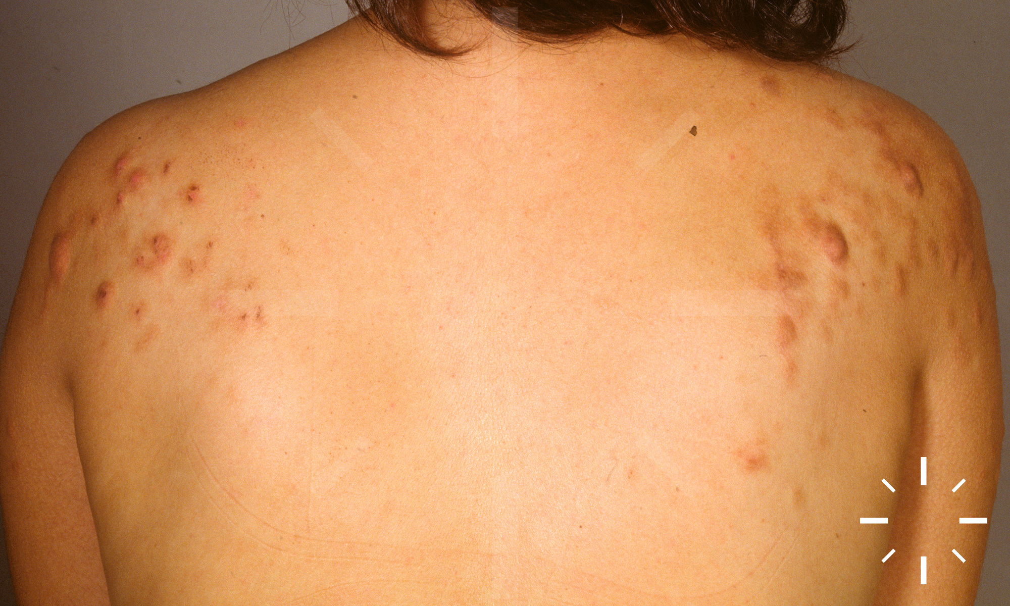
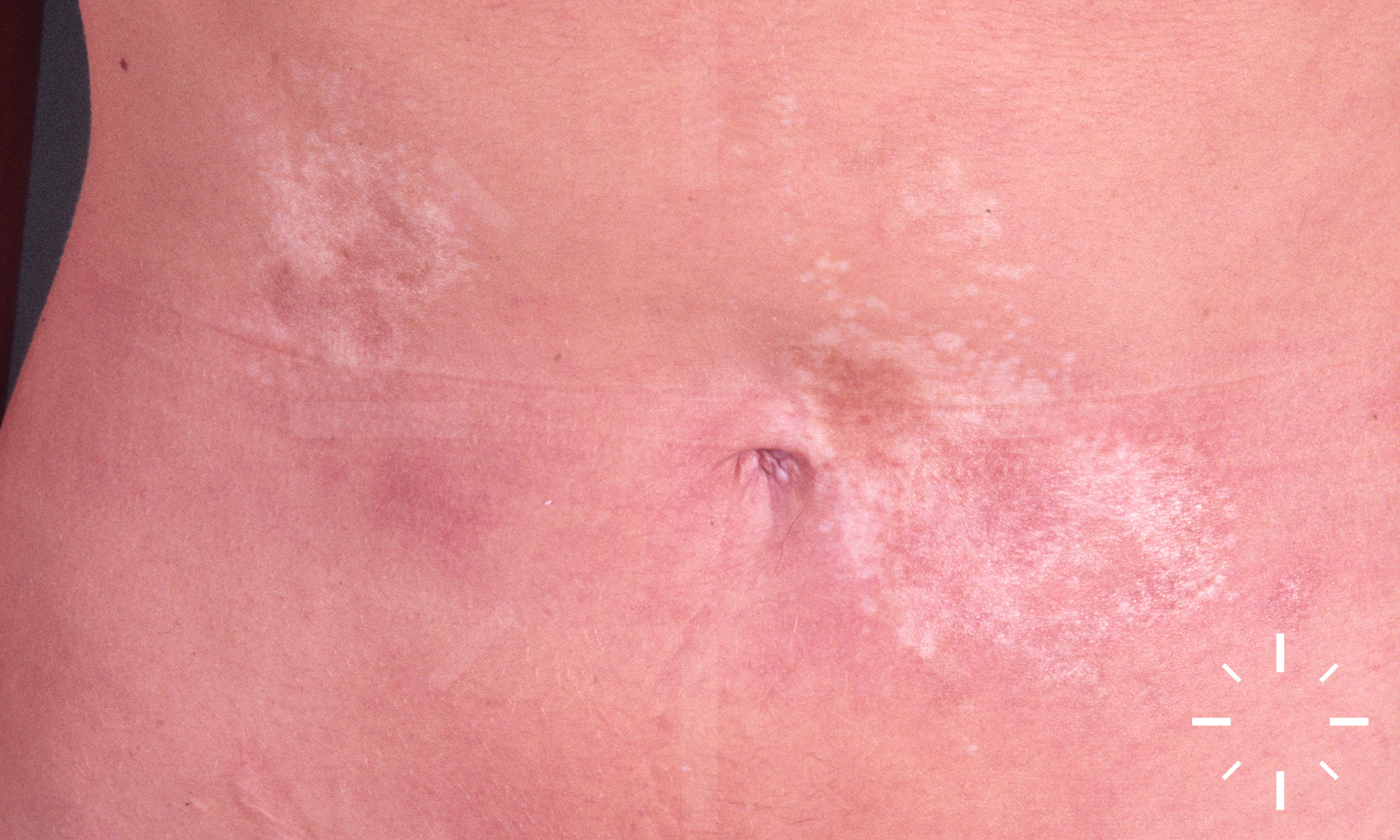
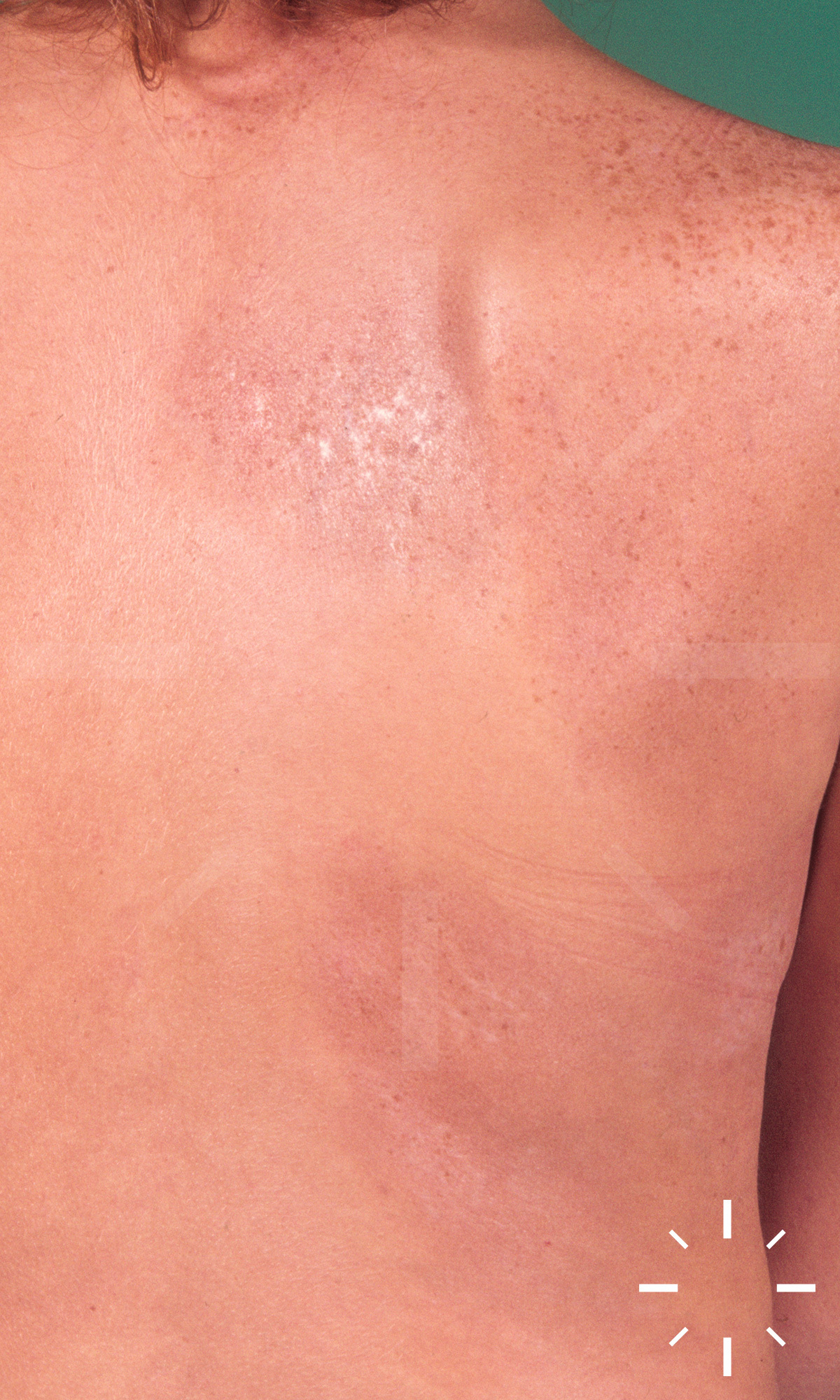
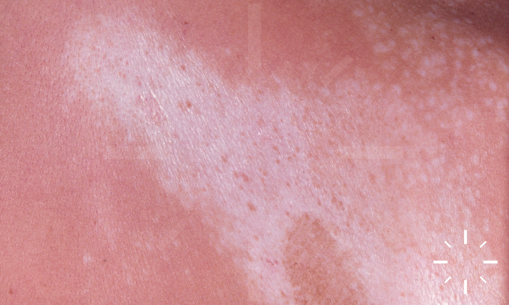
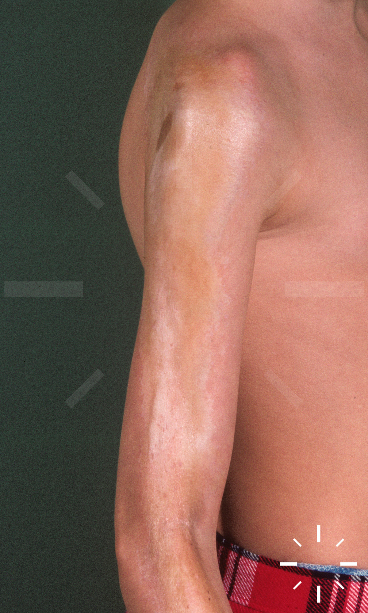
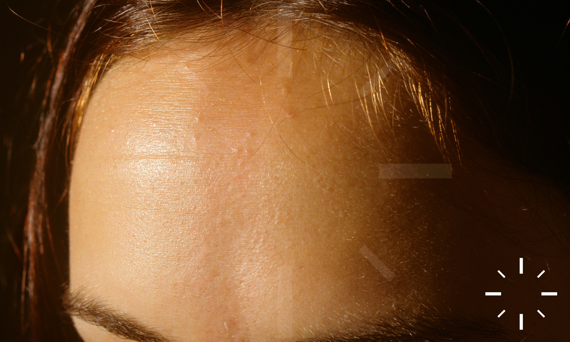
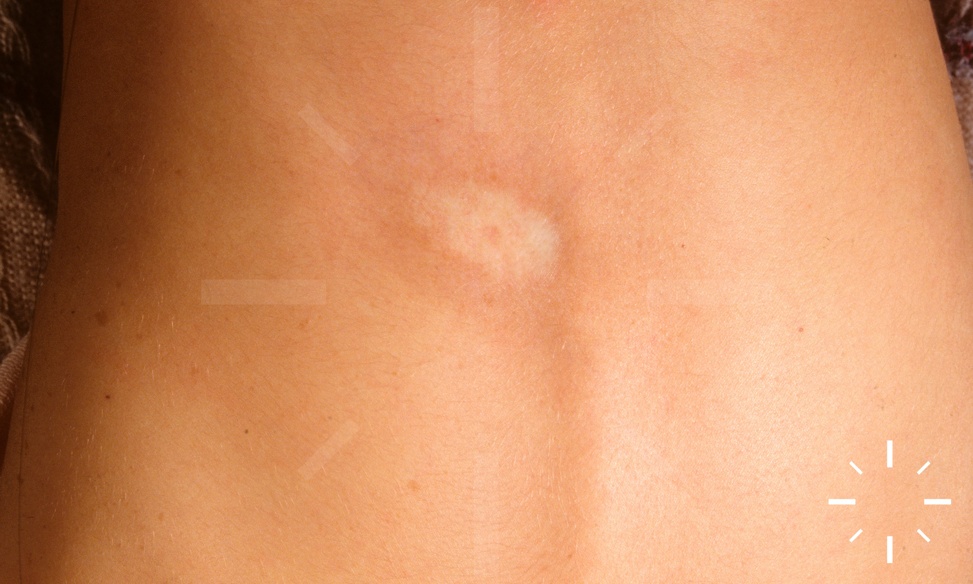
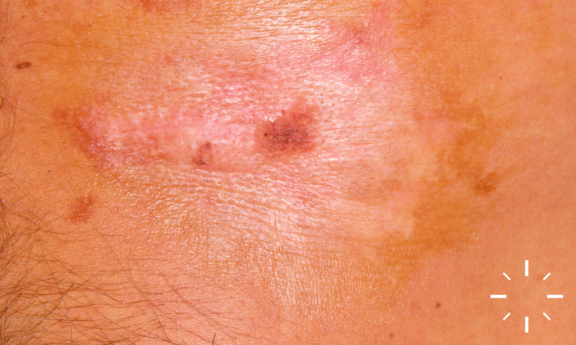
Morphea

Last Updated: 2023-07-07

Author(s): Anzengruber F., Navarini A.

ICD11: EB61.0

Localised Scleroderma, circumscripted scleroderma

A self-limited autoimmune disease belonging to the group of collagenoses, characterised by fibrosclerosis of the skin. Both the involvement of internal organs and the transition to systemic scleroderma are a rarity.

  • Age peak: infancy 2-8 years and between 20-50 years
  • F:M = 2.6 - 6:1
  • 85% of all affected individuals are Caucasian
  • Association to chronic polyarthritis or lupus erythematosus

  • Type I: Plaque-like scleroderma (approx. 70% of adults, approx. 30% of children)
    • Localisation: mainly the trunk area
    • Subform: Atrophodermia idiopathica et progressiva (Pierini-Pasini type)
  • Type II: Linear scleroderma: (10-30% of adults, approx. 65% of children)
    • Localisation: mainly extremities and face
      • 1a: localised, 1b: generalised type
      • 2: scleroderma en coup de sabre
  • Type III: Subcutaneous, profound scleroderma (ca.12% of children)

Special forms:

  • Pansclerotic disabling disease of children
  • Sclerofascia
  • Eosinophilic fasciitis (Shulman type)
  • Hemiatrophia faciei progressiva (Romberg type)

  • Initially, there are centrifugally growing whitish, brownish or red plaques. An indurated, ivory-coloured atrophic zone is often visible centrally. The lesions are bordered by a blue-red erythema rim, the so-called Lilac Ring, in case of progression. 
  • Depending on the form, subcutaneous, fascia, muscle, periosteum and skeletal tissue are affected during progression.
  • Division according to S1- Guideline "Circumscribed Scleroderma"
  • Limited circumstrictive forms of scleroderma (type I)
    • Plaque-type morphea (with 26%, the 2nd most common scleroderma form in children)
    • Morphea guttata (is called "extragenital lichen sclerosus et atrophicus")
    • Atrophodermia Pierini-Pasini (Atrophodermia idiopathica et progressiva)
  • Generalised forms of scleroderma
    • Generalised morphea (for this, at least 3 different anatomical localisations must be affected)
    • Disabling pansclerotic morphea
    • Eosinophilic fasciitis
  • Linear forms of scleroderma (type II)
    • Linear scleroderma of the "en coup de sabre" type: subcortical calcifications or brain atrophy, which can lead to e.g.: migraine, epilepsy.
    • Linear scleroderma
    • Progressive fascial hemiatrophy (Parry-Romberg syndrome): subcortical calcifications or brain atrophy, which can lead to e.g.: Migraine, epilepsy.
  • Deep form of scleroderma (type III)
    • Morphea profunda

Severity: depends on the localisation and extent of the skin lesions.

  • Clinic (is sufficient in most cases for diagnosis)
  • Biopsy
  • There are no serological progression parameters!
  • Laboratory (useful for differential diagnosis)
    • blood count (sometimes eosinophilia in linear forms)
    • creatinine kinase (e.g. of concomitant myositis)
    • Autoantibodies (ANA (positive in up to 80% even without systemic involvement), anti-Scl-70)
    • Borrelia serology (pseudoscleroderma in Borrelia infection)

Further diagnostics:

  • Modified Rodnan Skin Score (thickness of the skin is measured by palpation)
  • Cutometer (elasticity measurement)
  • Durometer (hardness test)
  • Quantification of induration:
    • 20 MHz sonography
    • Laser Doppler
  • Depth extension:
    • CT
    • MRI

  • Torso (58%)
  • Legs (24%)
  • Arms (12%)
  • Head (6%)

  • Early stage (inflammatory stage):
    • Perivascular and periadnexal lymphohistiocytic infiltrates in the reticular dermis and subcutis
    • Thickening of the dermal oedematous collagen bundles
    • Septal panniculitis
    • Round cell infiltrates at the dermo-subcutaneous junction
  • Late stage (sclerotic stage)
    • Rarefaction of adnexal glands (e.g. sweat glands)
    • Sclerosis of the dermis
    • More dermal connective tissue, less subcutaneous adipose tissue
    • Homogenised widened collagen fibre bundles run parallel to the skin surface
    • Narrowed vessels
    • Edematous adipose tissue septa

  • Joint contractures
  • Cosmetic impairment

None known.

Most of the time, progression stops after about 7 years.

  1. Fleischmajer R. Generalized Morphea. Arch Dermatol 1972;106:509.
  2. Diaz-Perez JL. Disabling pansclerotic morphea of children. Archives of Dermatology 1980;116:169-73.
  3. Uitto J, Santa Cruz DJ, Bauer EA, Eisen AZ. Morphea and lichen sclerosus et atrophicus. Journal of the American Academy of Dermatology 1980;3:271-9.
  4. Daniel Su WP, Person JR. Morphea profunda A new concept and a histopathologic study of 23 cases. The American Journal of Dermatopathology 1981;3:251-60.
  5. Connelly MG, Winkelmann RK. Coexistence of lichen sclerosus, morphea, and lichen planus. Journal of the American Academy of Dermatology 1985;12:844-51.
  6. Kencka D, Blaszczyk M, Jab, lstrok, nacute, ska S. Atrophoderma Pasini-Pierini Is a Primary Atrophic Abortive Morphea. Dermatology 1995;190:203-6.
  7. Cunningham BB, Landells IDR, Langman C, Sailer DE, Paller AS. Topical calcipotriene for morphea/linear scleroderma. Journal of the American Academy of Dermatology 1998;39:211-5.
  8. Seyger MMB, van den Hoogen FHJ, de Boo T, de Jong EMGJ. Low-dose methotrexate in the treatment of widespread morphea. Journal of the American Academy of Dermatology 1998;39:220-5.
  9. Nagai Y, Hattori T, Ishikawa O. Unilateral Generalized Morphea in Childhood. The Journal of Dermatology 2002;29:435-8.
  10. Sehgal VN, Srivastava G, Aggarwal AK, Behl PN, Choudhary M, Bajaj P. Localized scleroderma/morphea. International Journal of Dermatology 2002;41:467-75.
  11. Tollefson MM, Witman PM. En coup de sabre morphea and Parry-Romberg syndrome: A retrospective review of 54 patients. Journal of the American Academy of Dermatology 2007;56:257-63.
  12. Christen-Zaech S, Hakim MD, Afsar FS, Paller AS. Pediatric morphea (localized scleroderma): Review of 136 patients. Journal of the American Academy of Dermatology 2008;59:385-96.
  13. Patel AR, Pavletic SZ, Turner ML, Cowen EW. The Isomorphic Response in Morphealike Chronic Graft-vs-Host Disease. Arch Dermatol 2008;144.
  14. Kroft EBM, de Jong EMGJ, Evers AWM. Psychological Distress in Patients With Morphea and Eosinophilic Fasciitis. Arch Dermatol 2009;145.
  15. Kroft EBM, Groeneveld TJ, Seyger MMB, de Jong EMGJ. Efficacy of Topical Tacrolimus 0.1% in Active Plaque Morphea. American Journal of Clinical Dermatology 2009;10:181-7.
  16. Leitenberger JJ, Cayce RL, Haley RW, Adams-Huet B, Bergstresser PR, Jacobe HT. Distinct Autoimmune Syndromes in Morphea. Arch Dermatol 2009;145.
  17. Hansen CB, Callen JP. Connective tissue panniculitis: lupus panniculitis, dermatomyositis, morphea/scleroderma. Dermatologic Therapy 2010;23:341-9.
  18. Saxton-Daniels S, Jacobe HT. An Evaluation of Long-term Outcomes in Adults With Pediatric-Onset Morphea. Arch Dermatol 2010;146.
  19. Budamakuntla L, Malvankar D. Extensive morphea profunda with autoantibodies and benign tumors: A rare case report. Indian Dermatol Online J 2012;3:208.
  20. Lutz V. High Frequency of Genital Lichen Sclerosus in a Prospective Series of 76 Patients With Morphea. Arch Dermatol 2012;148:24.
  21. Schanz S, Henes J, Ulmer A, et al. Response Evaluation of Musculoskeletal Involvement in Patients With Deep Morphea Treated With Methotrexate and Prednisolone: A Combined MRI and Clinical Approach. American Journal of Roentgenology 2013;200:W376-W82.
  22. Polcari I, Moon A, Mathes EF, Gilmore ES, Paller AS. Headaches as a Presenting Symptom of Linear Morphea en Coup de Sabre. PEDIATRICS 2014;134:e1715-e9.
  23. Jacobe, H. (2016). Treatment of morphea (localized scleroderma) in adults. Uptodate.com. Retrieved 31 May 2016, from http://www.uptodate.com/contents/treatment-of-morphea-localized-scleroderma-in-adults?source=search_result&search=localized+scleroderma&selectedTitle=3%7E40
  24. Höger, P. (2016). AWMF: Detail. Awmf.org. Retrieved 31 May 2016, from http://www.awmf.org/leitlinien/detail/ll/027-060.html