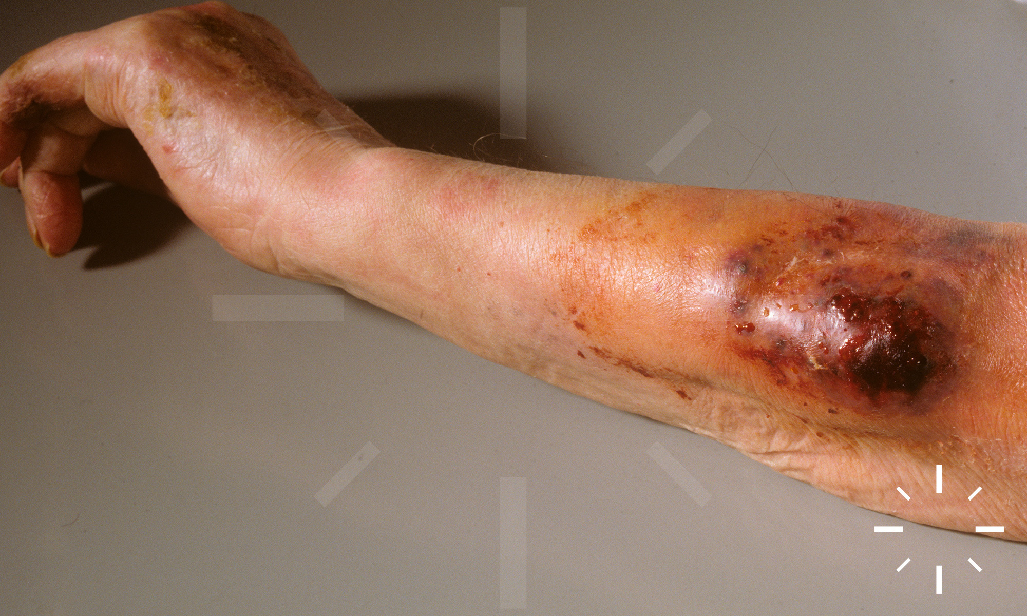
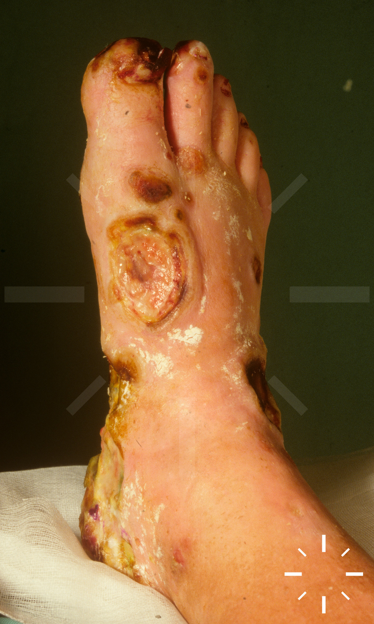
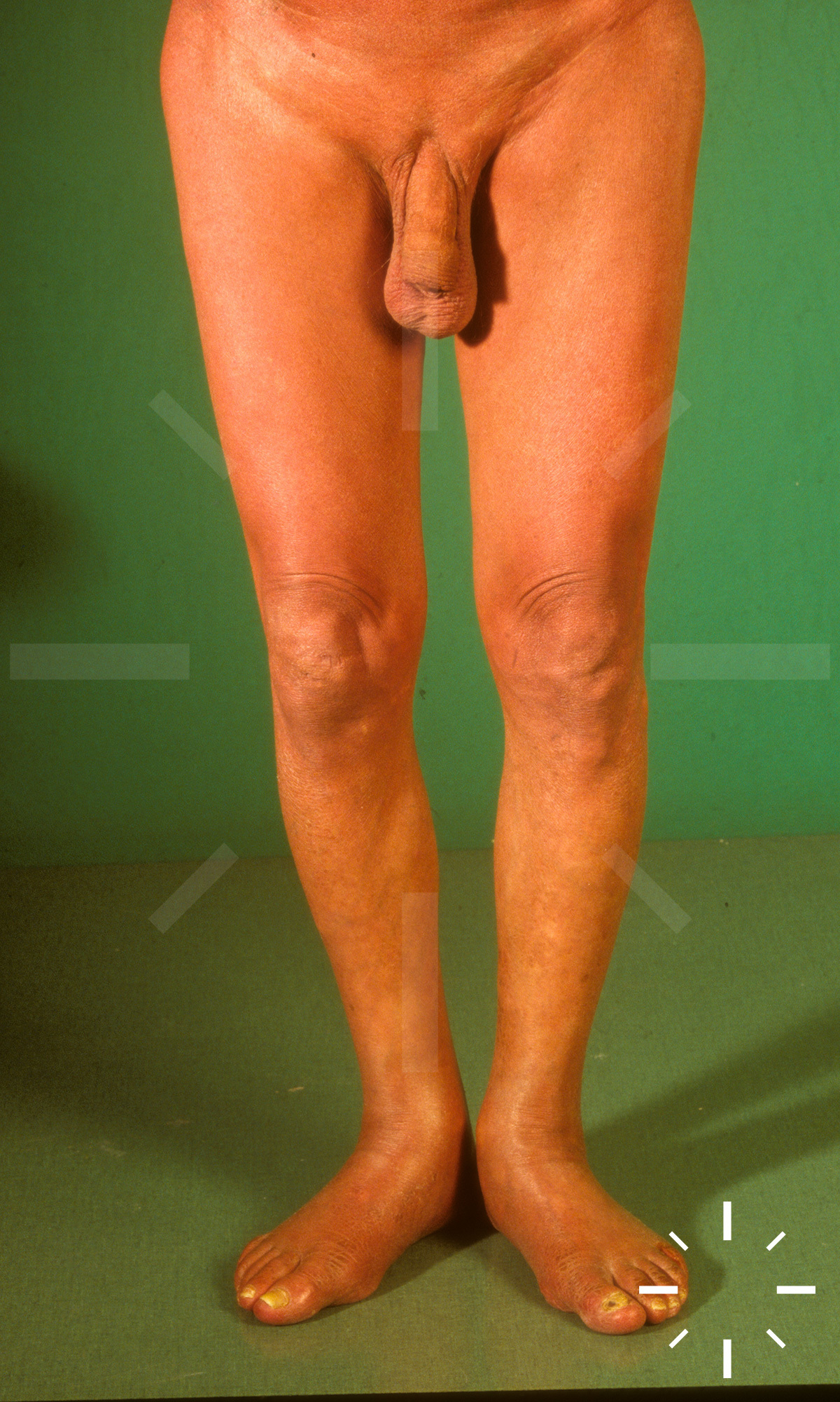
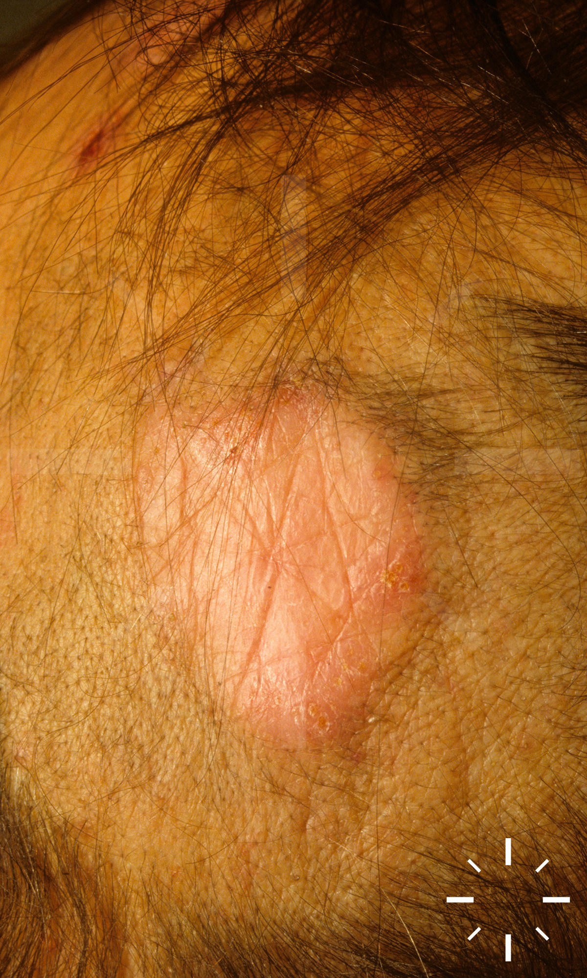
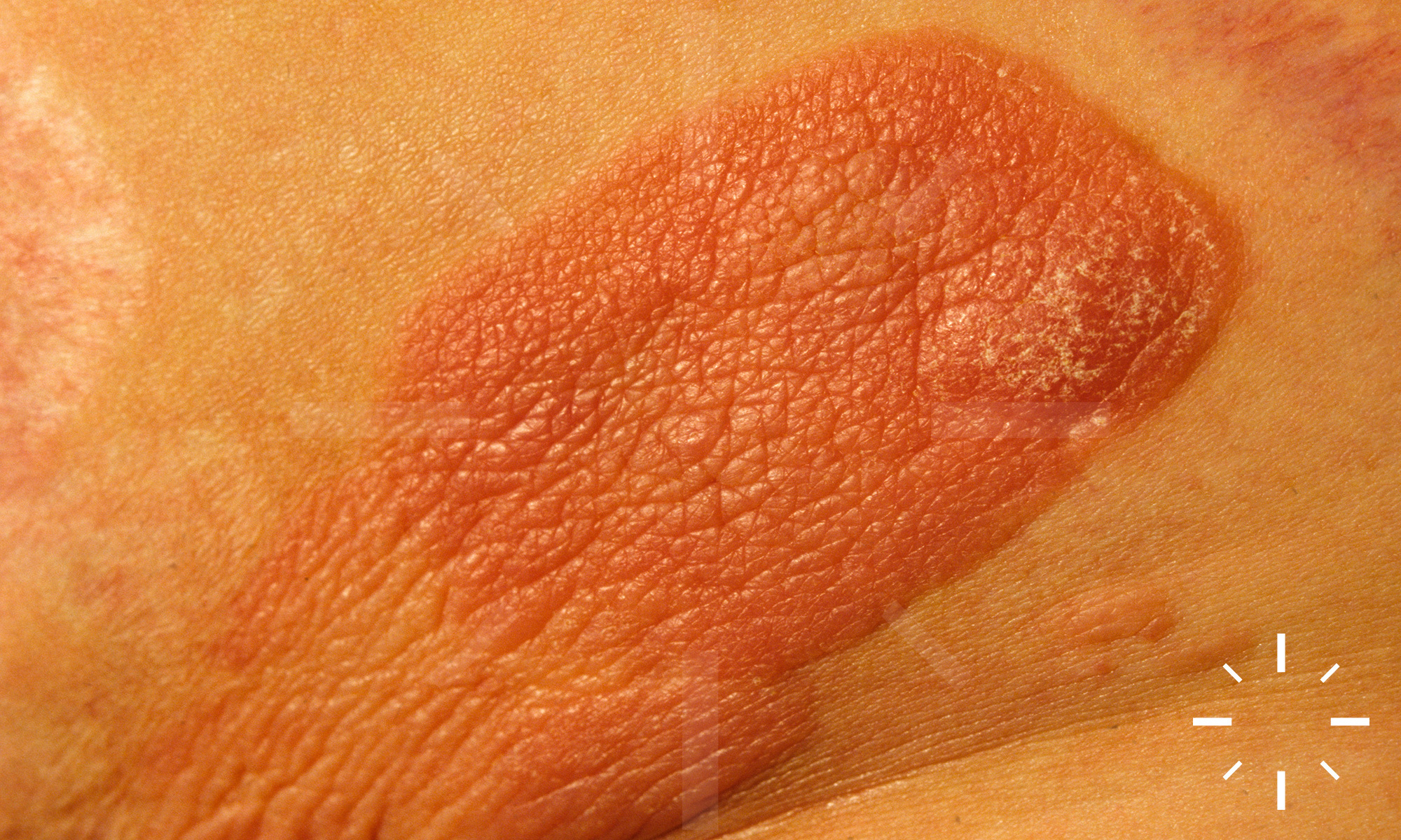
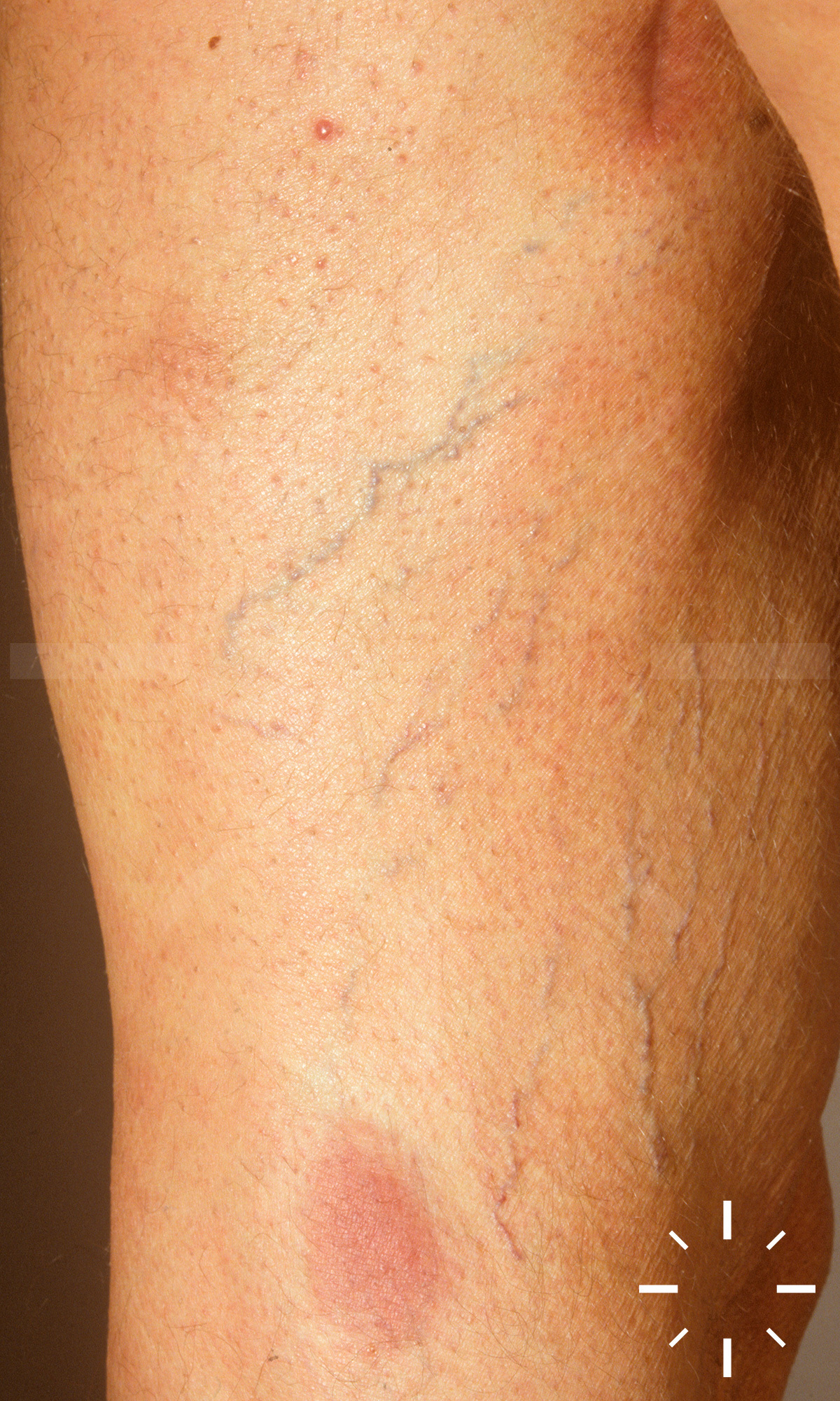
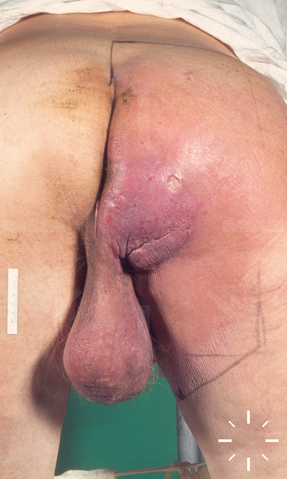
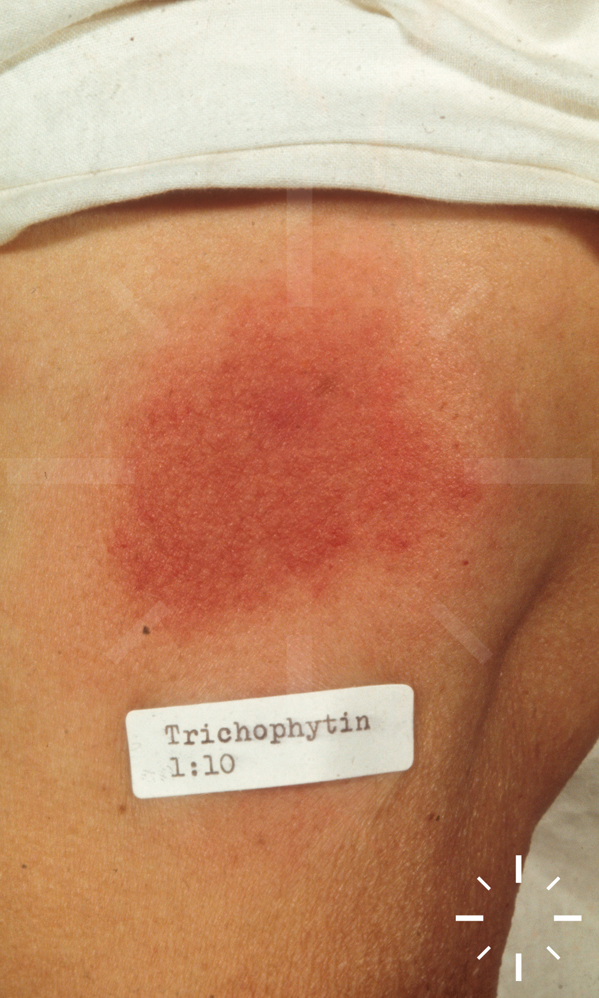
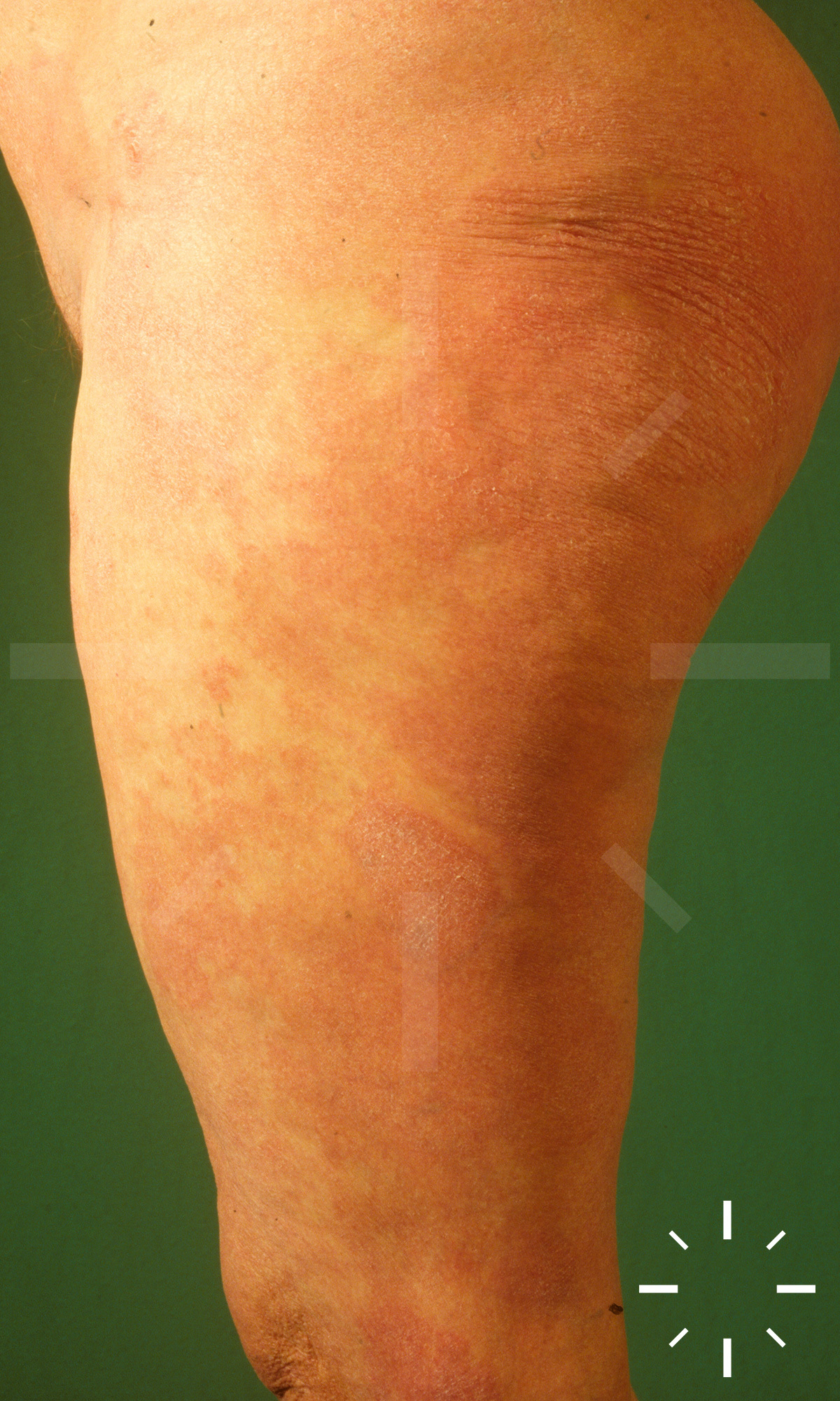
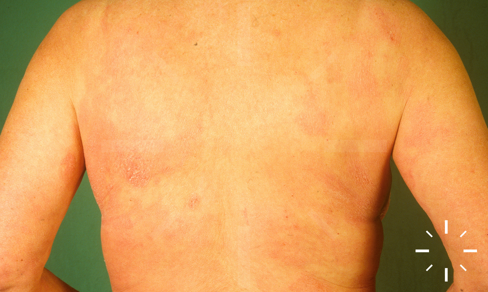
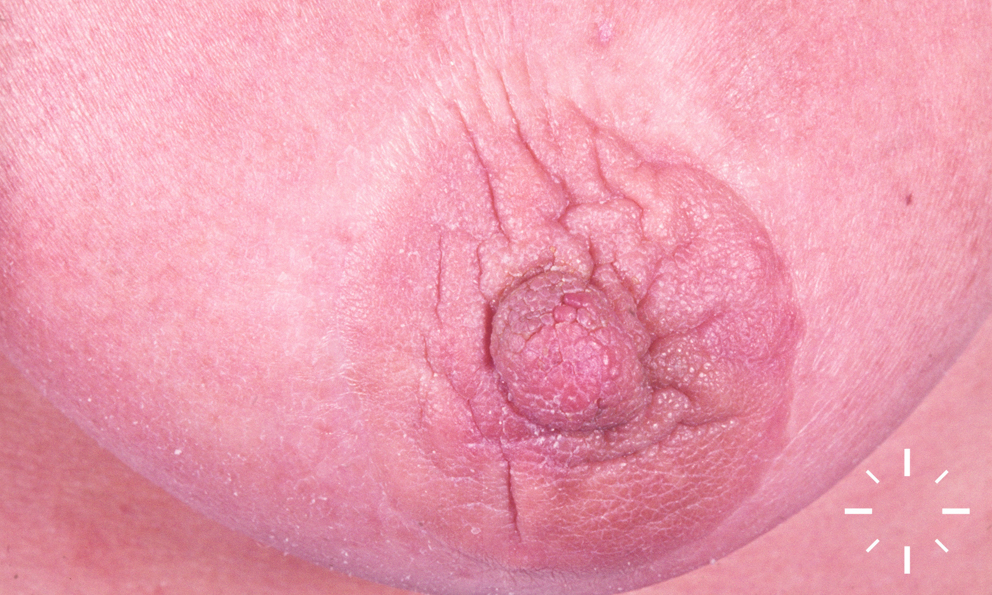
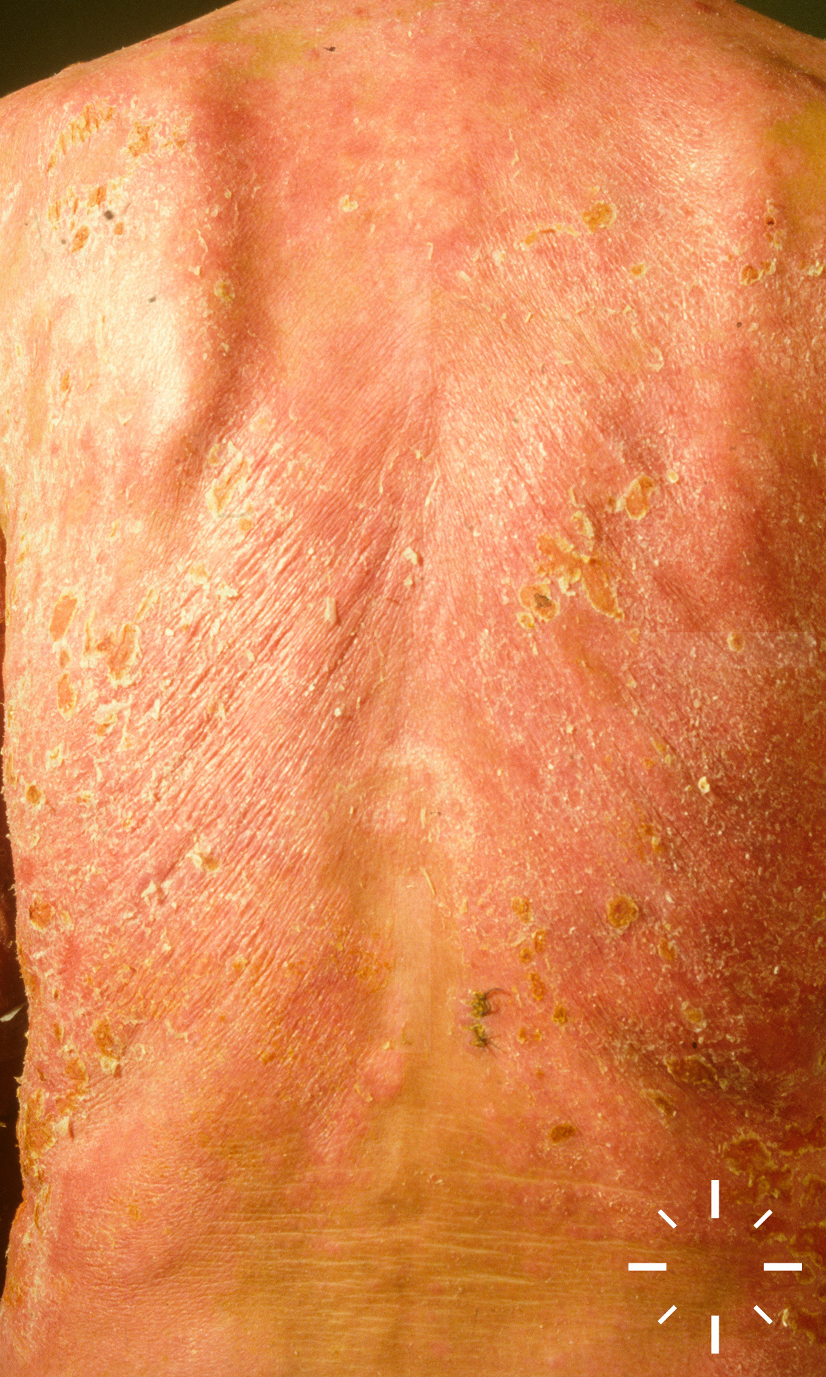
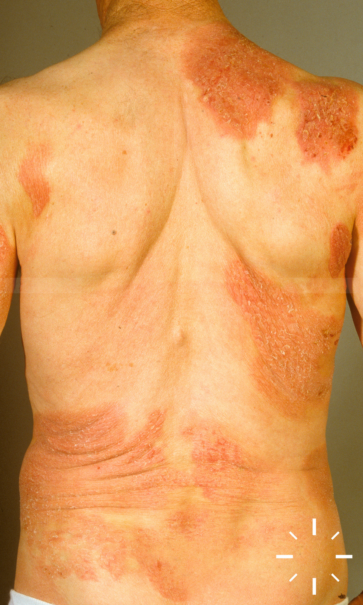
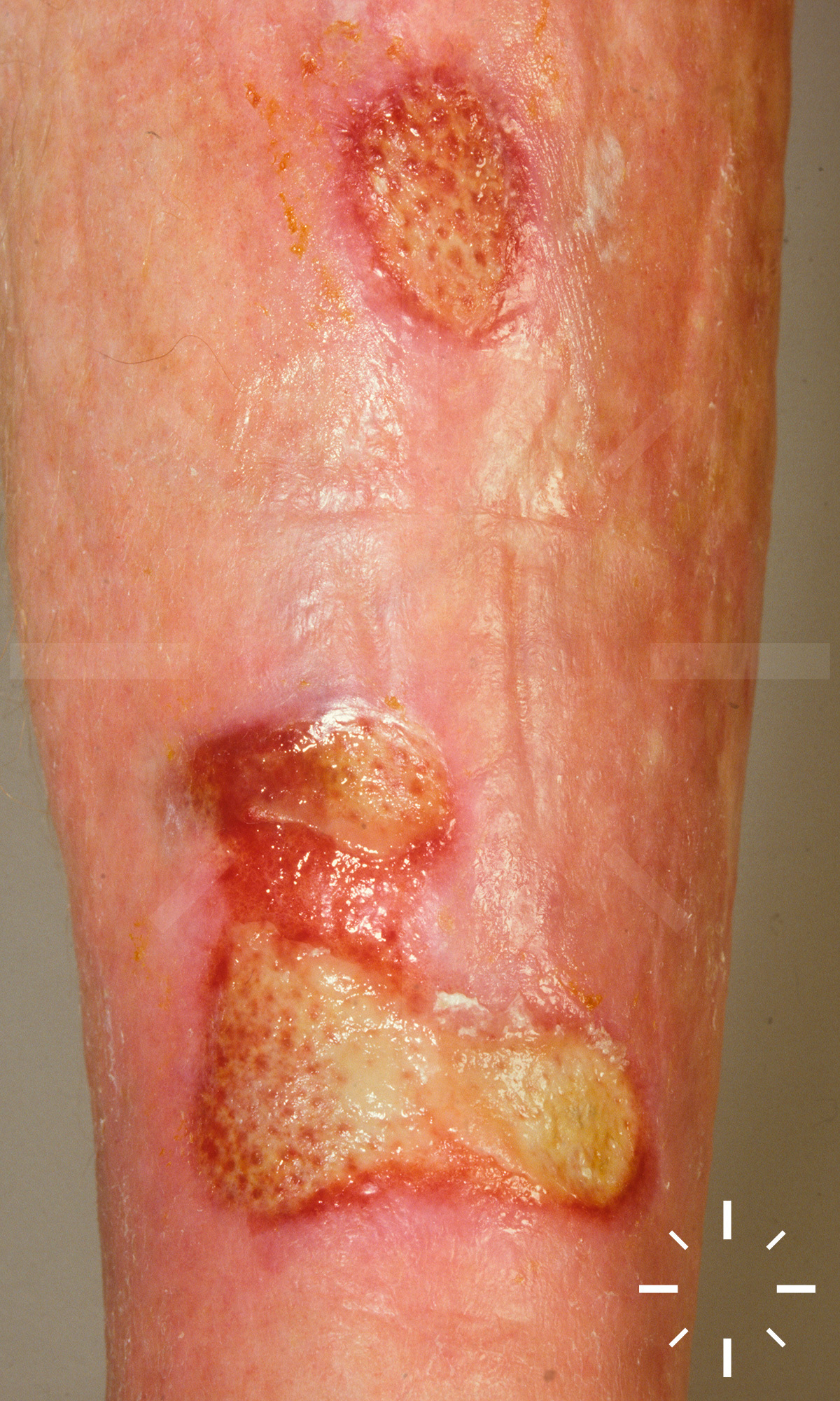
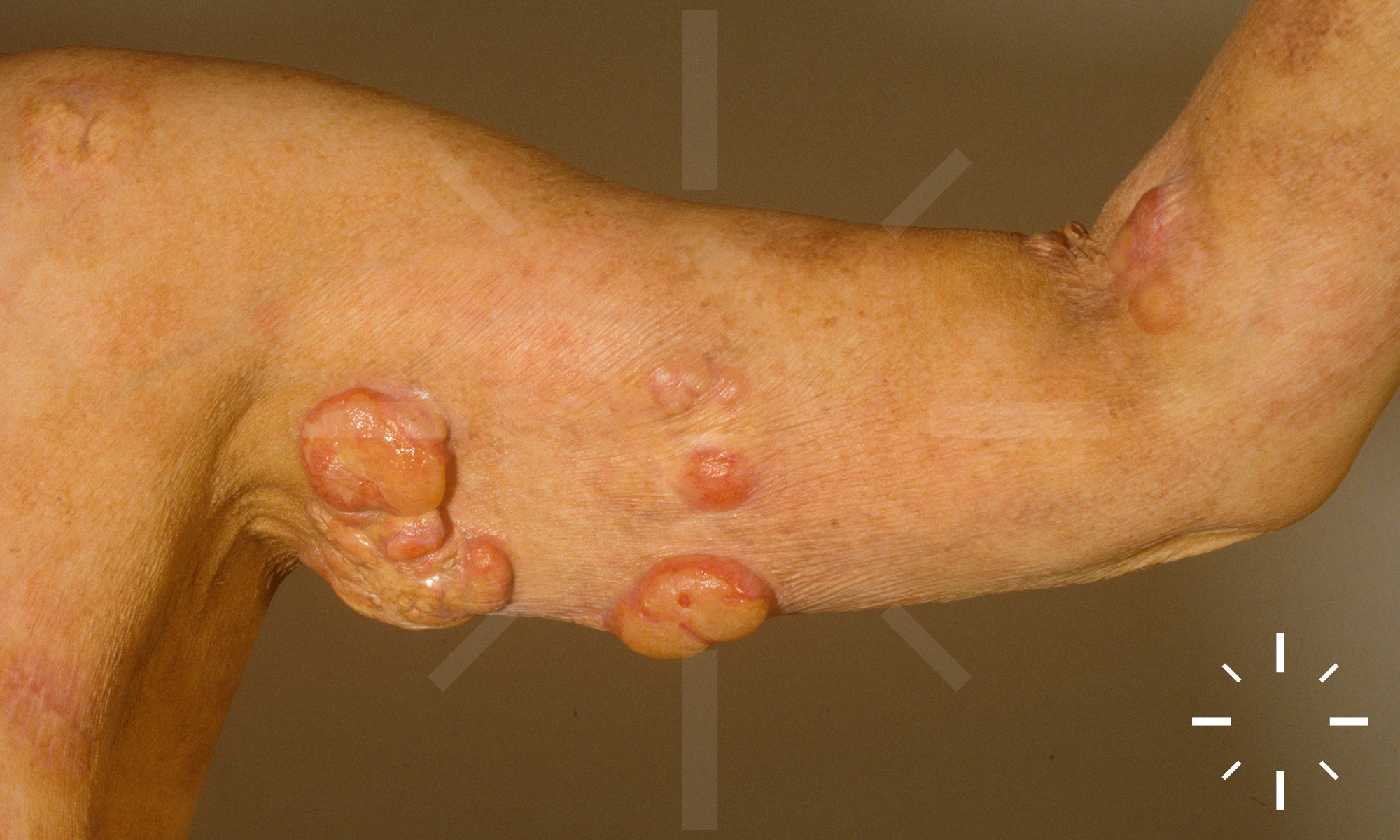
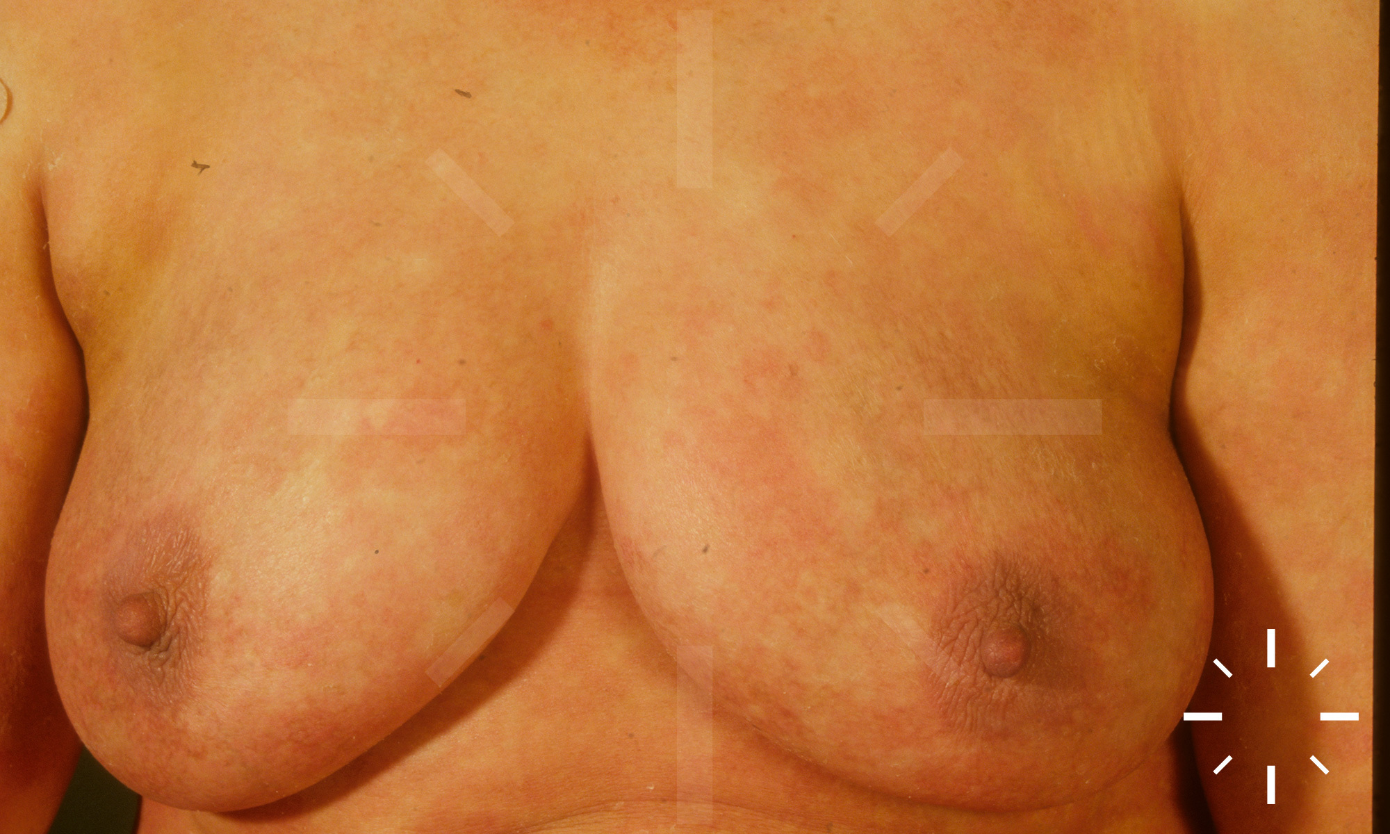
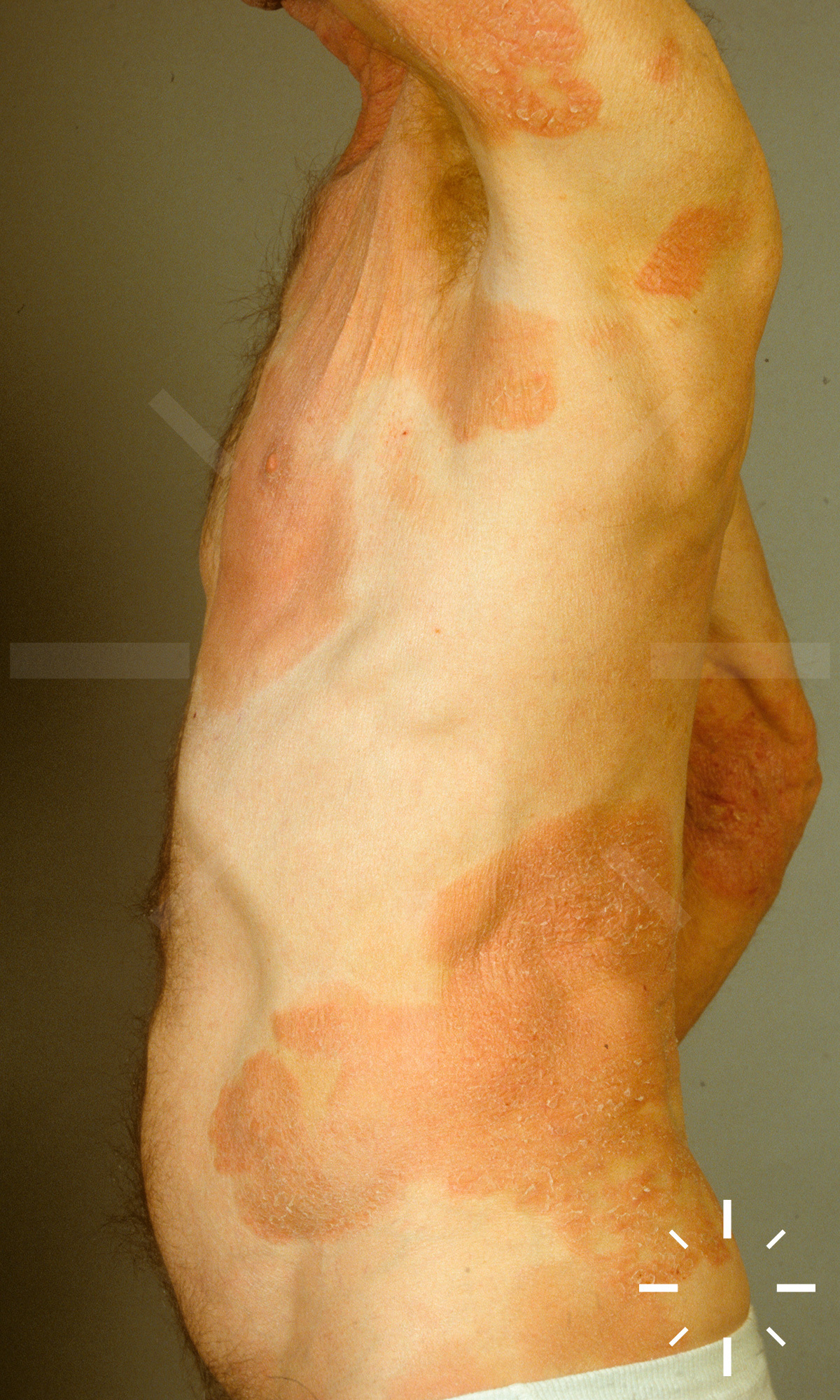
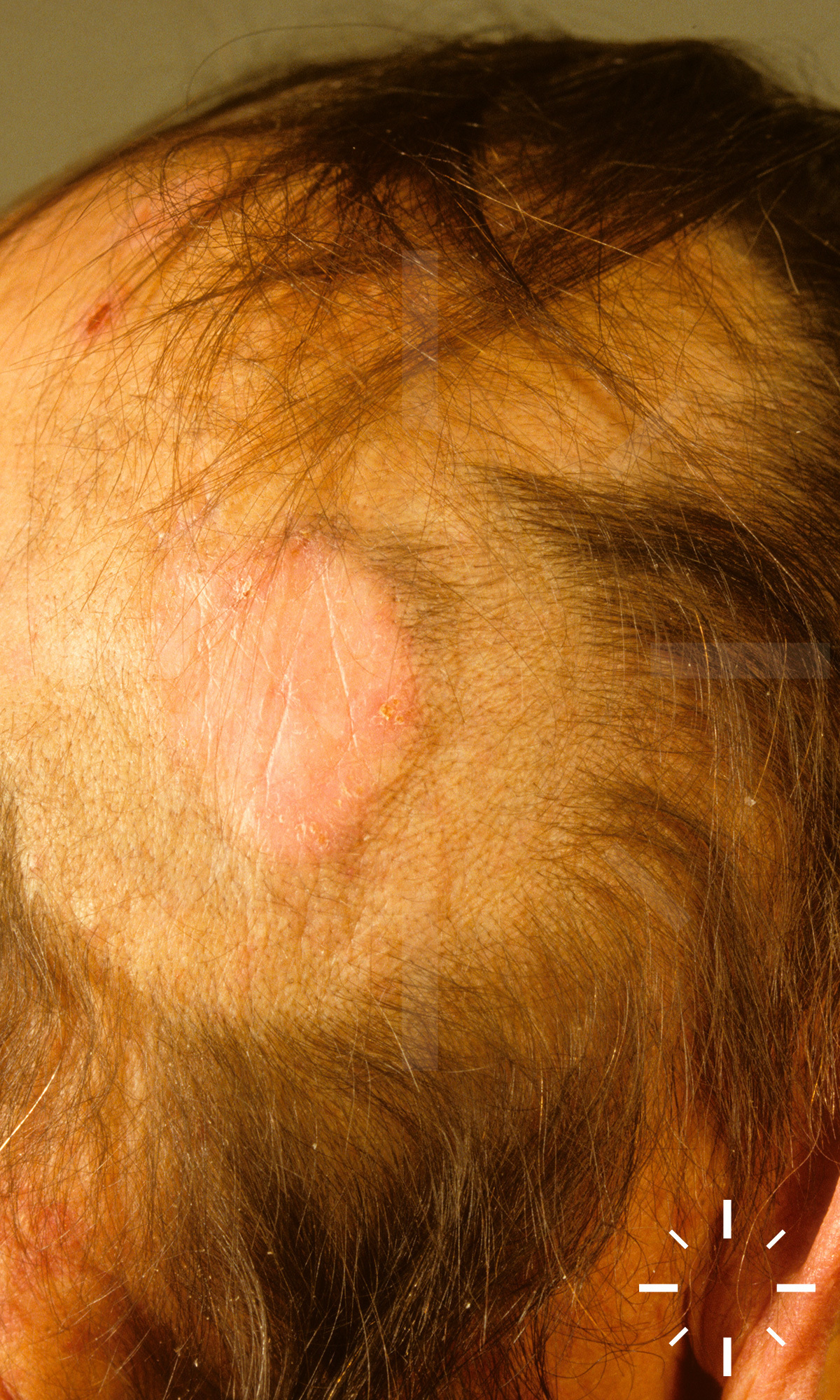
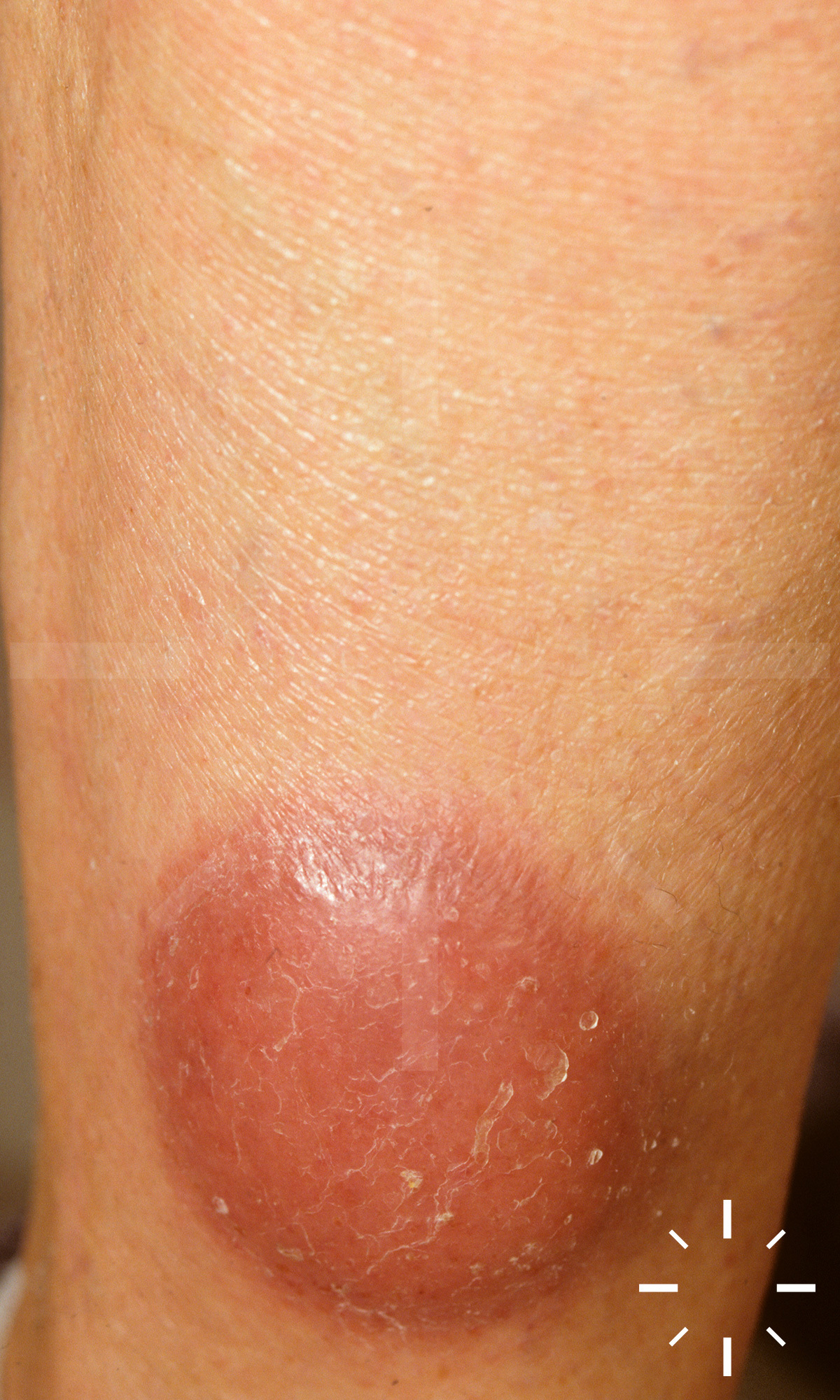
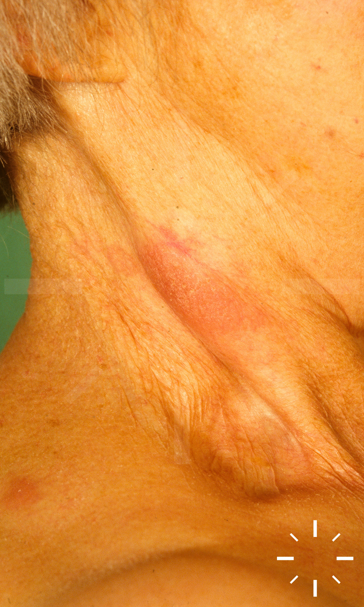
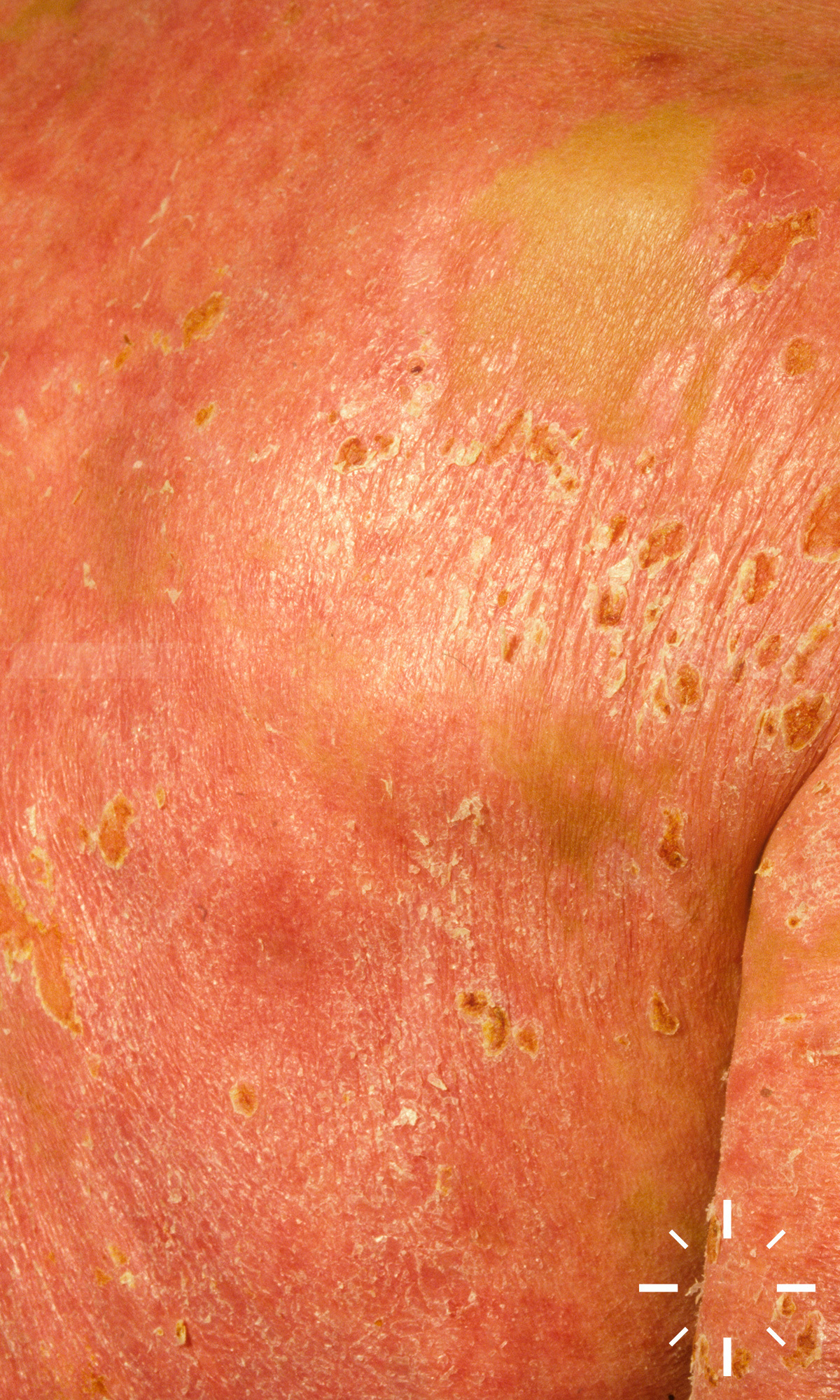
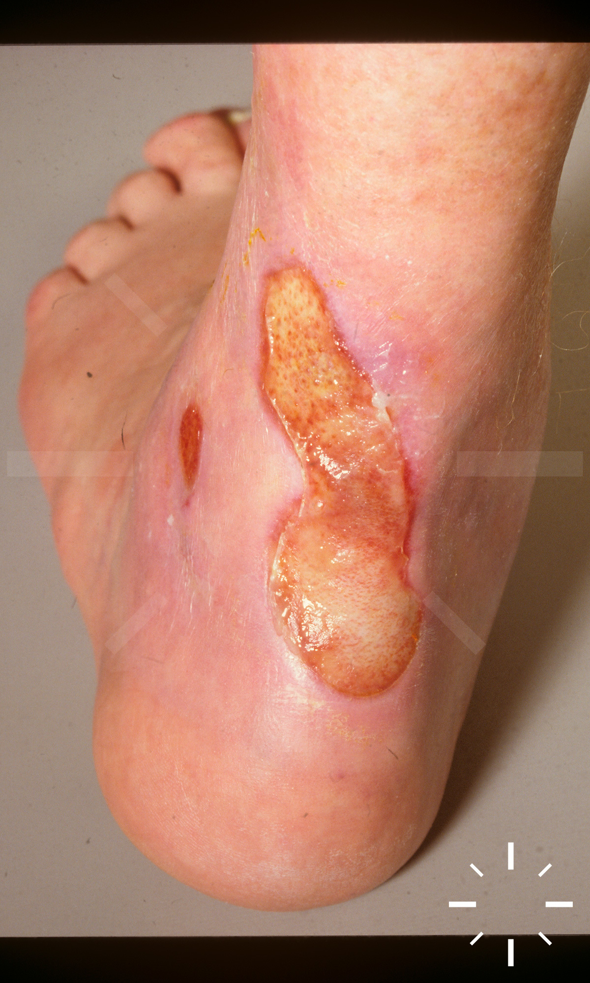
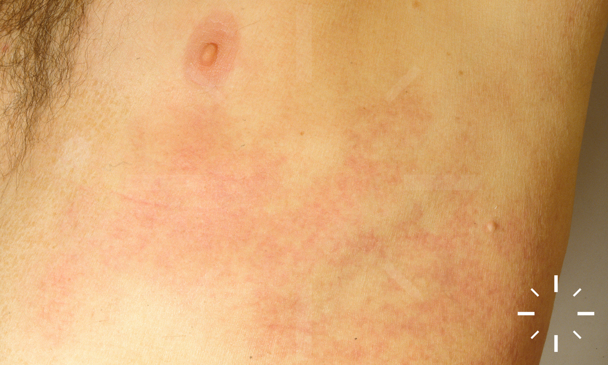
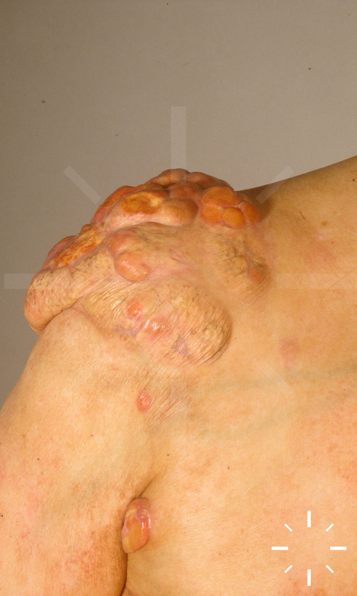
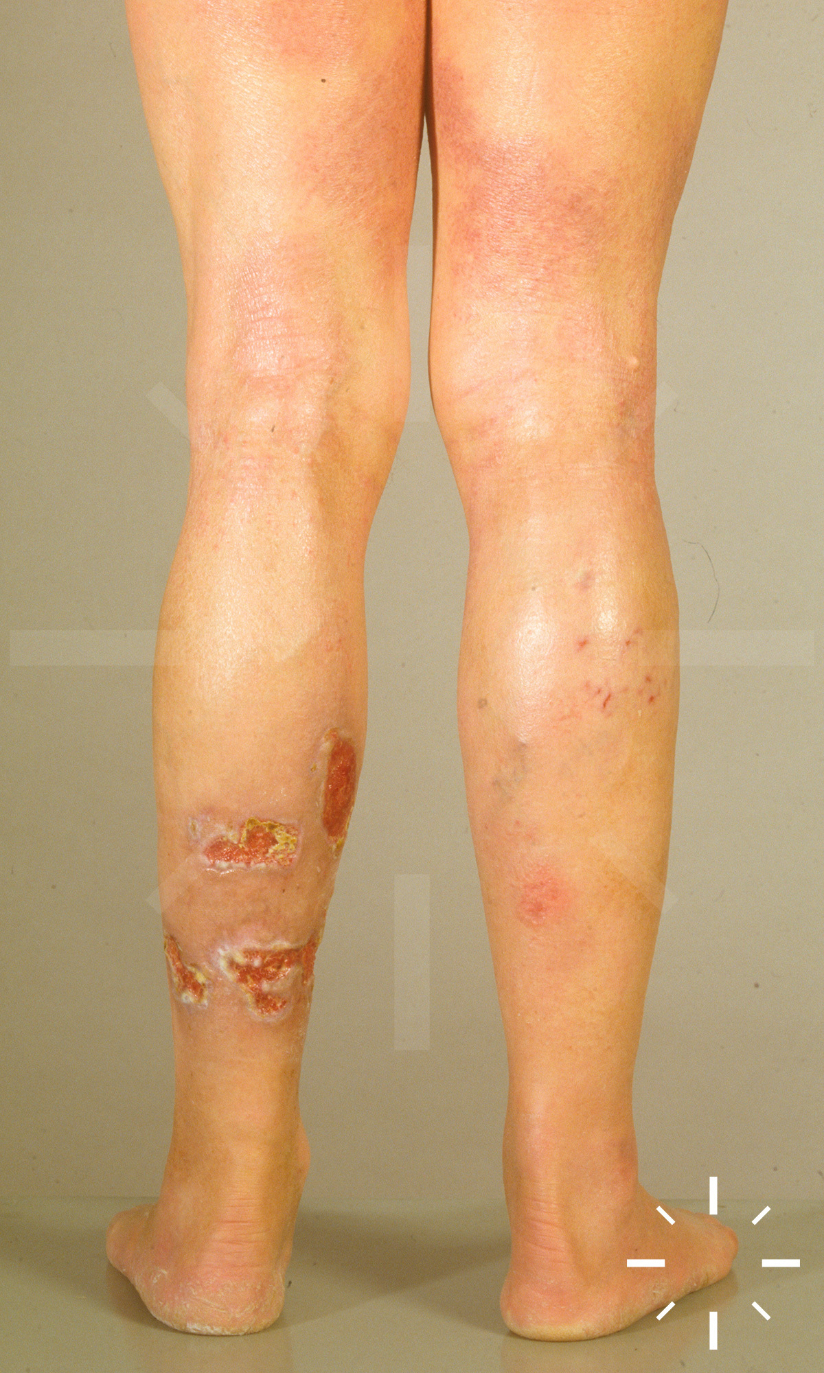
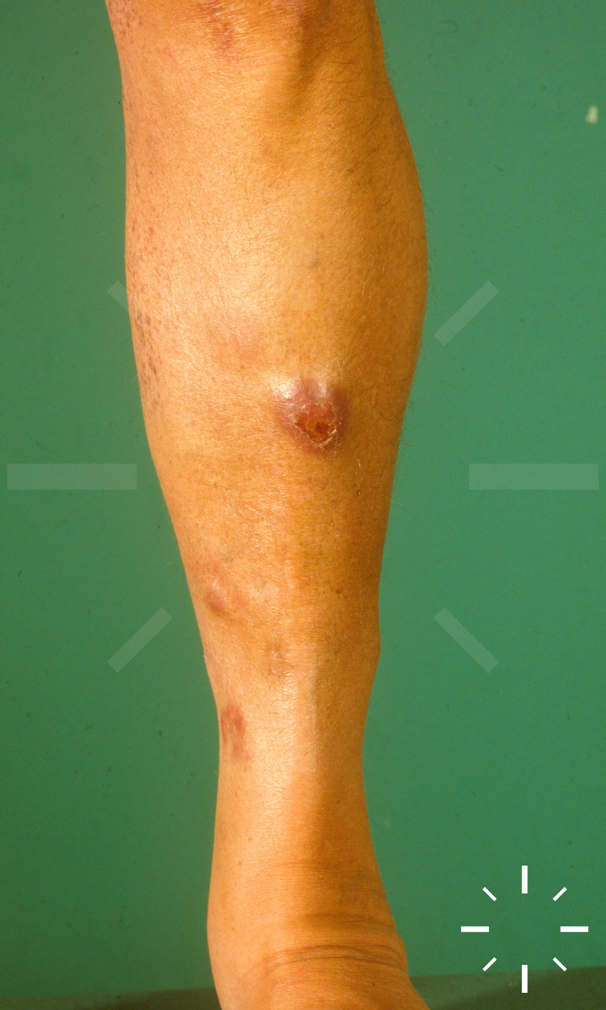
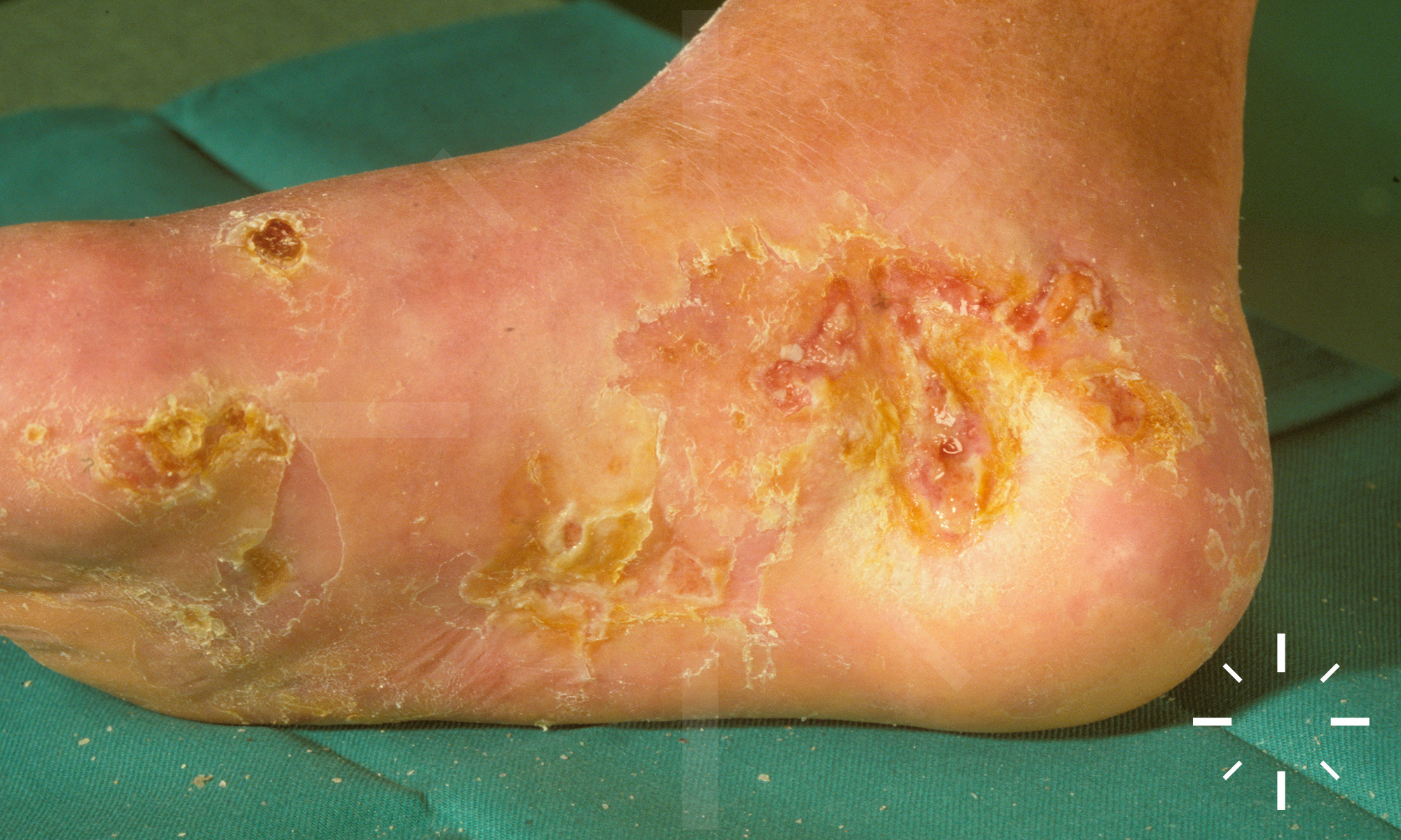
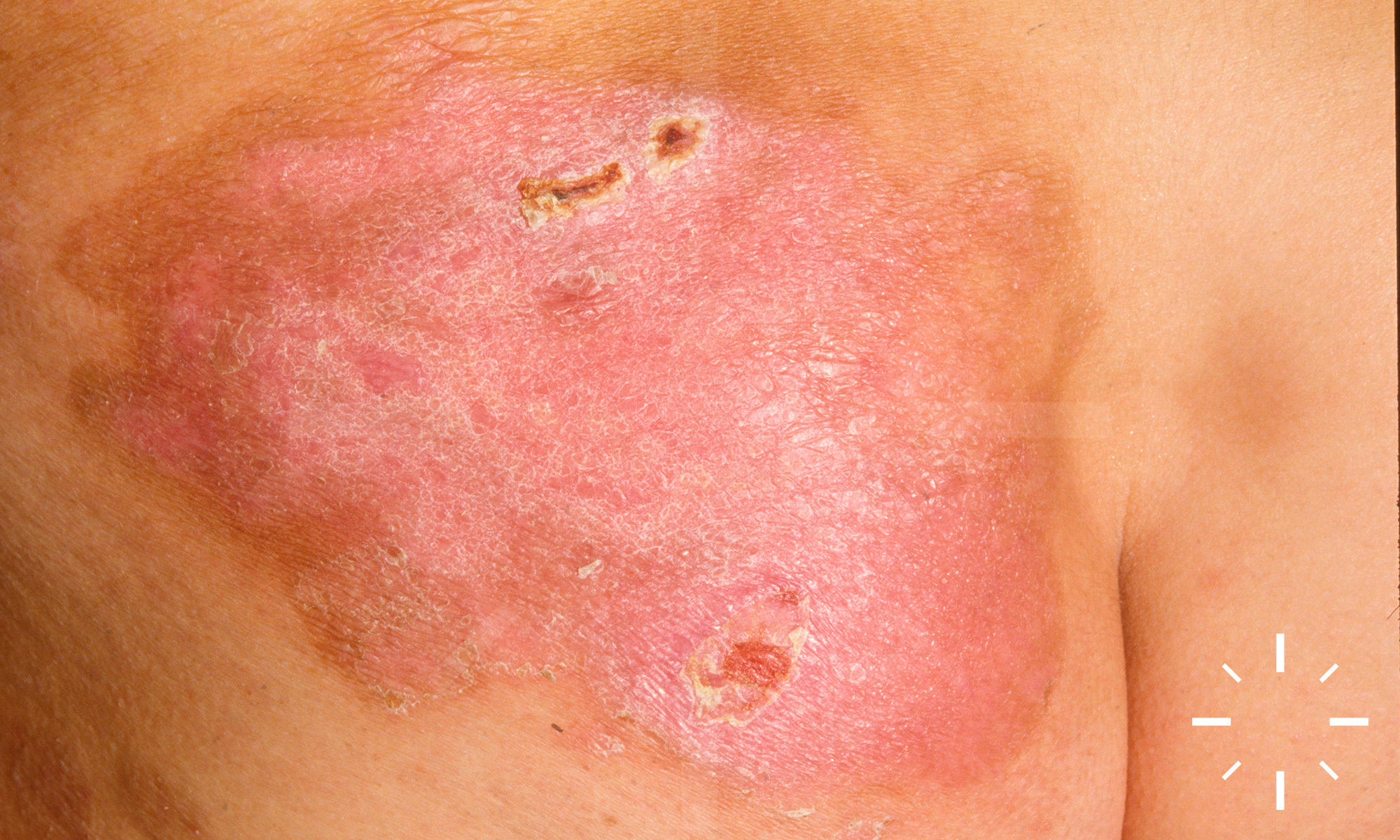
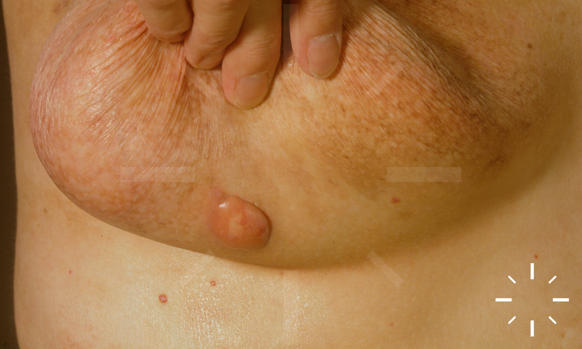
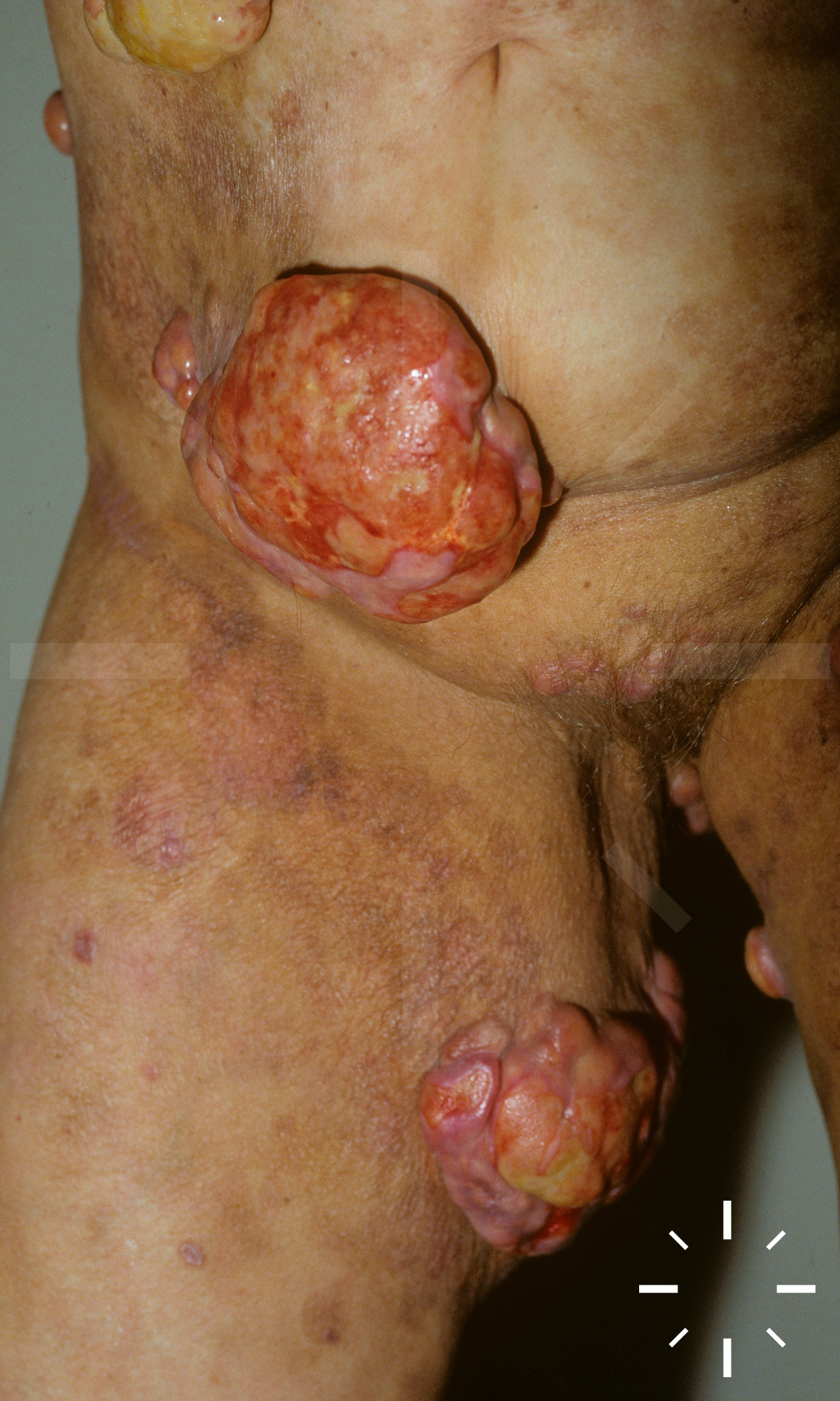
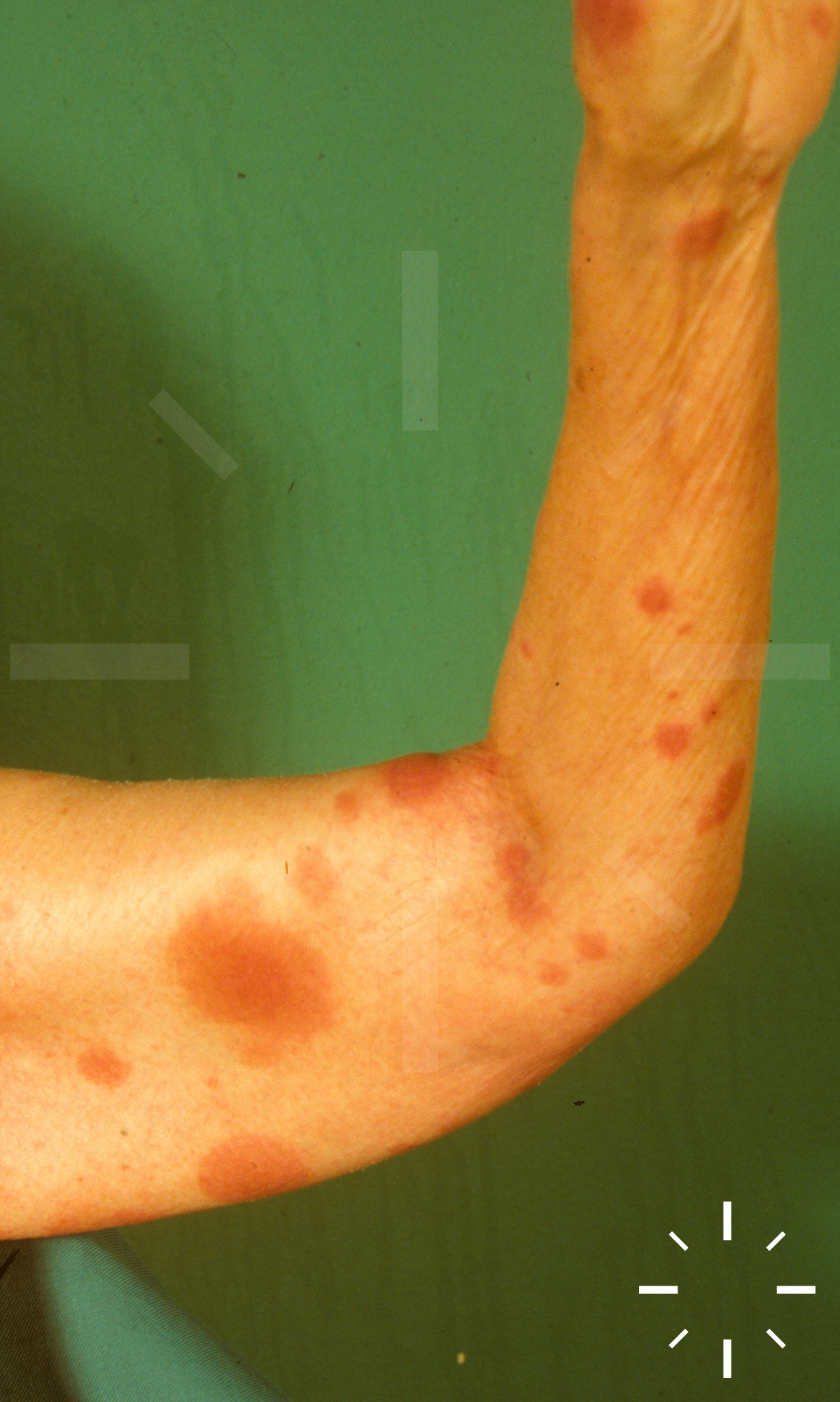
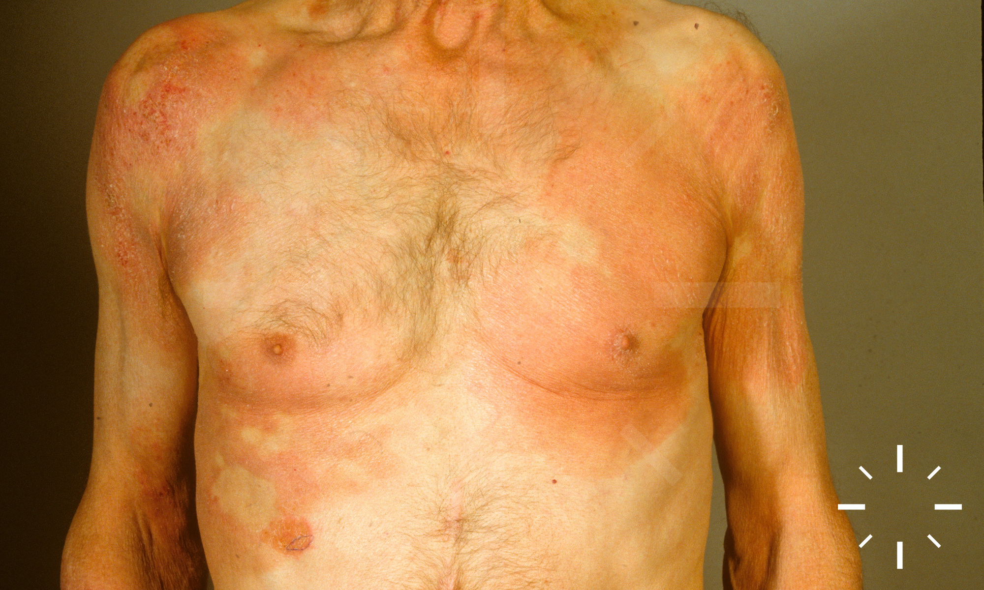
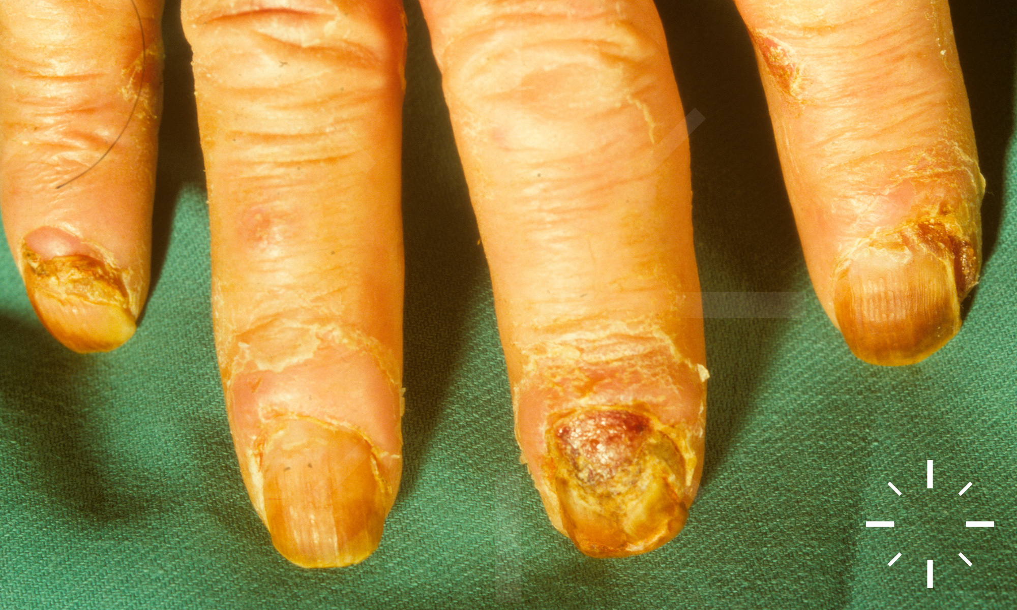
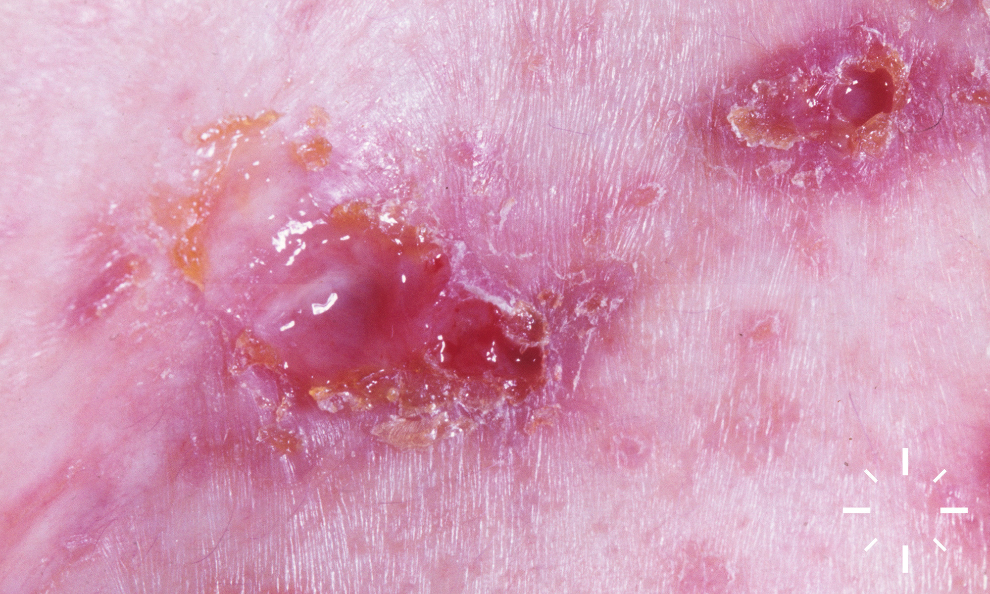
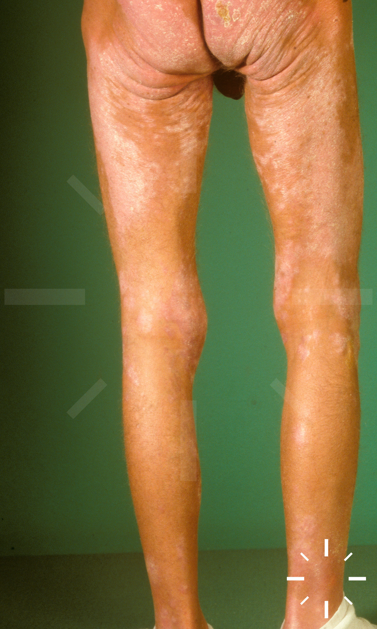
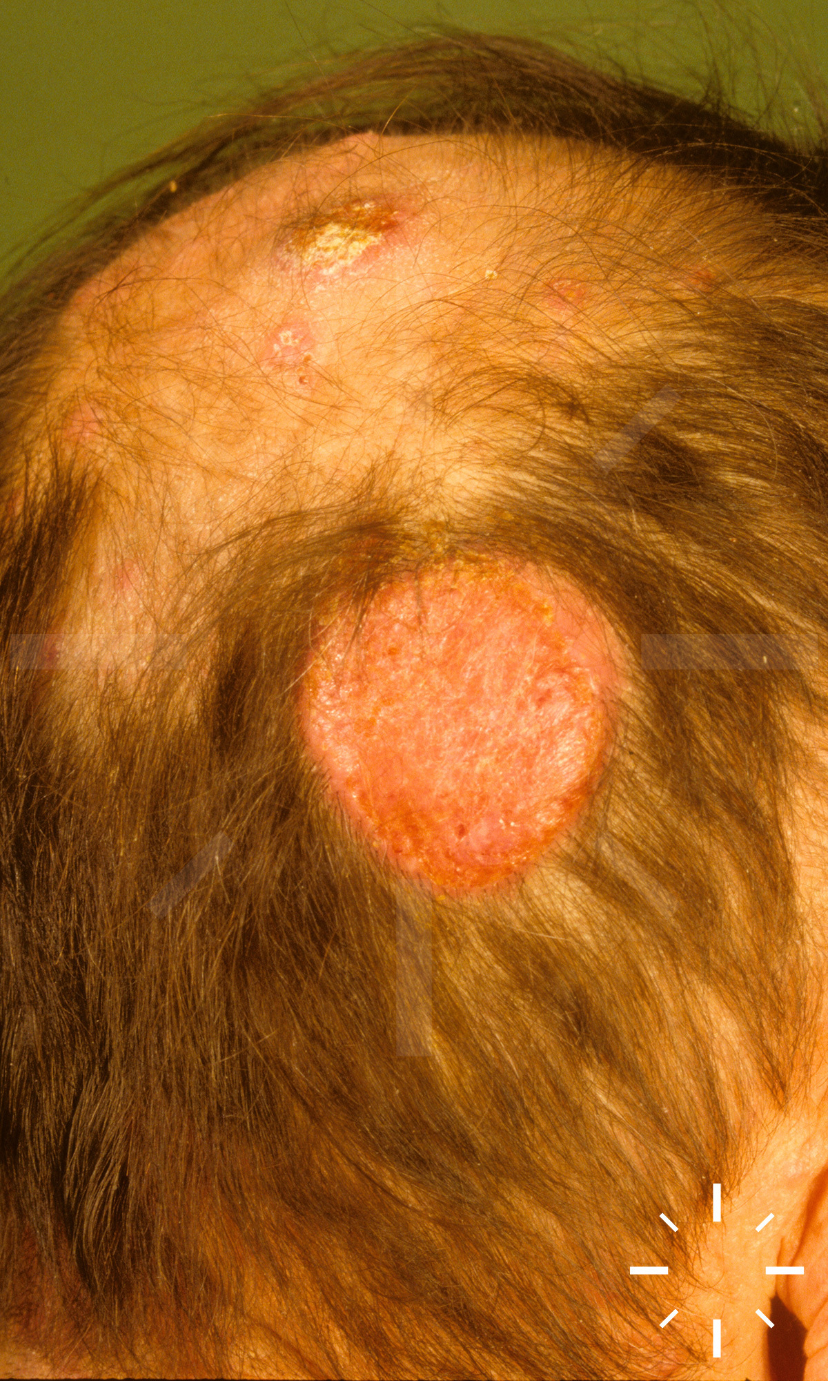
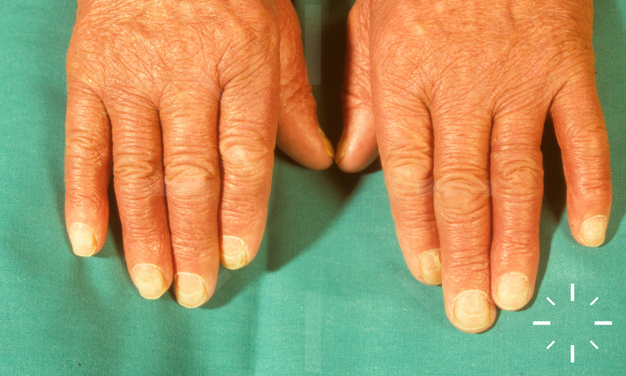
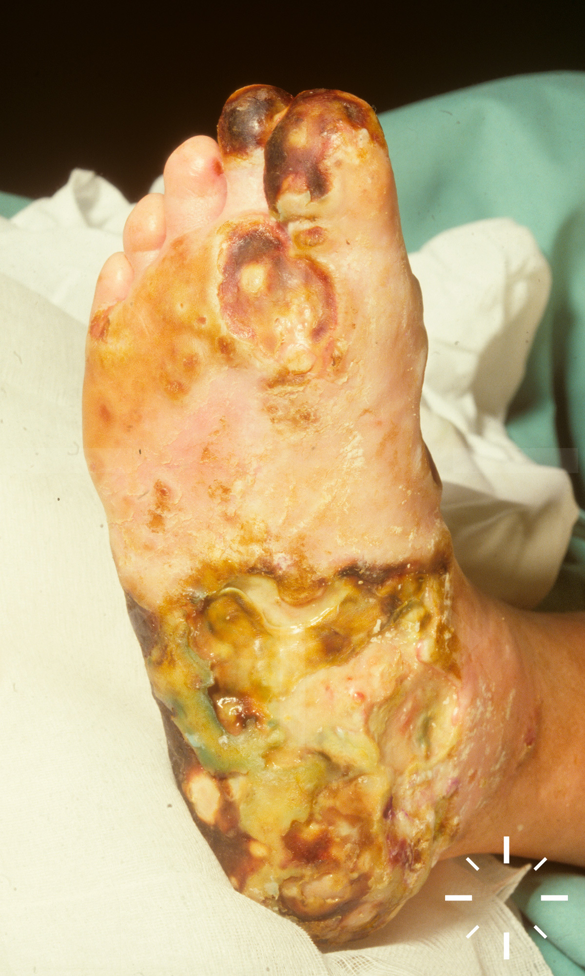
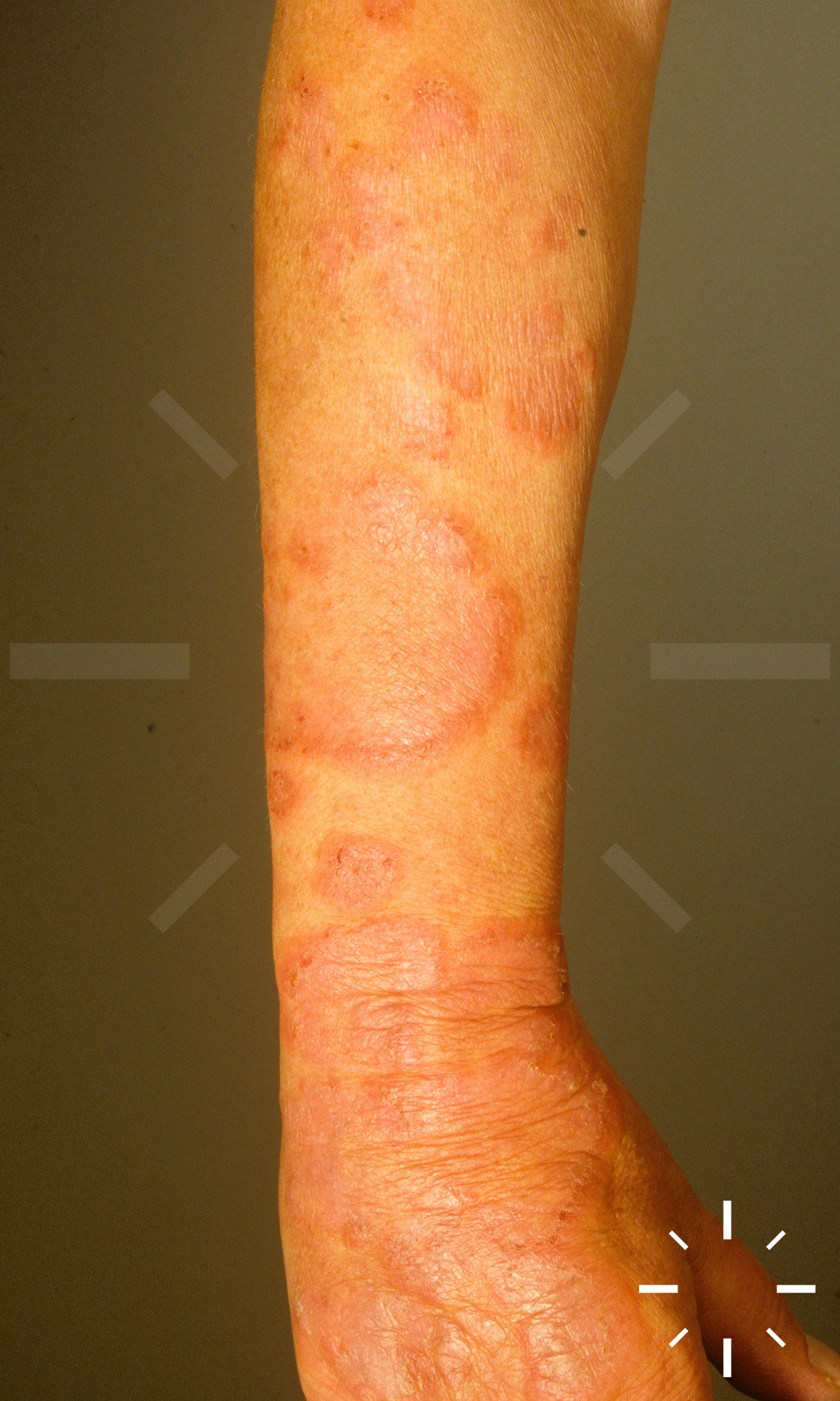
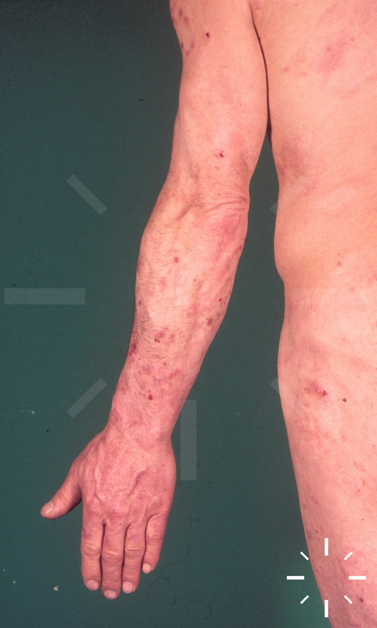
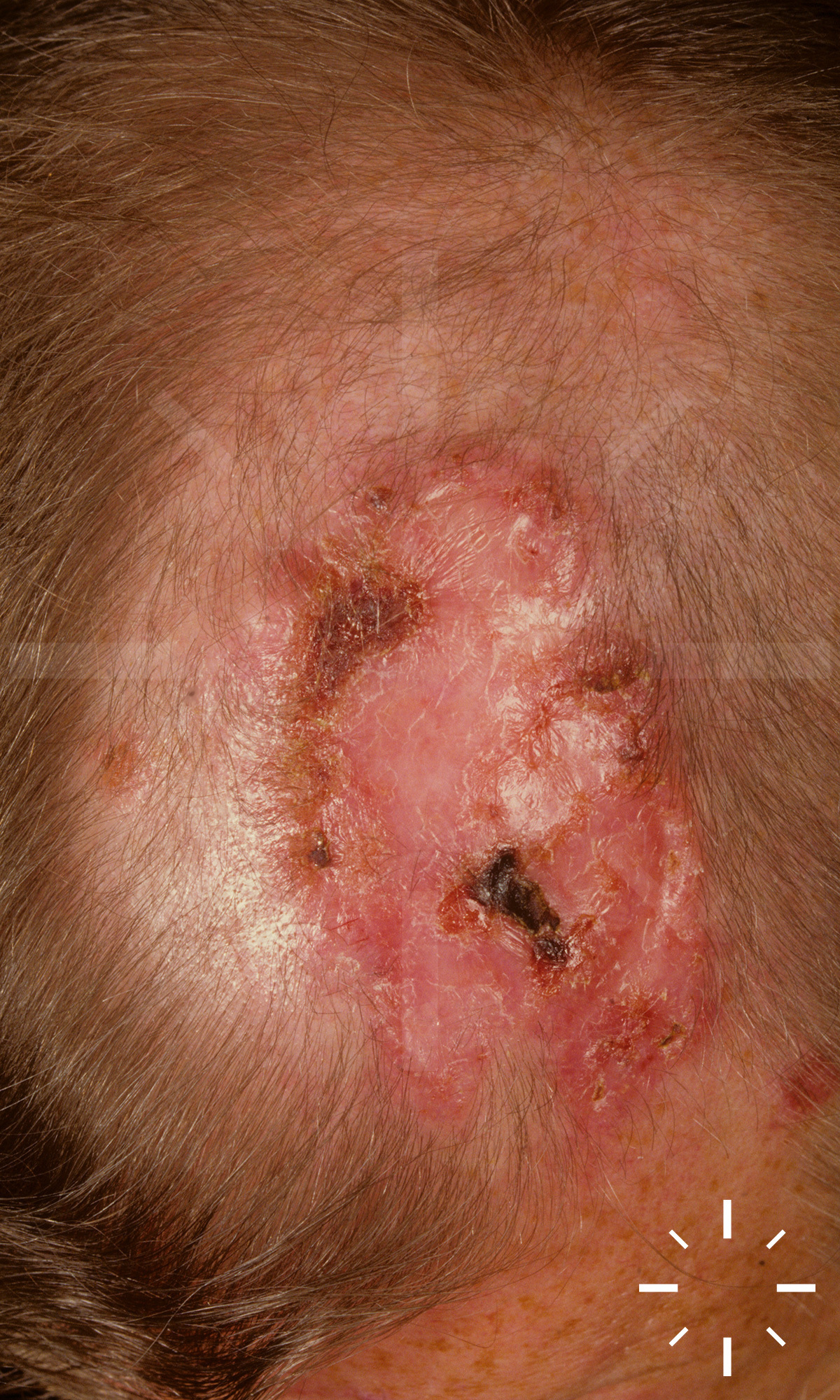
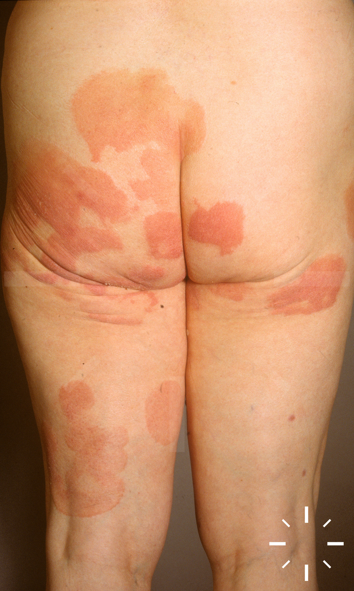
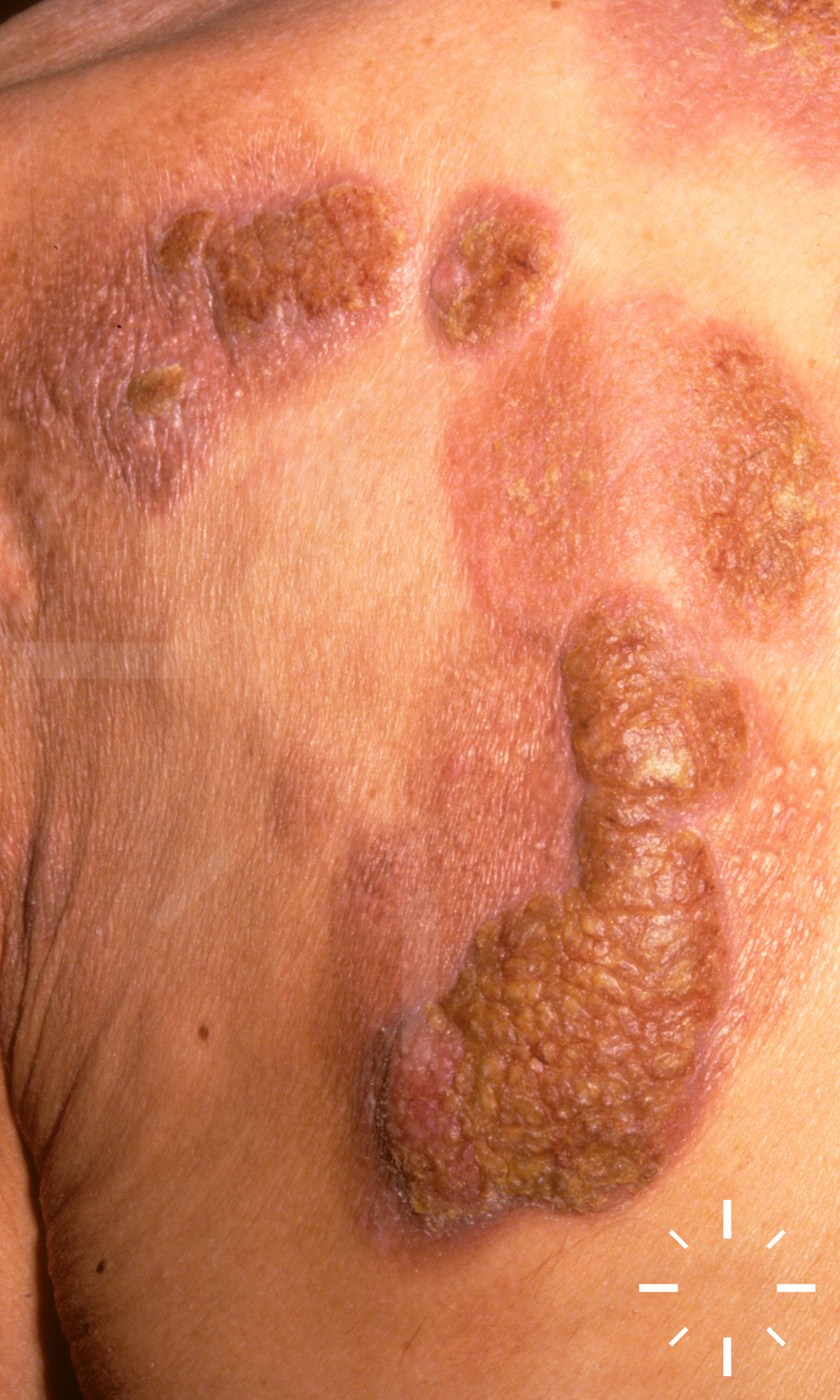
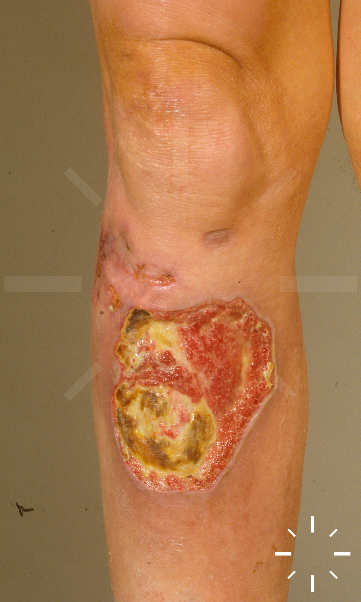
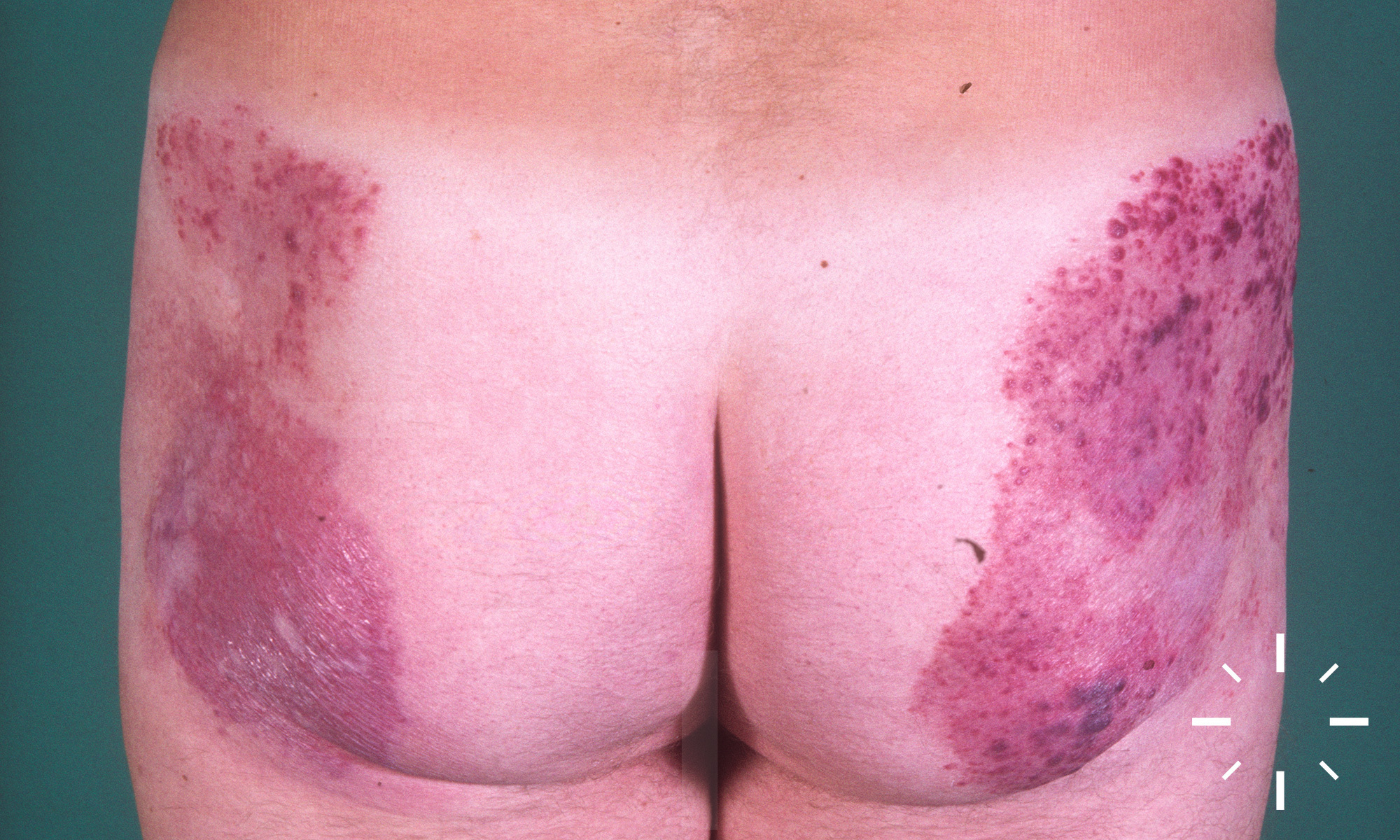
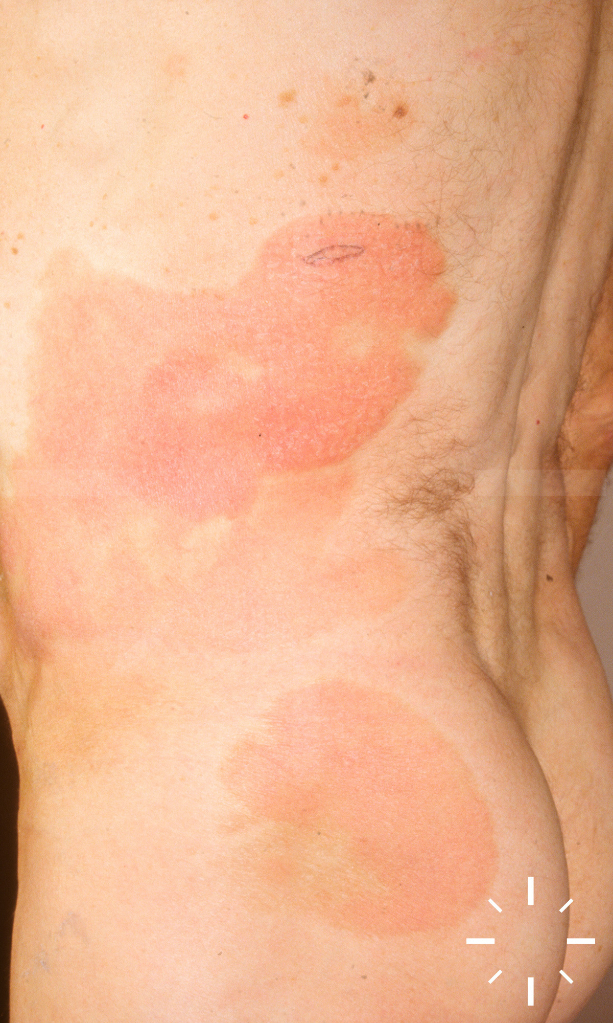
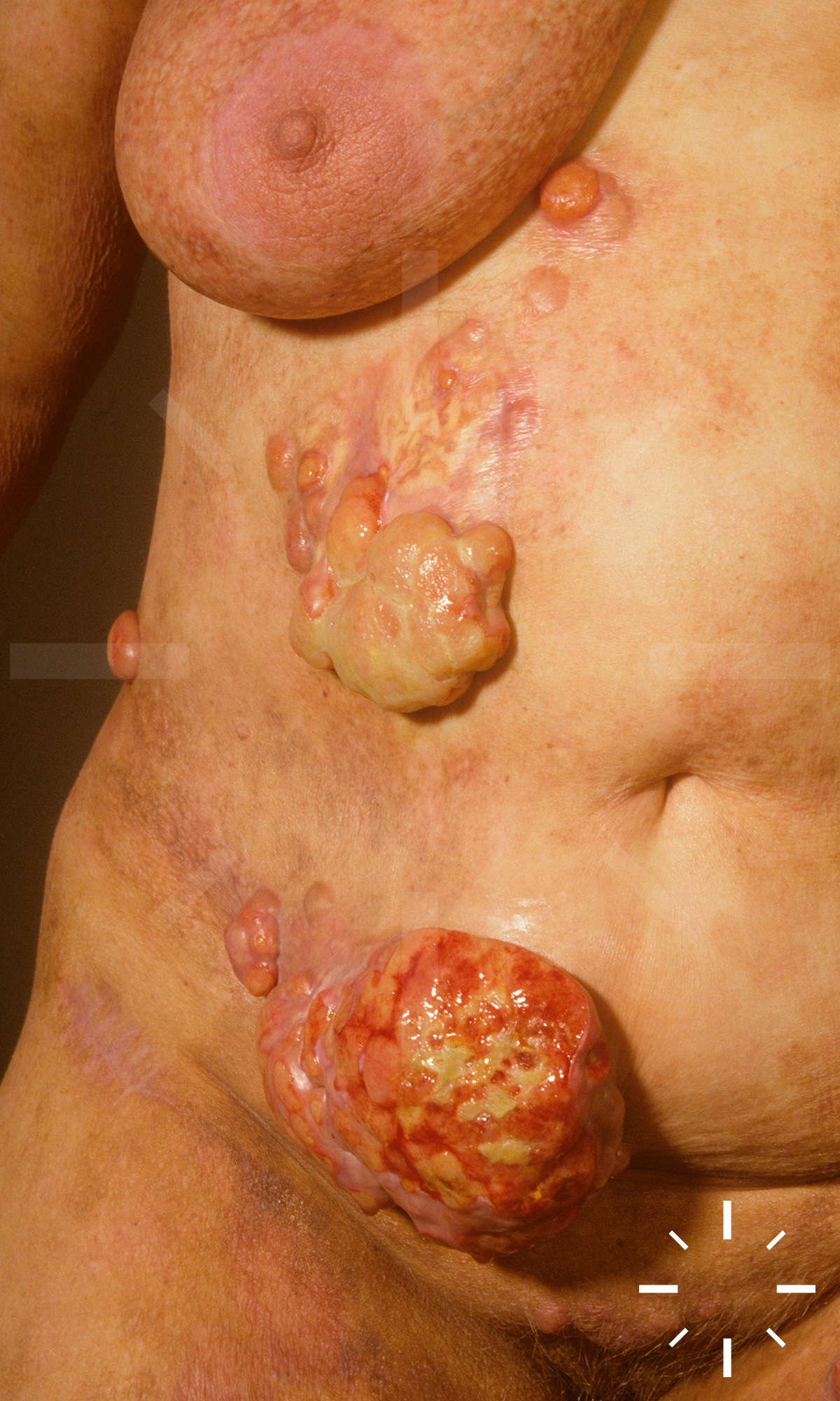
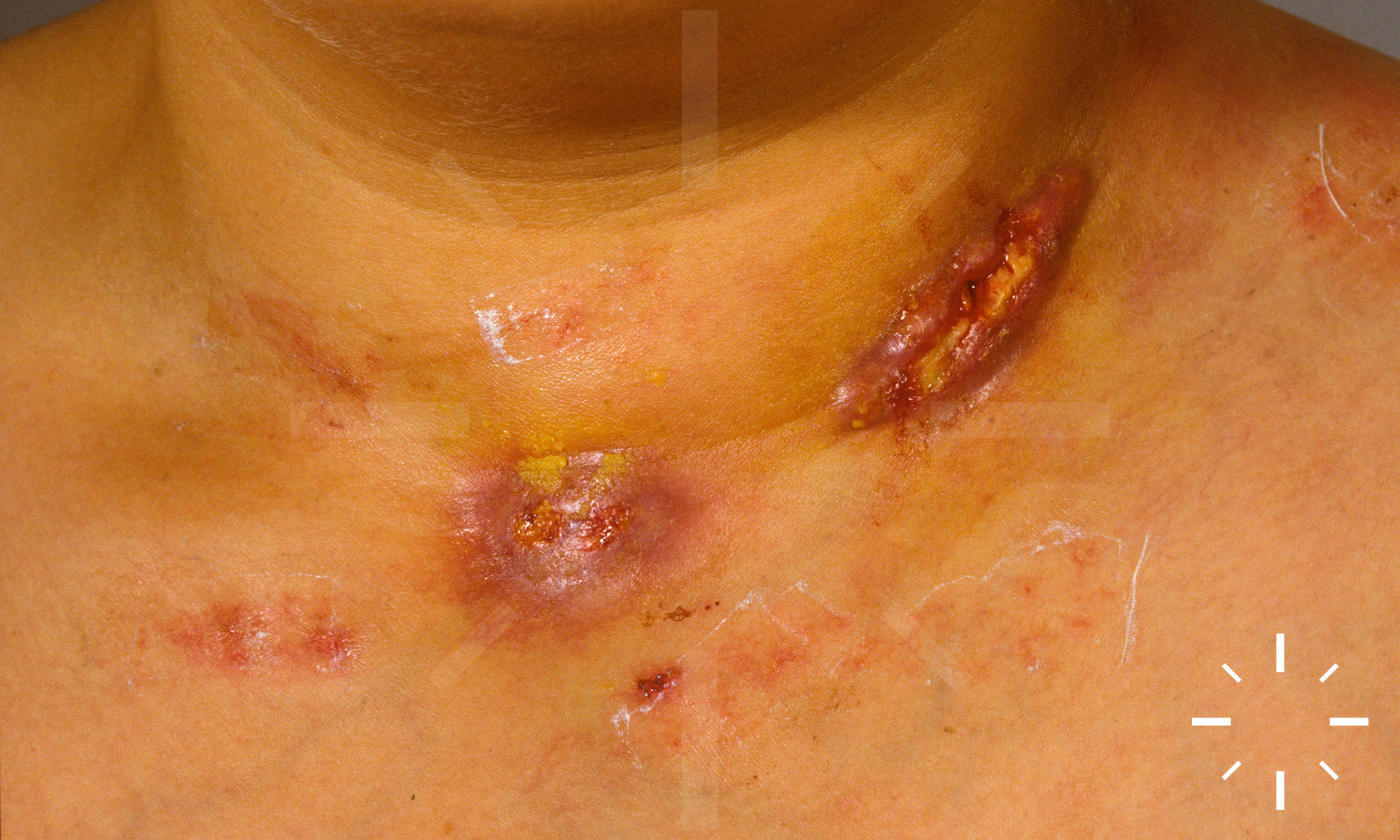
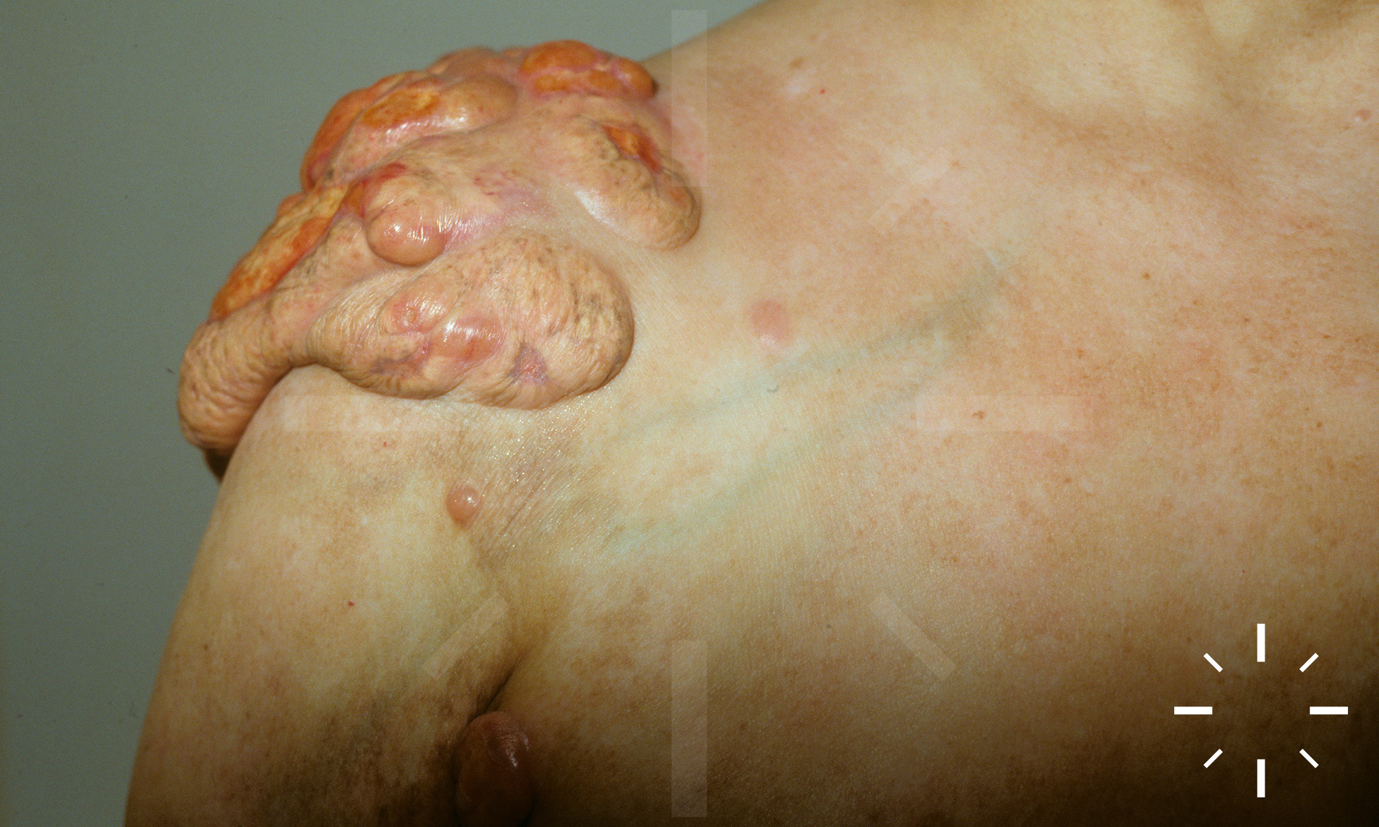
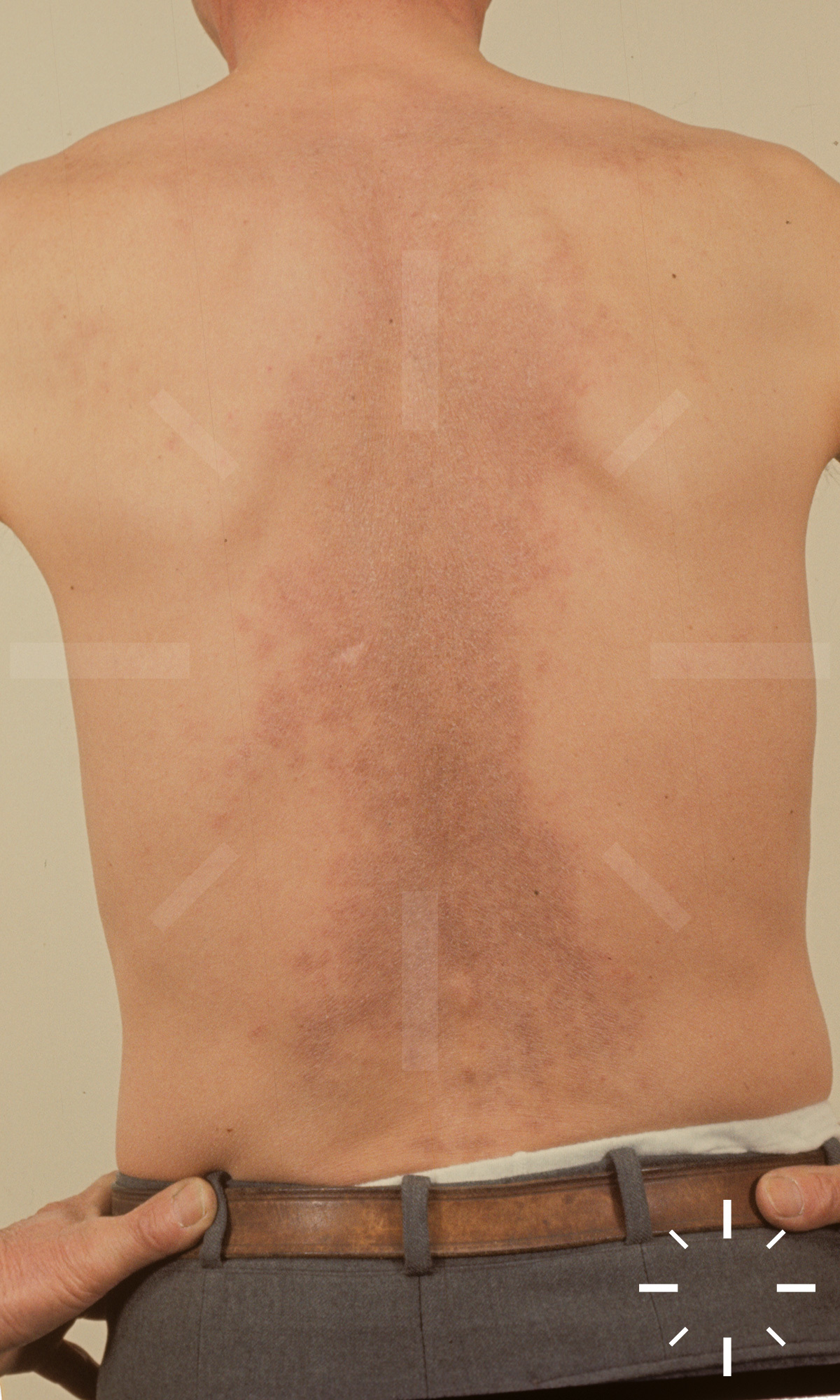
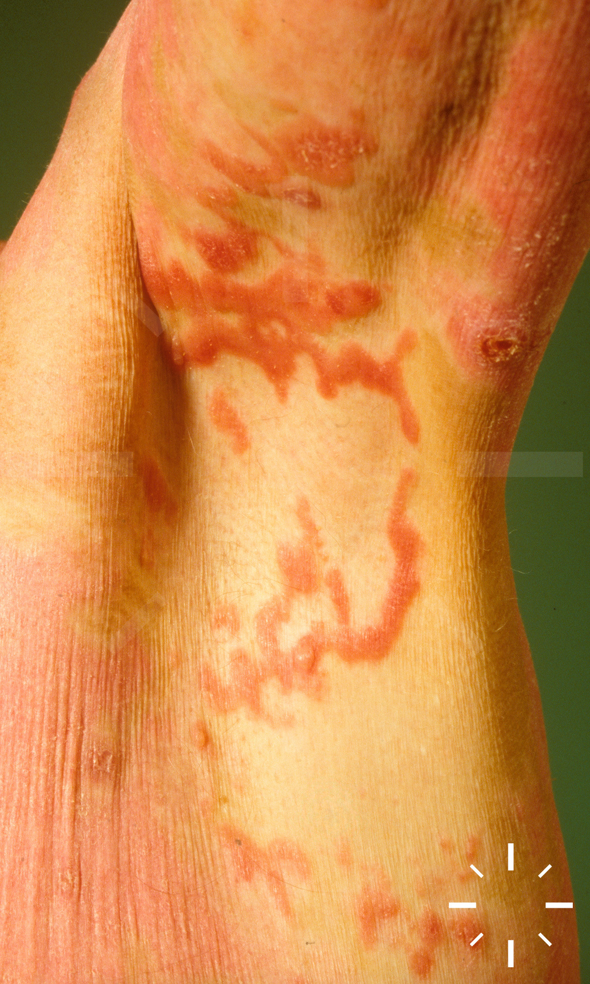
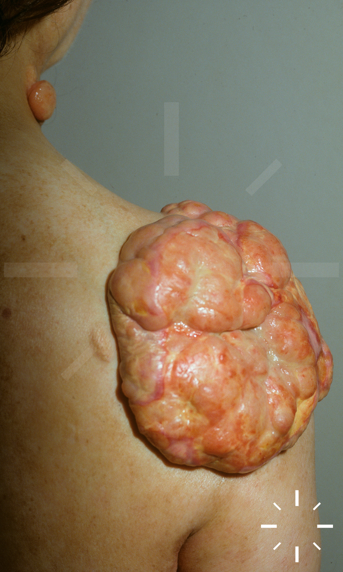
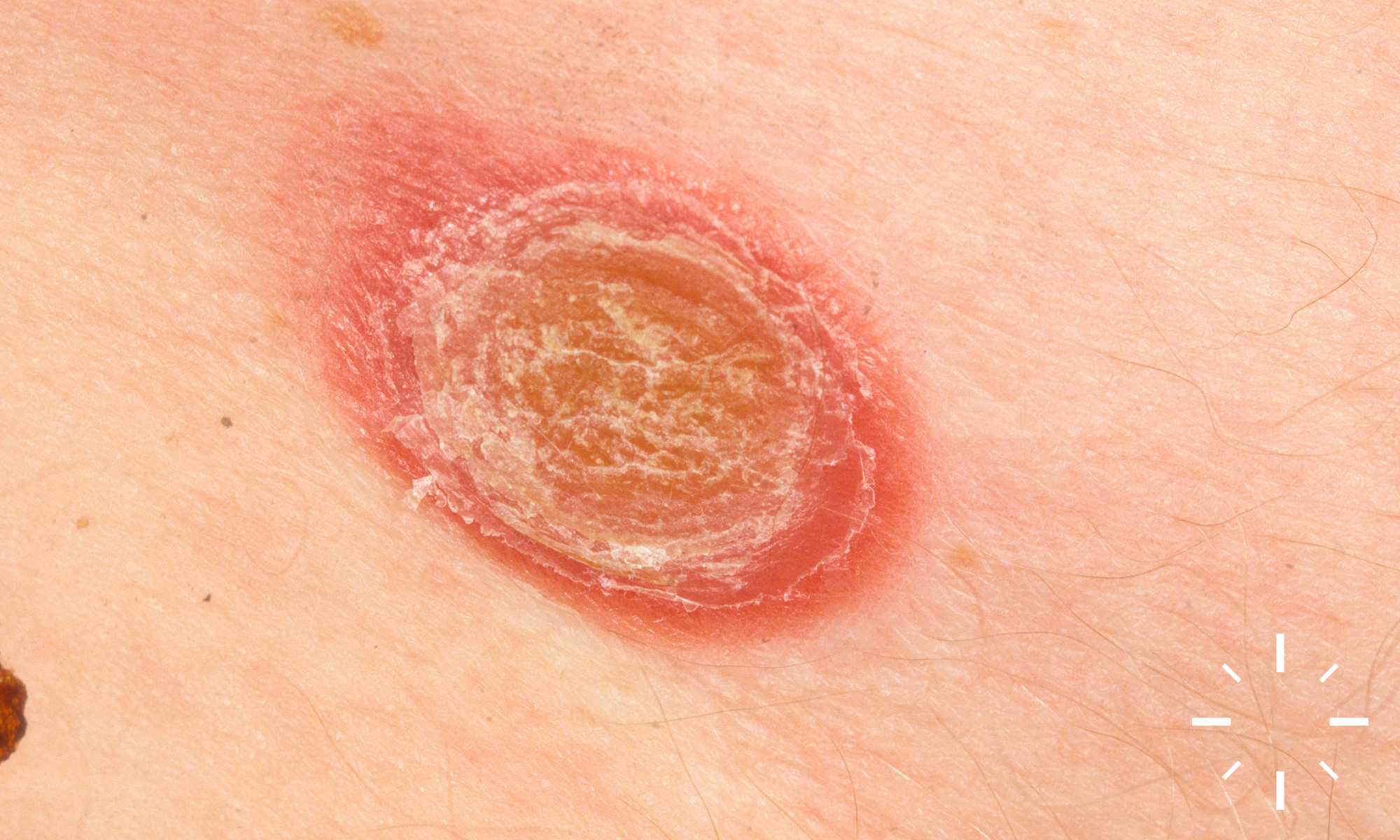
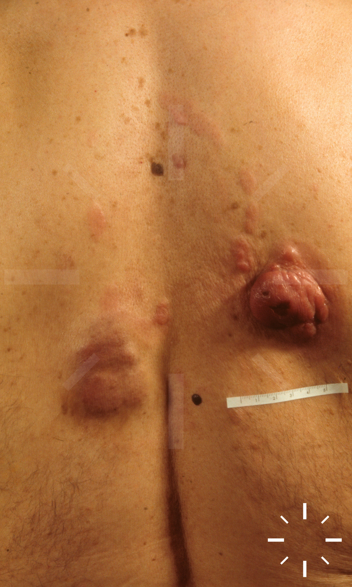
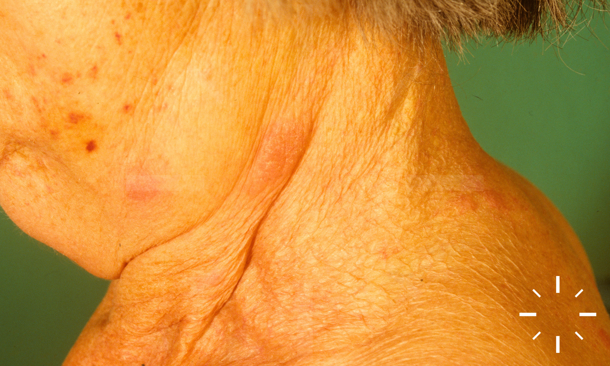
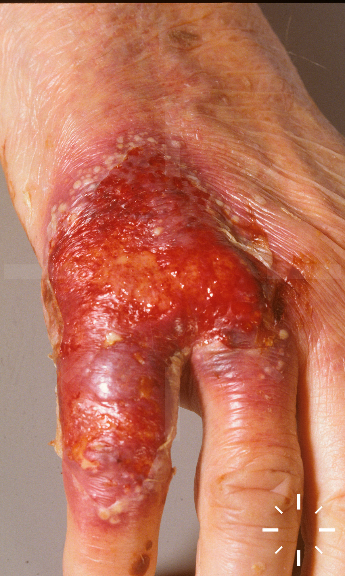
Mycosis fungoides

Zuletzt aktualisiert: 2022-11-16

Autor(en): Anzengruber F.

ICD11: 2B01

MF

Häufigstes primär kutanes T-Zell-Lymphom mit niedriger Malignität.

  • Inzidenz: 0,4-0,5/100.000/Jahr
  • Ab dem 40 Lj. auftretend
  • Männer : Frauen = 2 : 1

  • Hauptform
  • Varianten:
    • Follikulotrope MF
    • Pagetoide Retikulose
    • Granulomatous slack skin
  • Seltene Varianten:
    • Syringotrope MF
    • Ichtyhyosiforme MF
    • Pustulöse MF
    • Interstitielle Mf
    • Vegetierend-papillomatöse MF
    • Bullöse MF
    • Hyperkeratotisch-verruköse MF
    • Erythrodermische Mycosis fungoides
    • Palmoplantare MF
    • Poikilodermatitische MF
    • Hyper- und hypopigmentierte Mycosis fungoides

Die Pathogenese ist weitgehend unklar. Eine mögliche Theorie ist die persistierende Stimulation durch Viren, Bakterien oder anderen externen Einflüssen. Dadurch komme es zu einer Stimulation und Proliferation eines Lymphozytenklons mit chromosomaler Instabilität.

  • Patch-Stadium
    • Erythematöse, scharf begrenzte, pityriasiforme oder psoriasiforme, teils konfluierende Makulae.
  • Plaque-Stadium
    • Erythematöse bis violett-bräunliche, lichenifizierte, teils mit einer Alopezie einhergehende Plaques
  • Tumor-Stadium
    • Exophytisch, teils pilzförmige, erythematös-livide Tumoren. Oftmals zeigt sich eine Erythrodermie

  • Anamnese
    • Dauer?
    • Zeitliche Progression
    • B-Symptomatik?
  • Klinik
    • Palpation aller Lymphknotenstationen, Leber und Milz. 
  • Labor
    • BSG/CRP, Differentialblutbild (oft Lymphozytose und Eosinophilie), Leberenzyme, Kreatinin, LDH, Elektrolyte
    • FACS-Analyse, CD4/CD8 Ratio, Bestimmung der CD4+CD7- Zellen
      • ggf. Immunelektrophorese
      • ggf. HTLV-Serologie (v.a. bei Pat. aus dem Ausland)
      • ggf. Borrelienserologie
    • IgE-Spiegel
      • Häufig erhöht
    • Lymphozytendifferenzierung
      • FACS-Analyse: CD4+-Zellen↑, manchmal CD8+ Zellen↑
    • Sézary -Zellen (Zellen mit großem, eingebuchtetem Kern von 6–10 μm Größe und in Elektronenmikroskopie Zerebriformer Kern sowie Glykogengranula im Zytoplasma)?
      • Bei > 1000 Zellen/μL starker Hinweis auf Sézary-Syndrom
  • Biopsie
    • Falls nur eine Biopsie, sollte die am stärksten indurierte Stelle gewählt werden
    • Immunphänotypisierung
    • Molekularbiologisch: Klonales Rearrangment von T Zell-Rezeptor-Genen.

Vor allem der Rumpf sowie die Beugeseiten der Beine und Arme sind betroffen. Kann deck-chair sign (Aussparung der Falten) positiv sein (DD Papuloerythroderma Ofuji).

Patch-, Plaquestadium:

  • Parakeratose, ggf. Akanthose, Exozytose, atypische Lymphozyten mit zerebriformen Kernen, Epidermotropismus, Pautrier-Mikroabszesse und "lining up" an der dermalen-epidermalen Junktion.

Tumorstadium:

  • Noduläre atypisch lymphozytäre Infiltrate. Epidermotropismus und Pautrier-Mikroabszesse ungewöhnlich

Häufig überlagerte Infektionen, da durch die Erkrankung eine Immunsuppression entsteht. 

Keine möglich.

Stadienabhängig von sehr gut im Stadium I (kaum Unterschiede zu nicht betroffenen Personen) zu infaust bei III bis IV.

  1. Willemze et al. Primary cutaneous lymphomas: ESMO Clinical practice guidelines for diagnosis, treatment and follow-up. Annals of Oncology 2018. https://doi.org/10.1093/annonc/mdy133
  2. Mycosis Fungoides, in An Illustrated Guide to Skin Lymphoma. Wiley-Blackwell. p. 9-38.
  3. Erratum in Olsen et al. Revisions to the staging and classification of mycosis fungoides and Sezary syndrome: a proposal of the International Society for Cutaneous Lymphomas (ISCL) and the cutaneous lymphoma task force of the European Organization of Research and Treatment of Cancer (EORTC). Blood. 2007;110:1713-1722. Blood, 2008. 111(9): p. 4830-4830.
  4. Beljaards, R.C., et al., Primary cutaneous T-cell lymphoma: Clinicopathological features and prognostic parameters of 35 cases other than mycosis fungoides and cd30-positive large cell lymphoma. J. Pathol., 1994. 172(1): p. 53-60.
  5. Beylot-Barry, M., et al., Is bone marrow biopsy necessary in patients with mycosis fungoides and Sezary syndrome? A histological and molecular study at diagnosis and during follow-up. Br J Dermatol, 2005. 152(6): p. 1378-1379.
  6. Bleehen, S.S. and D.N. Slater, (25) Pityriasis lichenoides developing into mycosis fungoides. Br J Dermatol, 1986. 115(s30): p. 69-70.
  7. Boztepe, G., et al., Narrowband ultraviolet B phototherapy to clear and maintain clearance in patients with mycosis fungoides. Journal of the American Academy of Dermatology, 2005. 53(2): p. 242-246.
  8. Bunn, P.A., Systemic Therapy of Cutaneous T-Cell Lymphomas (Mycosis Fungoides and the Sezary Syndrome). Annals of Internal Medicine, 1994. 121(8): p. 592.
  9. de Unamuno Bustos, B., et al., Adult pityriasis lichenoides-like mycosis fungoides: a clinical variant of mycosis fungoides. International Journal of Dermatology, 2014. 53(11): p. 1331-1338.
  10. Demirkesen, C., et al., The clinical features and histopathologic patterns of folliculotropic mycosis fungoides in a series of 38 cases. J Cutan Pathol, 2014. 42(1): p. 22-31.
  11. Díez Recio, E., et al., Topical 5-aminolevulinic acid photodynamic therapy for the treatment of unilesional mycosis fungoides: a report of two cases and review of the literature. International Journal of Dermatology, 2008. 47(4): p. 410-413.
  12. Dummer, R., et al., Aktuelle Aspekte zur Pathogenese von Sézary-Syndrom und Mycosis fungoides. Der Hautarzt, 2001. 52(3): p. 189-192.
  13. Gerami, P., et al., Folliculotropic Mycosis Fungoides. Arch Dermatol, 2008. 144(6).
  14. Gómez Orbaneja, J., et al., Lymphomatoid contact dermatitis A syndrome produced by epicutaneous hypersensitivity with clinical features and a histopathologic picture similar to that of mycosis fungoides. Contact Dermatitis, 1976. 2(3): p. 139-143.
  15. Holcomb, M., M. Duvic, and J. Cutlan, Erythema gyratum repens-like eruptions with large cell transformation in a patient with mycosis fungoides. International Journal of Dermatology, 2011. 51(10): p. 1231-1233.
  16. Jackow, C.M., et al., Follicular mucinosis associated with scarring alopecia, oligoclonal T-cell receptor V? expansion, and Staphylococcus aureus: When does follicular mucinosis become mycosis fungoides? Journal of the American Academy of Dermatology, 1997. 37(5): p. 828-831.
  17. Joly, P., et al., Primary cutaneous medium and large cell lymphomas other than mycosis fungoides. An immunohistological and follow-up study on 54 cases. British Journal of Dermatology, 2006. 132(4): p. 506-512.
  18. Jones, G.W., et al., Total skin electron radiation in the management of mycosis fungoides: Consensus of the European Organization for Research and Treatment of Cancer (EORTC) Cutaneous Lymphoma Project Group. Journal of the American Academy of Dermatology, 2002. 47(3): p. 364-370.
  19. Kalayciyan, A., et al., Milia in regressing plaques of mycosis fungoides: provoked by topical nitrogen mustard or not? International Journal of Dermatology, 2004. 43(12): p. 953-956.
  20. Kaye, F.J., et al., A Randomized Trial Comparing Combination Electron-Beam Radiation and Chemotherapy with Topical Therapy in the Initial Treatment of Mycosis Fungoides. New England Journal of Medicine, 1989. 321(26): p. 1784-1790.
  21. Kempf, W., et al., Granulomatous Mycosis Fungoides and Granulomatous Slack Skin. Arch Dermatol, 2008. 144(12).
  22. Kim, Y.H., Management with topical nitrogen mustard in mycosis fungoides. Dermatologic Therapy, 2003. 16(4): p. 288-298.
  23. Kim, Y.H., et al., TNM classification system for primary cutaneous lymphomas other than mycosis fungoides and Sezary syndrome: a proposal of the International Society for Cutaneous Lymphomas (ISCL) and the Cutaneous Lymphoma Task Force of the European Organization of Research and Treatment of Cancer (EORTC). Blood, 2007. 110(2): p. 479-484.
  24. Klemke, C.-D., et al., Clonal T cell receptor γ-chain gene rearrangement by PCR-based GeneScan analysis in the skin and blood of patients with parapsoriasis and early-stage mycosis fungoides. J. Pathol., 2002. 197(3): p. 348-354.
  25. Lazar, A.P., et al., Parapsoriasis and mycosis fungoides: The Northwestern University experience, 1970 to 1985. Journal of the American Academy of Dermatology, 1989. 21(5): p. 919-923.
  26. Lindae, M.L., Poikilodermatous mycosis fungoides and atrophic large-plaque parapsoriasis exhibit similar abnormalities of T-cell antigen expression. Archives of Dermatology, 1988. 124(3): p. 366-372.
  27. Lipsker, D., The Pigmented and Purpuric Dermatitis and the Many Faces of Mycosis fungoides. Dermatology, 2003. 207(3): p. 246-247.
  28. Nagase, K., et al., CD4/CD8 Double-negative Mycosis Fungoides Mimicking Erythema Gyratum Repens in a Patient with Underlying Lung Cancer. Acta Dermato Venereologica, 2014. 94(1): p. 89-90.
  29. Pimpinelli, N., et al., Defining early mycosis fungoides. Journal of the American Academy of Dermatology, 2005. 53(6): p. 1053-1063.
  30. Prince, H.M., S. Whittaker, and R.T. Hoppe, How I treat mycosis fungoides and Sezary syndrome. Blood, 2009. 114(20): p. 4337-4353.
  31. Resnik, K.S. and E.C. Vonderheid, Home UV phototherapy of early mycosis fungoides: Long-term follow-up observations in thirty-one patients. Journal of the American Academy of Dermatology, 1993. 29(1): p. 73-77.
  32. Rongioletti, F., et al., Follicular mucinosis: a clinicopathologic, histochemical, immunohistochemical and molecular study comparing the primary benign form and the mycosis fungoides-associated follicular mucinosis. Journal of Cutaneous Pathology, 2010. 37(1): p. 15-19.
  33. Sanchez, J.L. and B.A. Ackerman, The patch stage of mycosis fungoides Criteria for histologic diagnosis. The American Journal of Dermatopathology, 1979. 1(1): p. 5-26.
  34. Schappell, D.L., Treatment of Advanced Mycosis Fungoides and Sézary Syndrome With Continuous Infusions of Methotrexate Followed by Fluorouracil and Leucovorin Rescue. Arch Dermatol, 1995. 131(3): p. 307.
  35. Shabrawi-Caelen, L.E., et al., Hypopigmented Mycosis Fungoides. The American Journal of Surgical Pathology, 2002. 26(4): p. 450-457.
  36. Talpur, R., R. Bassett, and M. Duvic, Prevalence and treatment of Staphylococcus aureus colonization in patients with mycosis fungoides and Sézary syndrome. Br J Dermatol, 2008. 159(1): p. 105-112.
  37. Trautinger, F., et al., EORTC consensus recommendations for the treatment of mycosis fungoides/Sézary syndrome. European Journal of Cancer, 2006. 42(8): p. 1014-1030.
  38. uuml, et al., Follicular Mucinosis Associated with Mycosis Fungoides. Dermatology, 1991. 183(1): p. 66-67.
  39. Väkevä, L., et al., A Retrospective Study of the Probability of the Evolution of Parapsoriasis en Plaques into Mycosis Fungoides. Acta Dermato-Venereologica, 2005. 85(4): p. 318-323.
  40. van Doorn, R., E. Scheffer, and R. Willemze, Follicular Mycosis Fungoides, a Distinct Disease Entity With or Without Associated Follicular Mucinosis. Arch Dermatol, 2002. 138(2).
  41. van Doorn, R., et al., Oncogenomic analysis of mycosis fungoides reveals major differences with Sezary syndrome. Blood, 2008. 113(1): p. 127-136.
  42. Whittaker, S.J. and F.M. Foss, Efficacy and tolerability of currently available therapies for the mycosis fungoides and Sezary syndrome variants of cutaneous T-cell lymphoma. Cancer Treatment Reviews, 2007. 33(2): p. 146-160.
  43. Wong, W.-R., et al., Generalized Granuloma annulare Associated with Granulomatous Mycosis fungoides. Dermatology, 2000. 200(1): p. 54-56.
  44. Wood, G.S., et al., Detection of Clonal T-Cell Receptor γ Gene Rearrangements in Early Mycosis Fungoides/Sezary Syndrome by Polymerase Chain Reaction and Denaturing Gradient Gel Electrophoresis (PCR/DGGE). Journal of Investigative Dermatology, 1994. 103(1): p. 34-41.
  45. Zackheim, H.S., Treatment of Mycosis Fungoides With Topical Nitrosourea Compounds. Arch Dermatol, 1972. 106(2): p. 177.
  46. Zackheim, H.S., et al., Percutaneous absorption of I,3-bis (2-chloroethyl)-I-nitrosourea (BCNU, carmustine) in mycosis fungoides. Br J Dermatol, 1977. 97(1): p. 65-67.
  47. Zackheim, H.S., M. Kashani-Sabet, and S. Amin, Topical Corticosteroids for Mycosis Fungoides. Arch Dermatol, 1998. 134(8).
  48. Stadler, R., et al., Short German guidelines: cutaneous lymphomas. J Dtsch Dermatol Ges, 2008. 6 Suppl 1: p. S25-31.