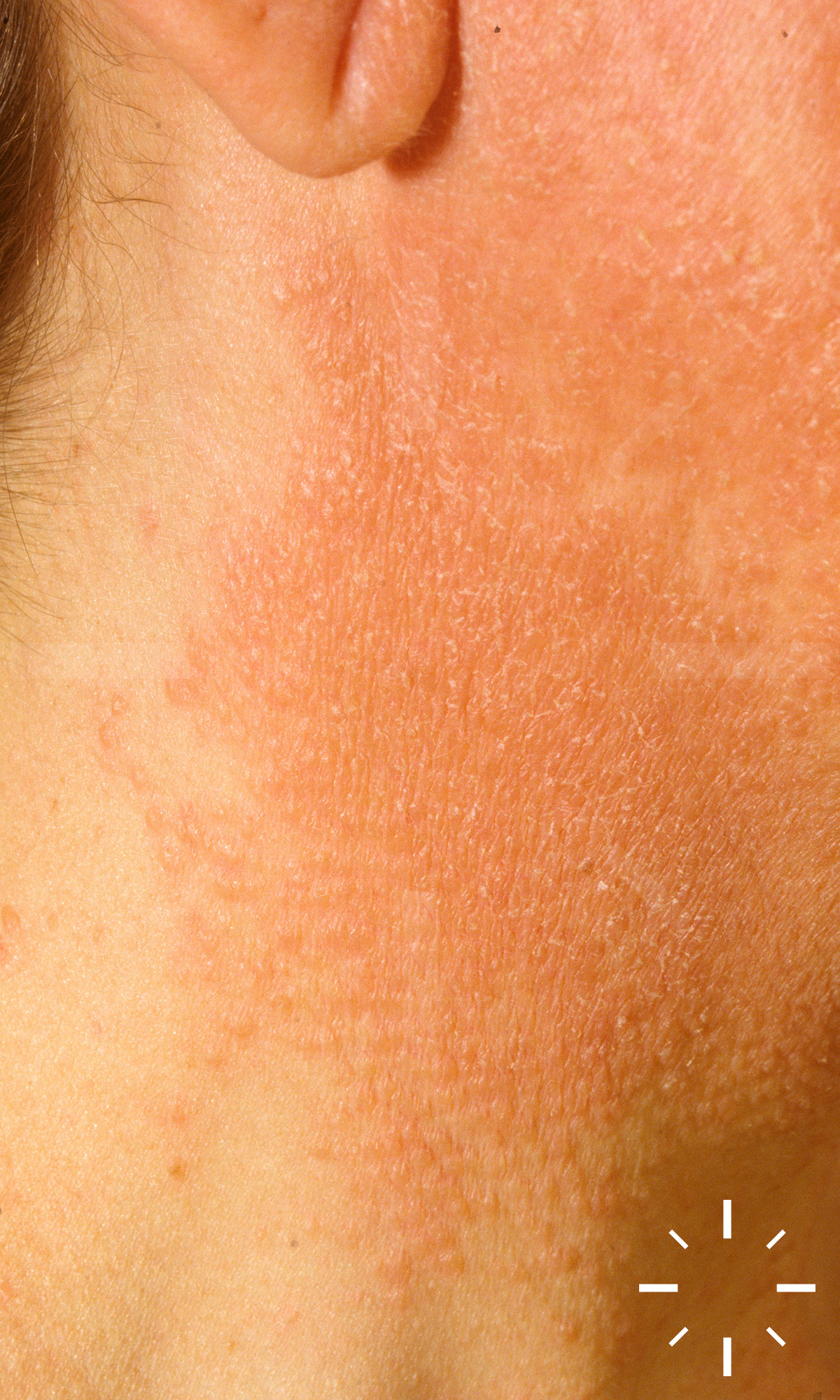
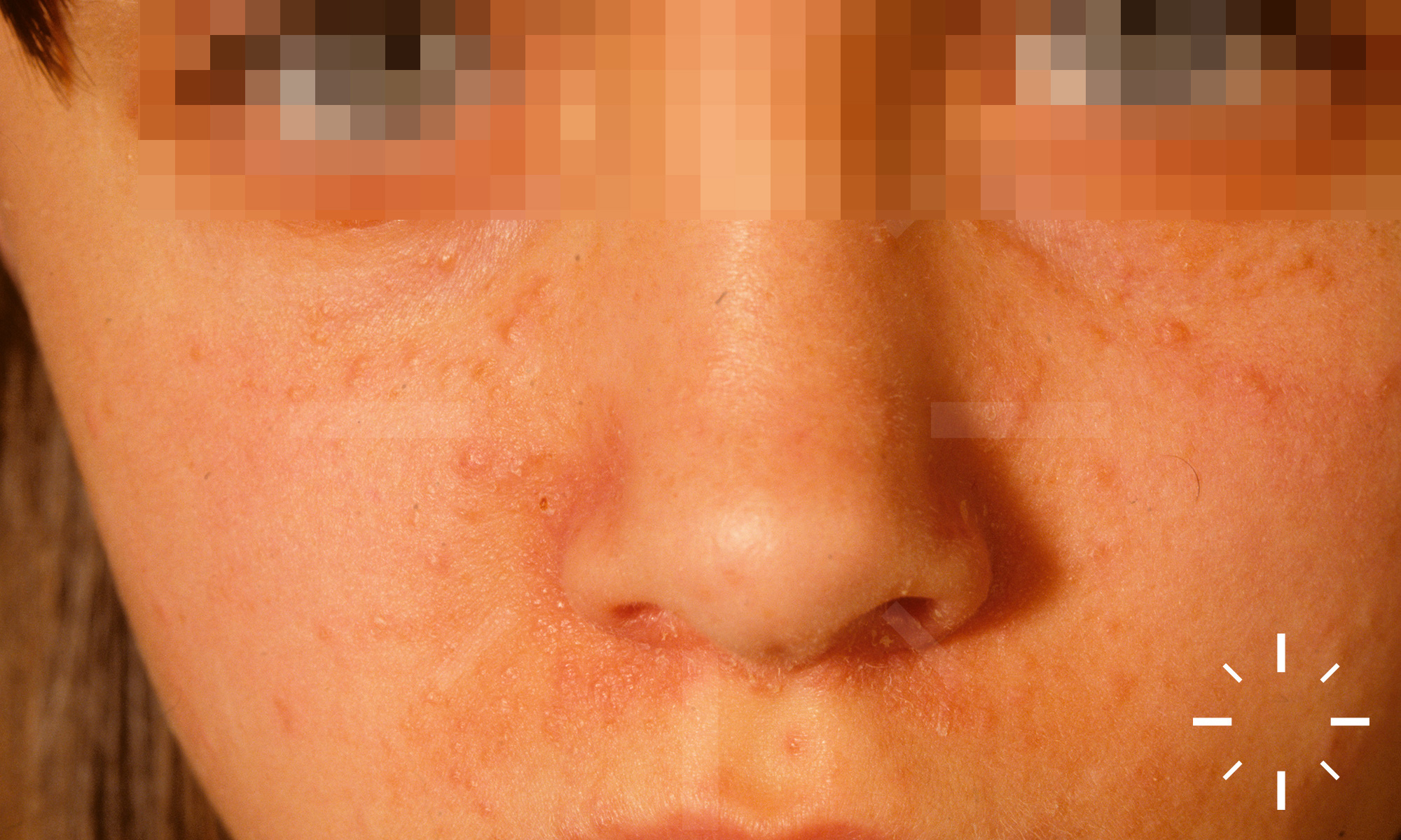
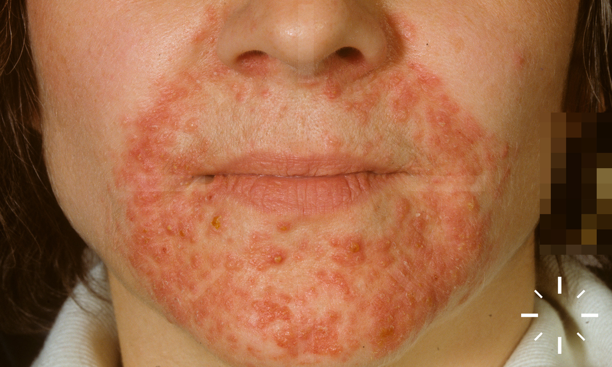
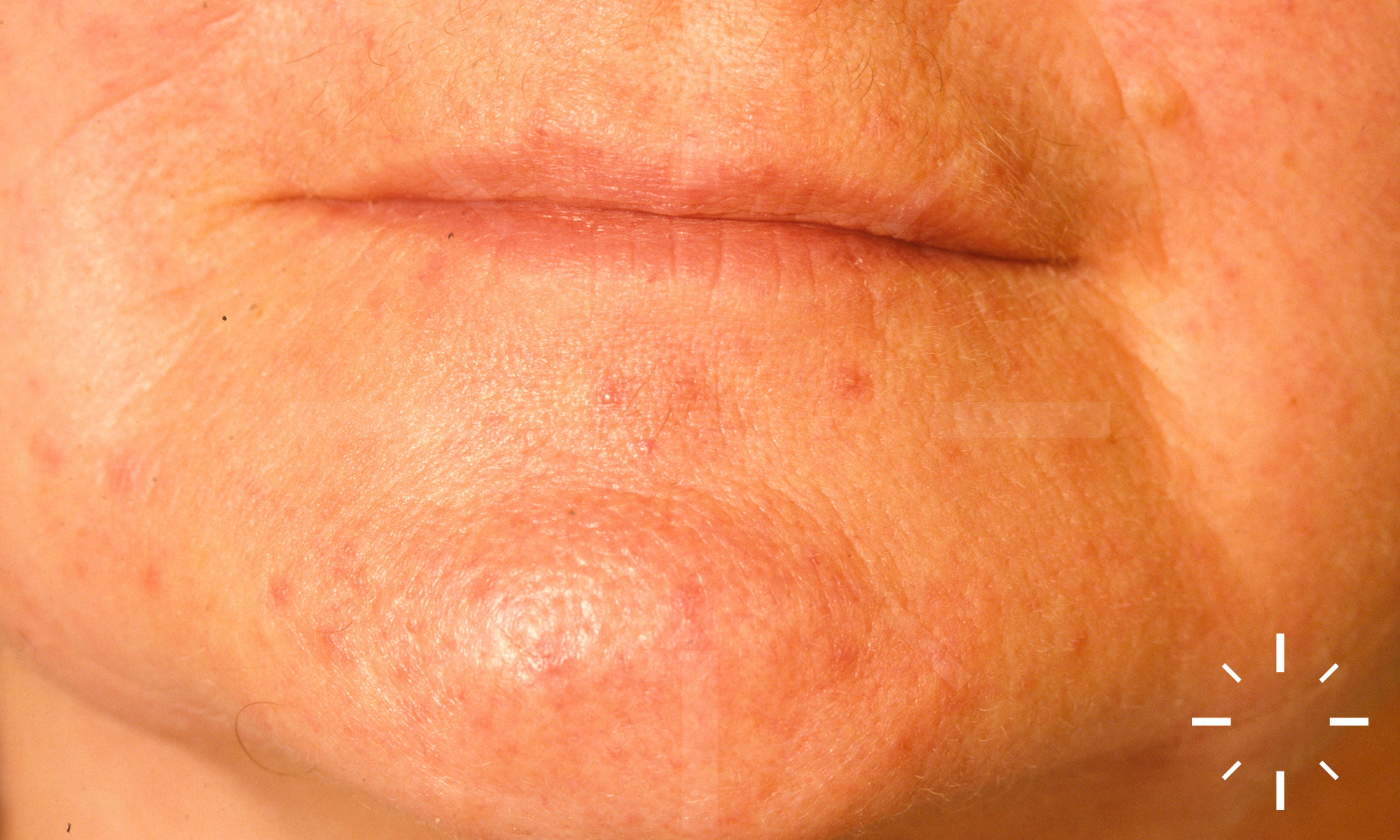
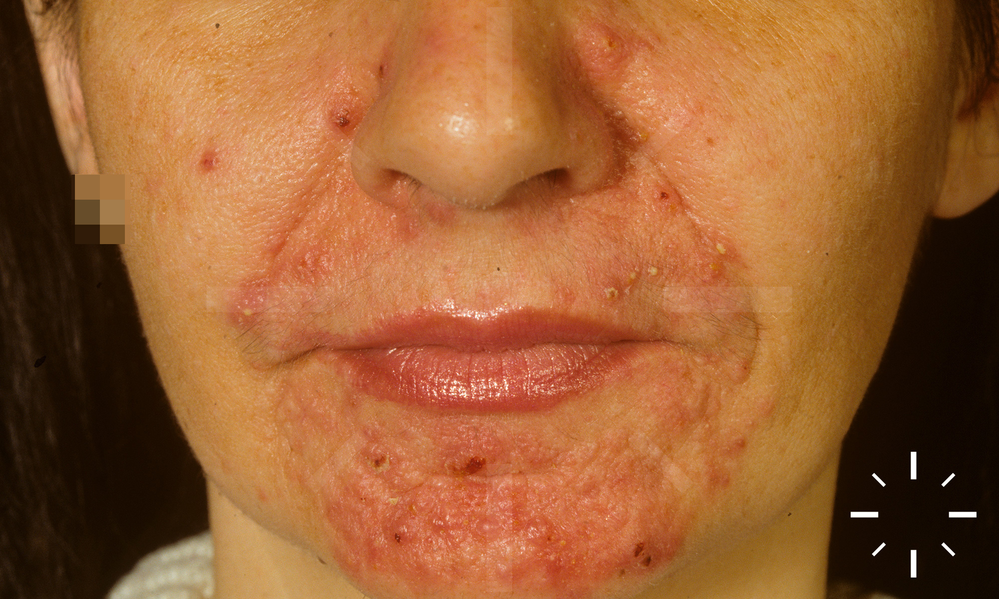
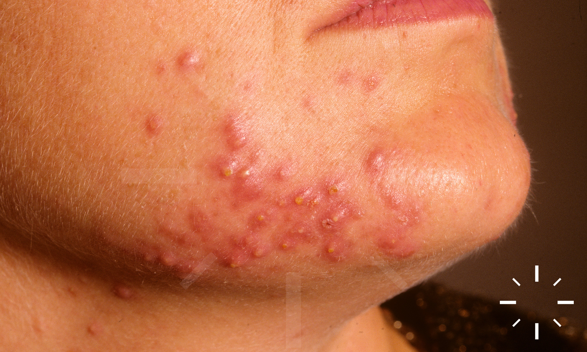
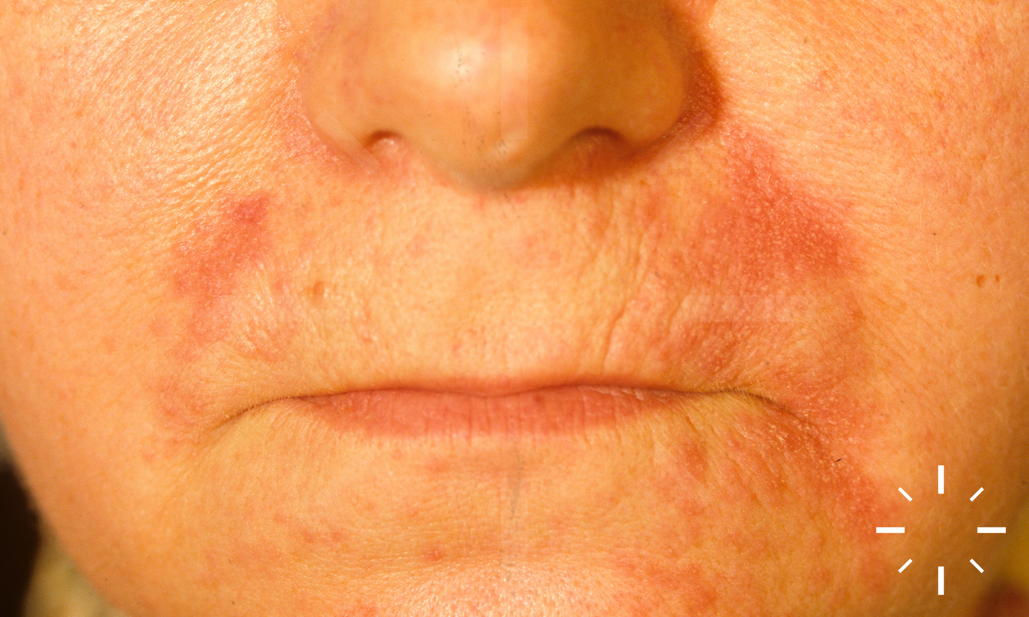
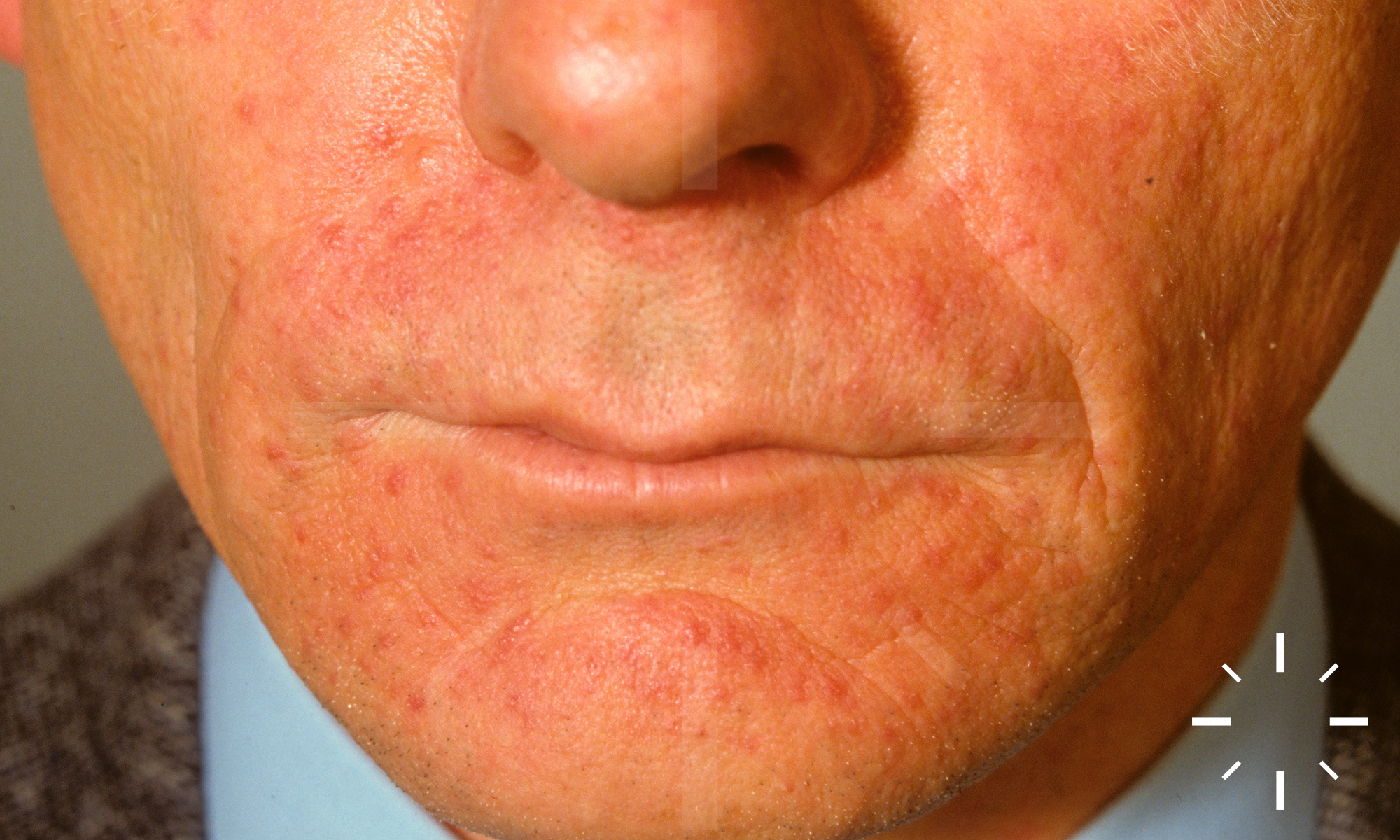
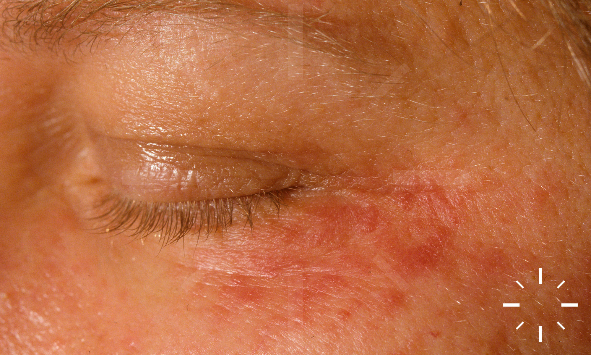
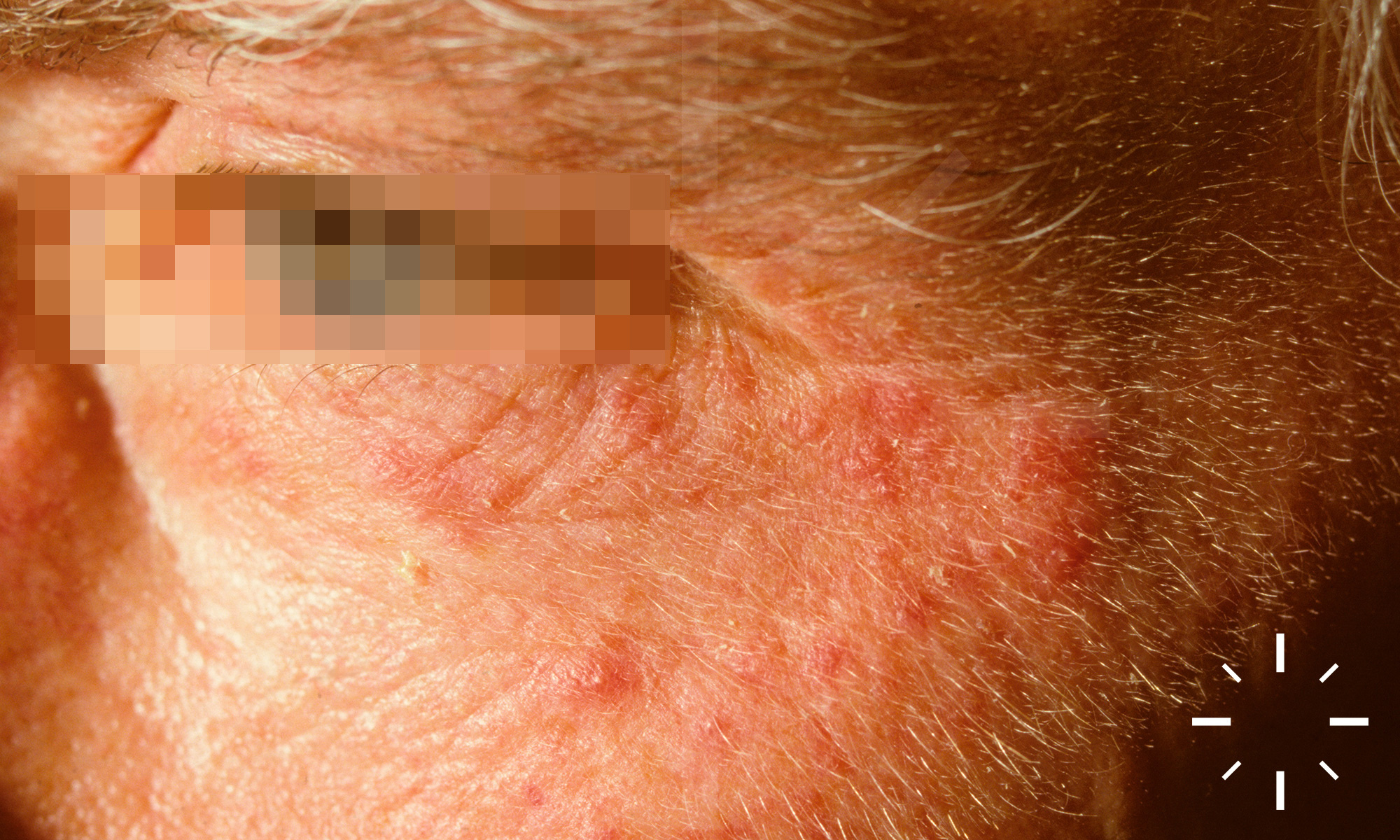
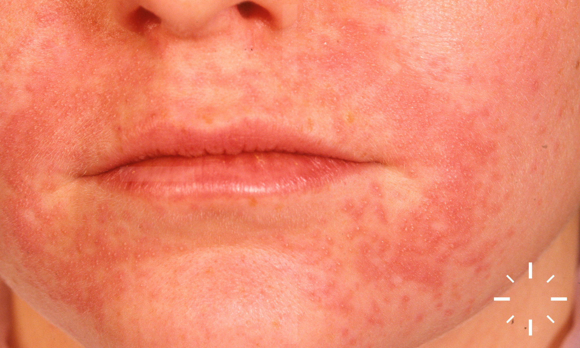
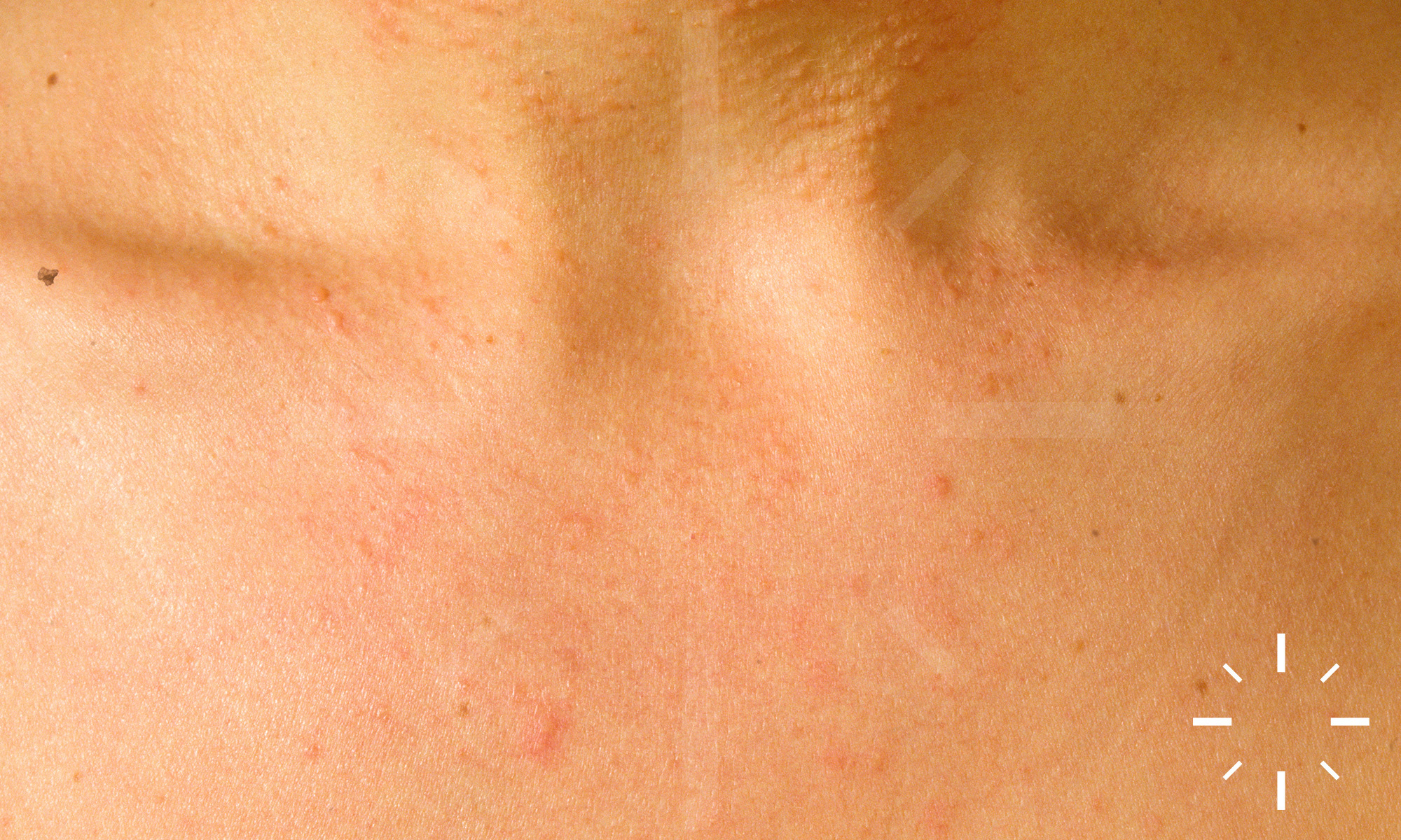
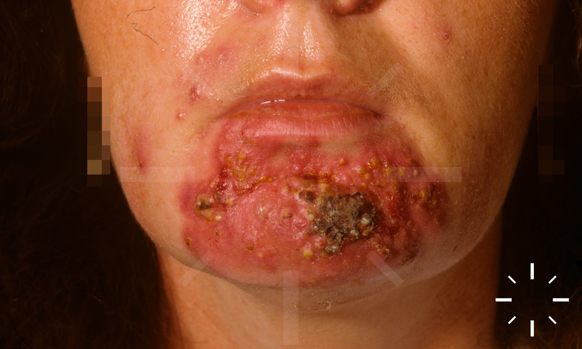
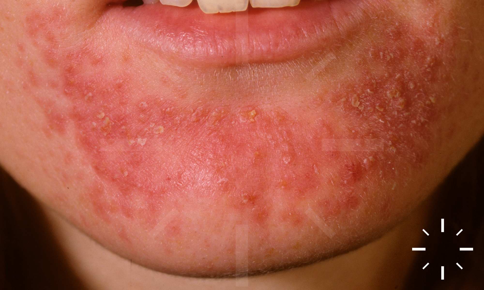
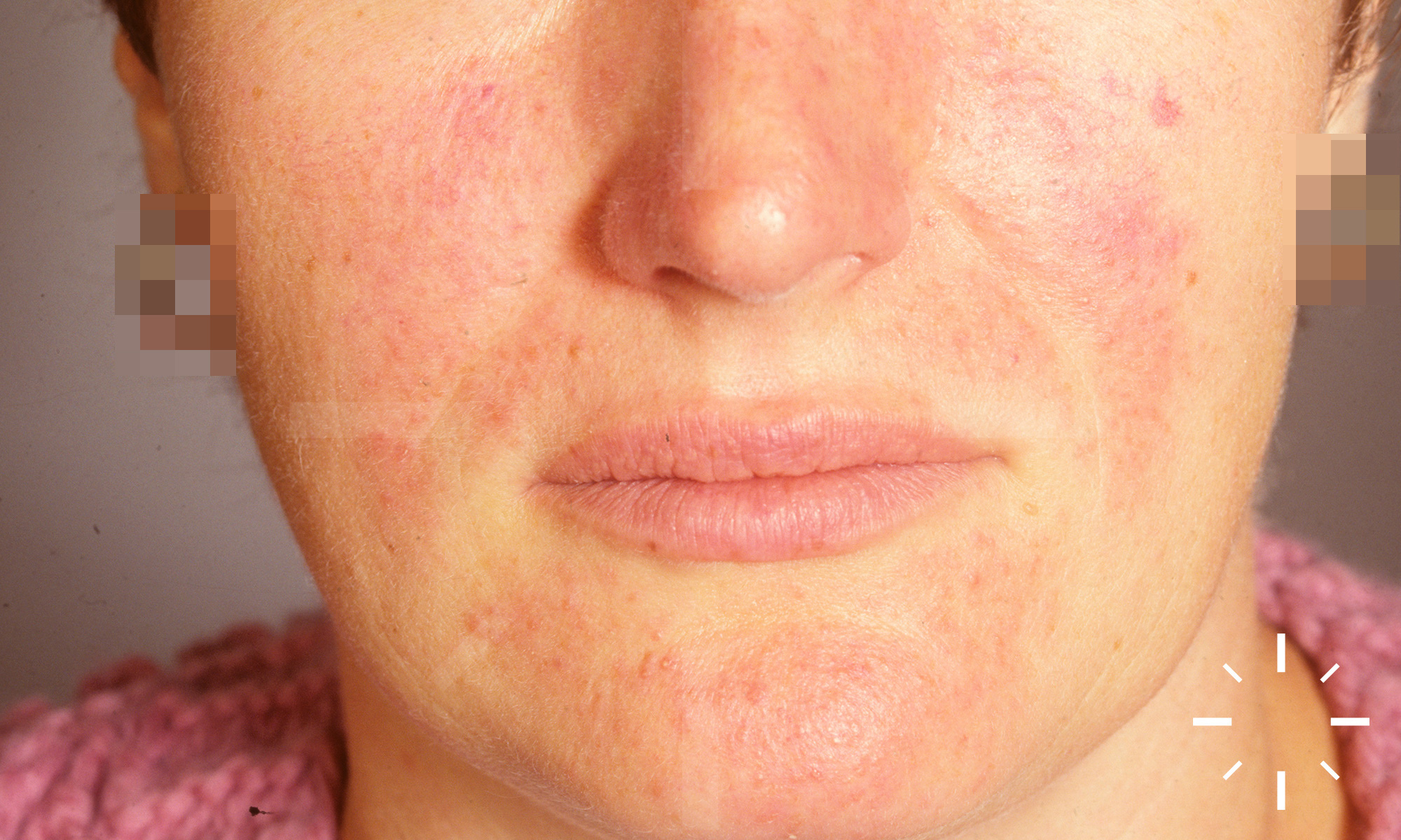
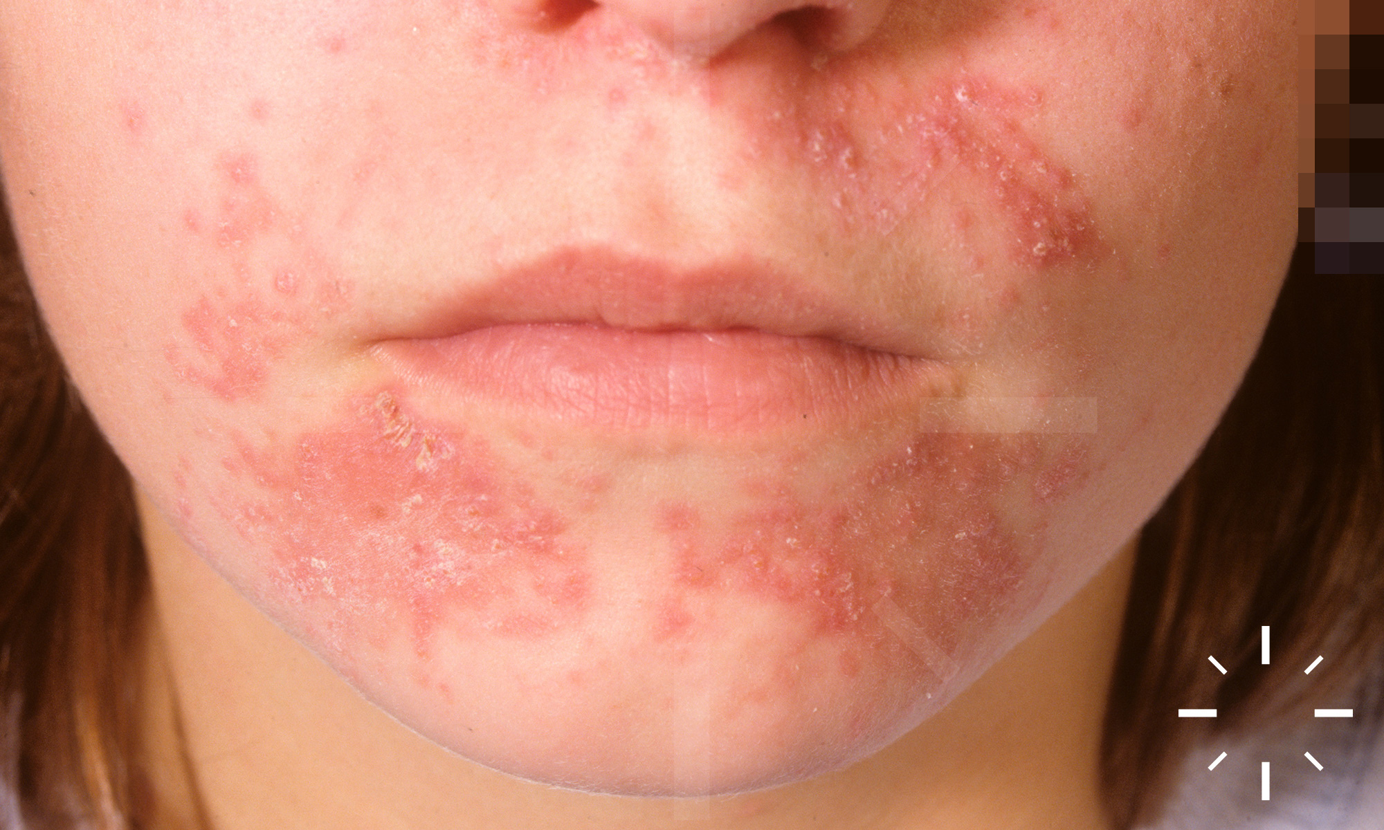
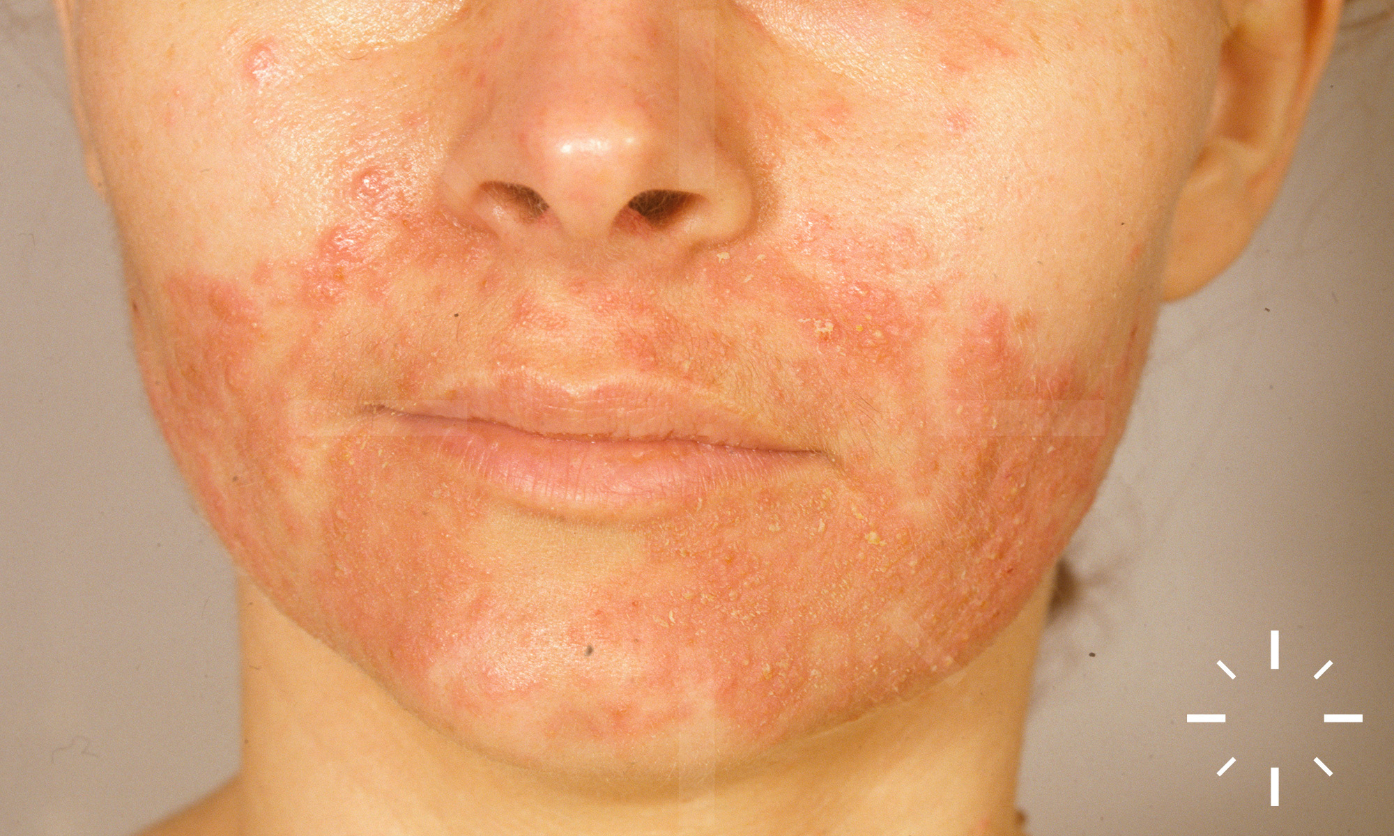
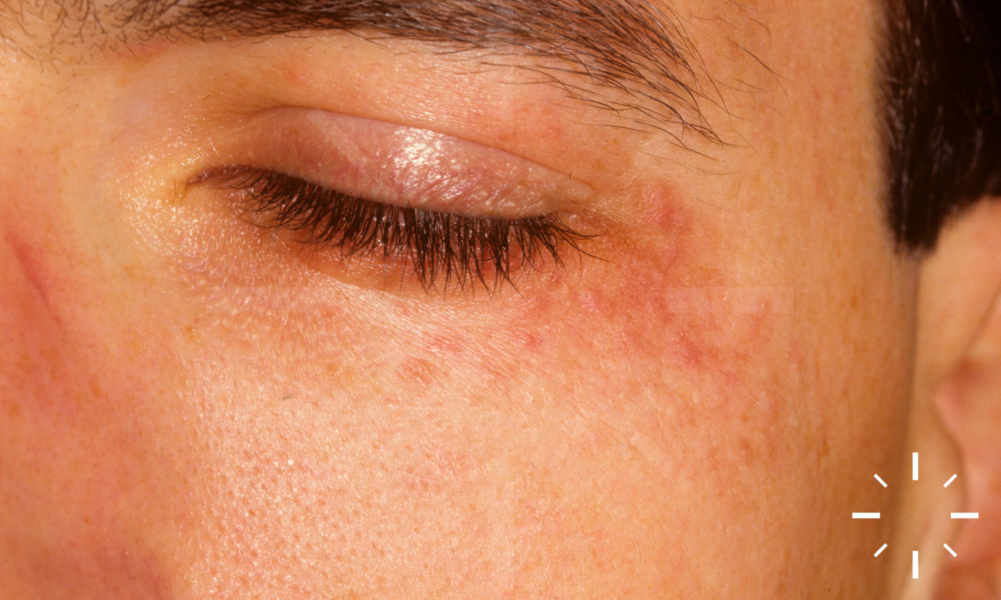
Perioral dermatitis

Last Updated: 2023-07-07

Author(s): Anzengruber F., Navarini A.

ICD11: ED90.1

  • Frumess and Lewis, 1957
  • Mihan and Ayres, 1964

Dermatitis perioralis, rosacea-like dermatitis, stewardess disease, periorificial dermatitis.

Reactive disease of the face, rosacea type. Itchy to painful skin changes on the face. Not infectious.

  • Incidence: approx. 1%/y.
  • Peak in age: 20-40 y.
  • Women >> men.
  • Professions with make-up compulsion more often affected ("stewardess disease")

None.

  • Caused by excessive use of skin care products, cosmetics, sun creams
  • Predisposed individuals include atopics
  • Vicious cycle when using topical glucocorticoids
  • UV light (is not a sole trigger, but can lead to worsening)

The disease is close to rosacea, but the distribution is different. The skin changes are always found around an orificium: perioral, perinasal and/or periorbital erythematous, often grouped, pruritic to burning, confluent, often slightly scaly papules, papulovesicles and pustules, sometimes also thin plaques.

  • Clinical picture
  • No laboratory tests necessary
  • In case of doubt, trial excision

  • Perioral, periocular, perinasal
  • Regular recess of about 1-2 mm of the skin directly adjacent to the orificium, because there are no vellus hair follicles in this zone, but they are necessary for follicular-bound disease

Follicular centred pathology. Epidermis spongiotic, lymphocytic perivascular infiltrates around follicular ostia. No demodex mites, telangiectasias, actinic elastosis as in rosacea.

If possible, abstain from facial care products.

  • Only when patients acknowledge the trigger does improvement occur
  • Frequently protracted
  • Poor adherence to cosmetic abstinence leads to recurrence

  1. Frieden IJ. Granulomatous Perioral Dermatitis in Children. Arch Dermatol 1989;125:369.
  2. Hafeez ZH. Perioral dermatitis: an update. International Journal of Dermatology 2003;42:514-7.
  3. Takiwaki H, Tsuda H, Arase S, Takeichi H. Differences between intrafollicular microorganism profiles in perioral and seborrhoeic dermatitis. Clin Exp Dermatol 2003;28:531-4.
  4. Dirschka T, Tronnier H, Folster-Holst R. Epithelial barrier function and atopic diathesis in rosacea and perioral dermatitis. Br J Dermatol 2004;150:1136-41.
  5. Wilkinson DS, Kirton V, Wilkinson JD. Perioral dermatitis: a 12-year review. British Journal of Dermatology 2006;101:245-57.
  6. Weston WL, Morelli JG. IDENTICAL TWINS WITH PERIORAL DERMATITIS. Pediatric Dermatology 2009;15:144-.