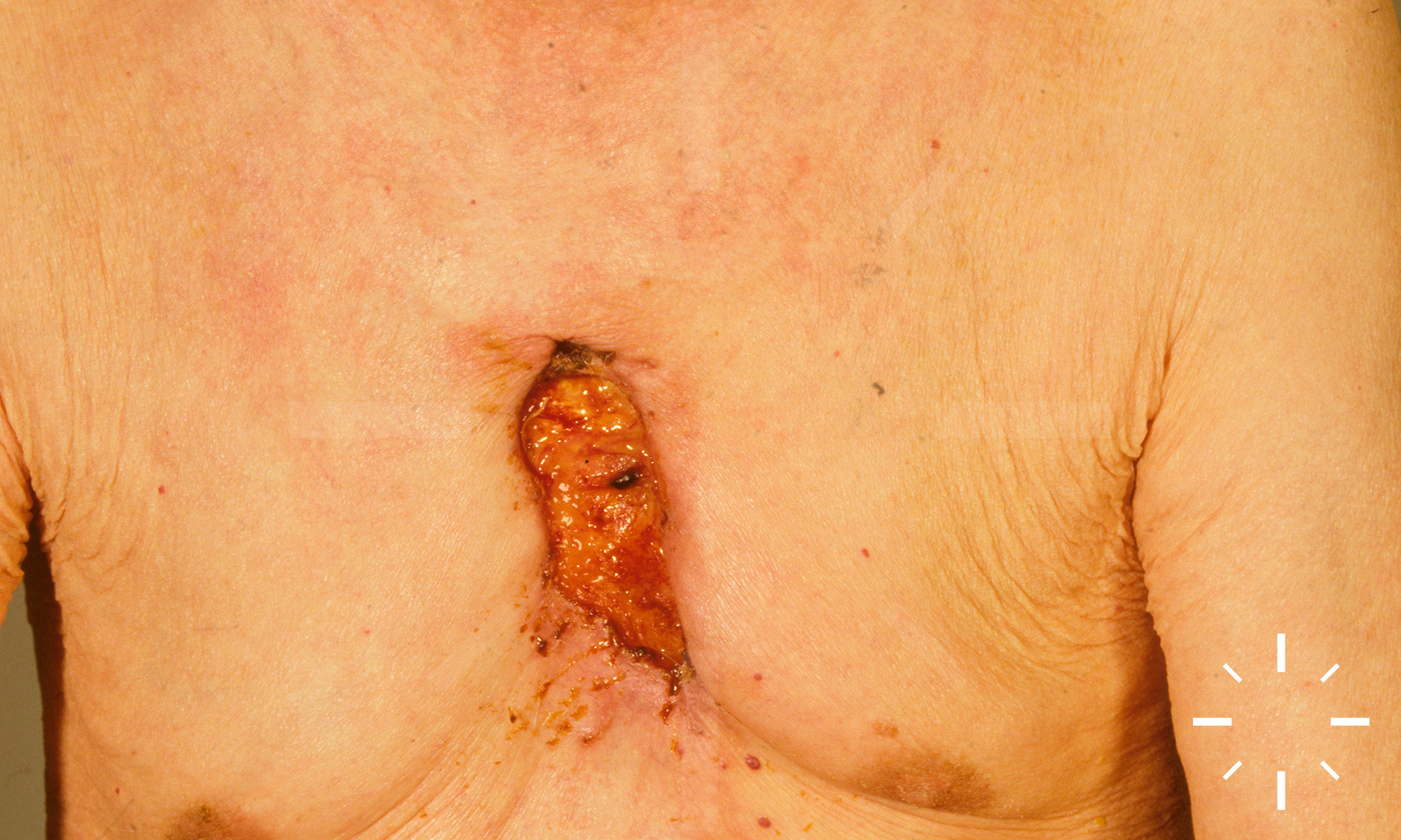
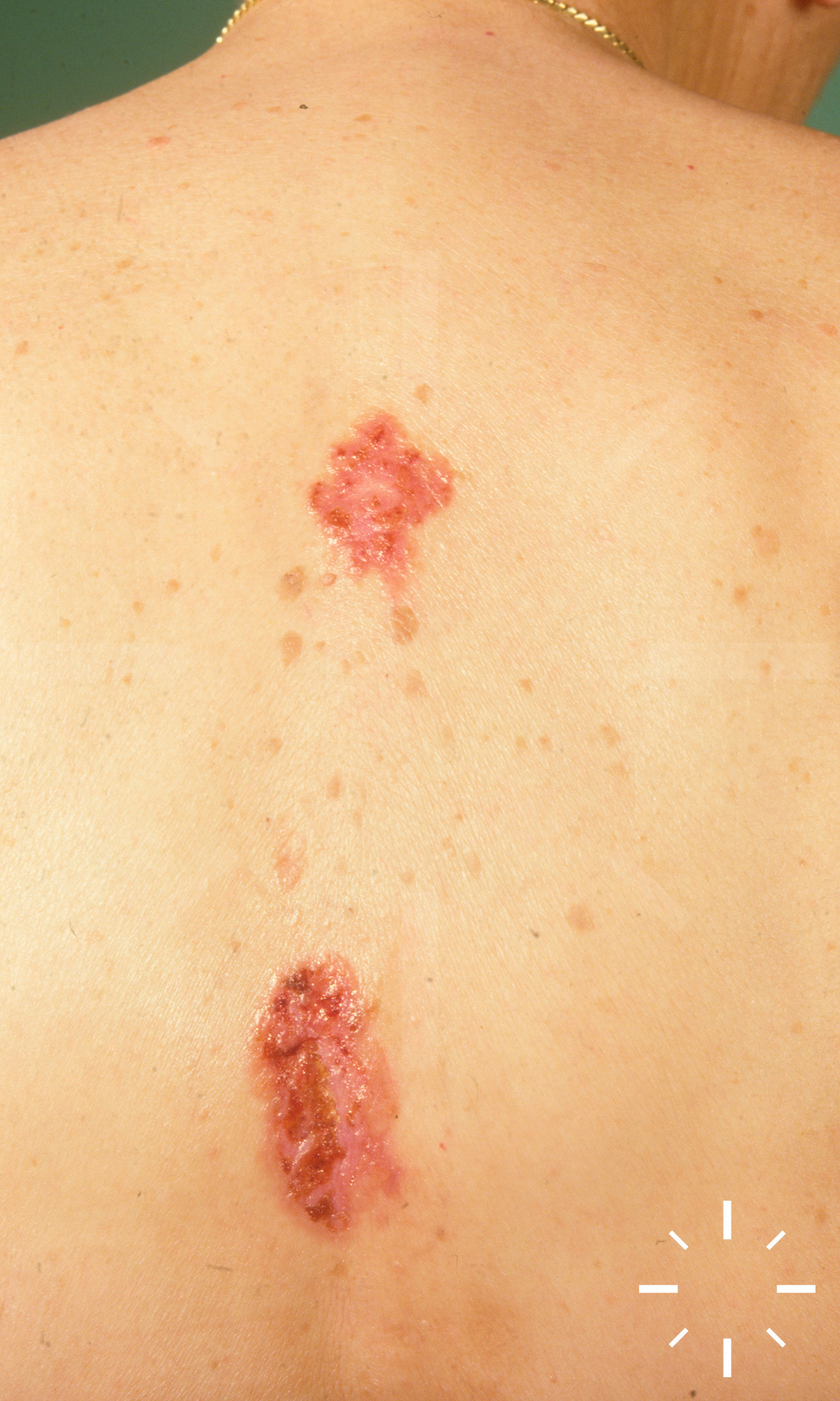
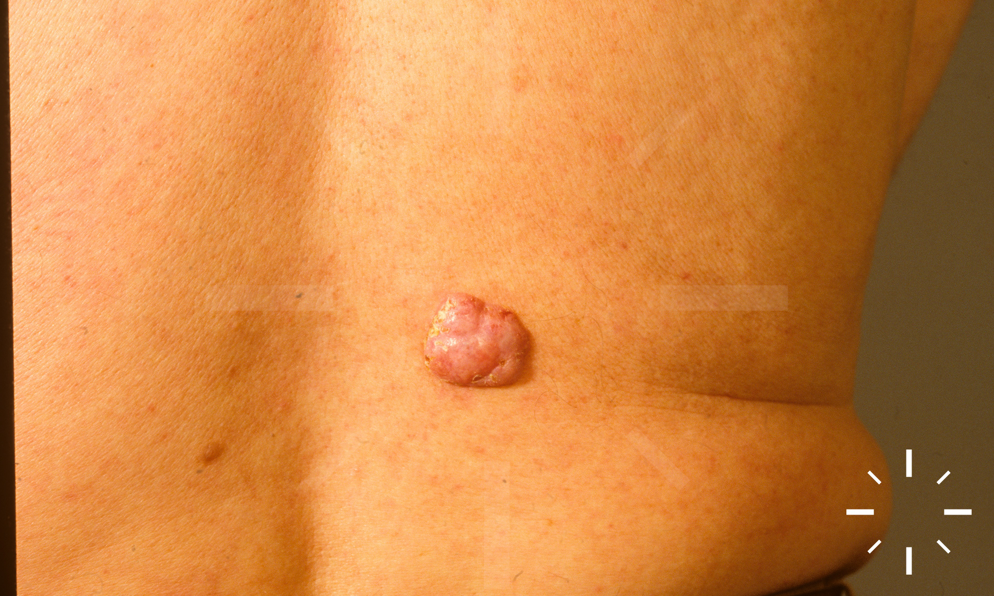
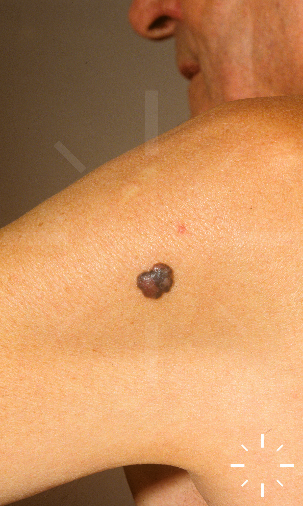
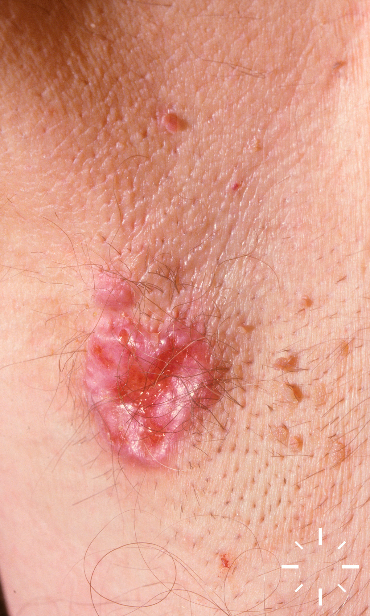
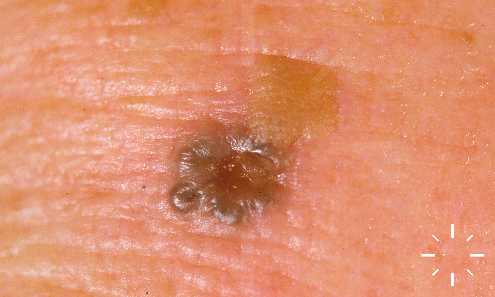
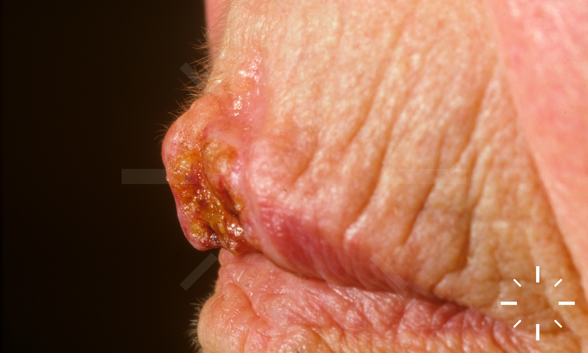
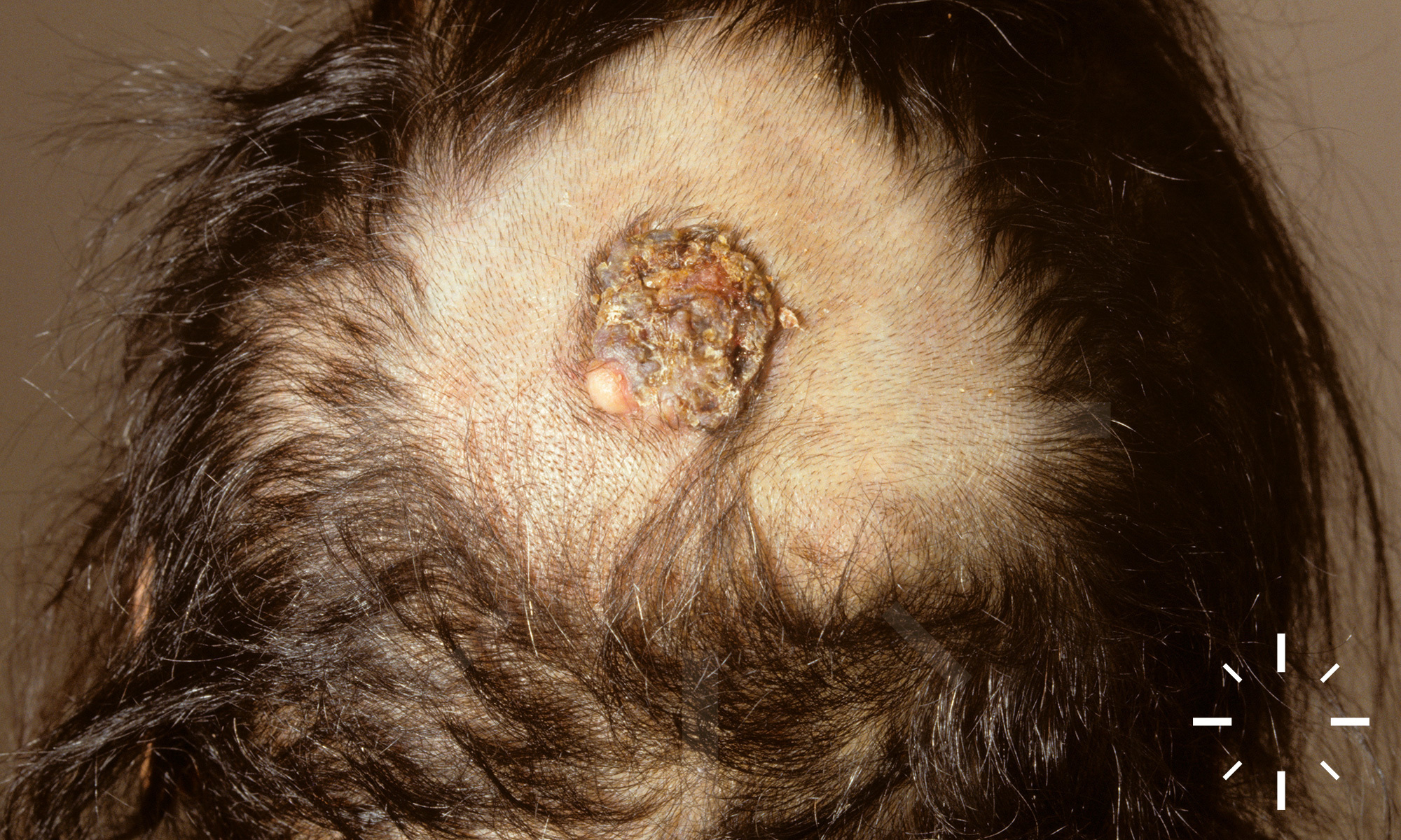
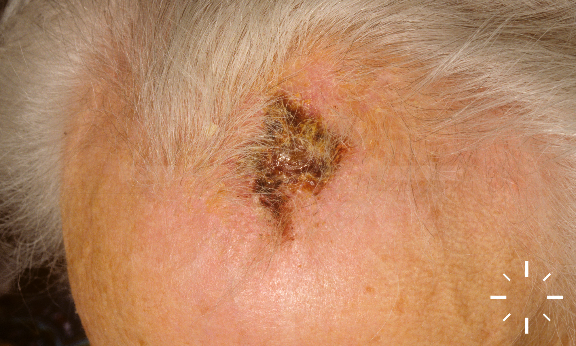
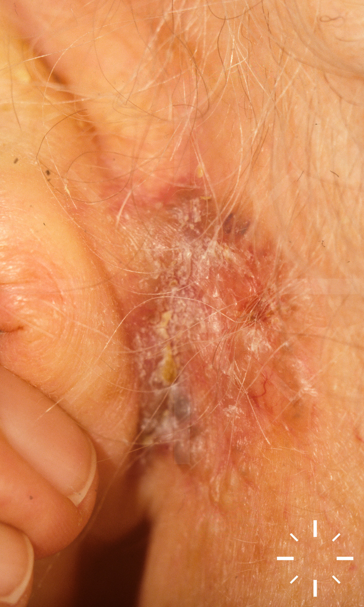
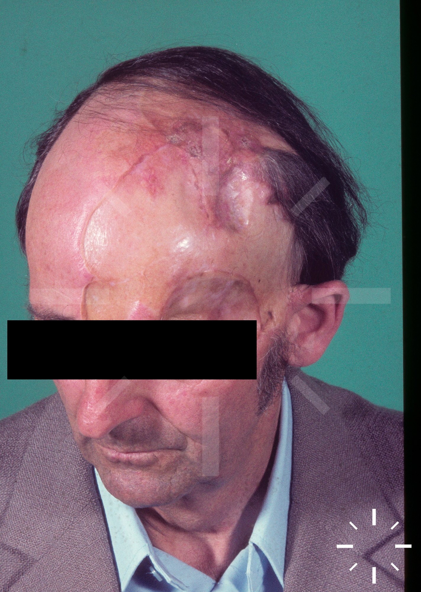
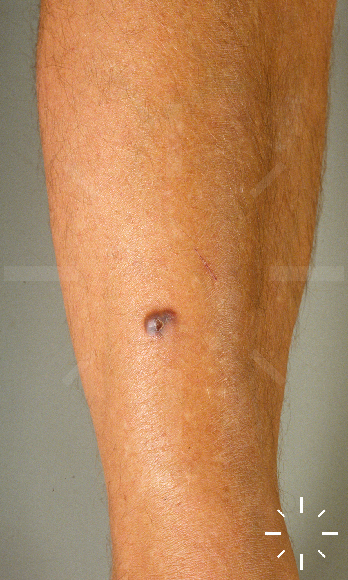
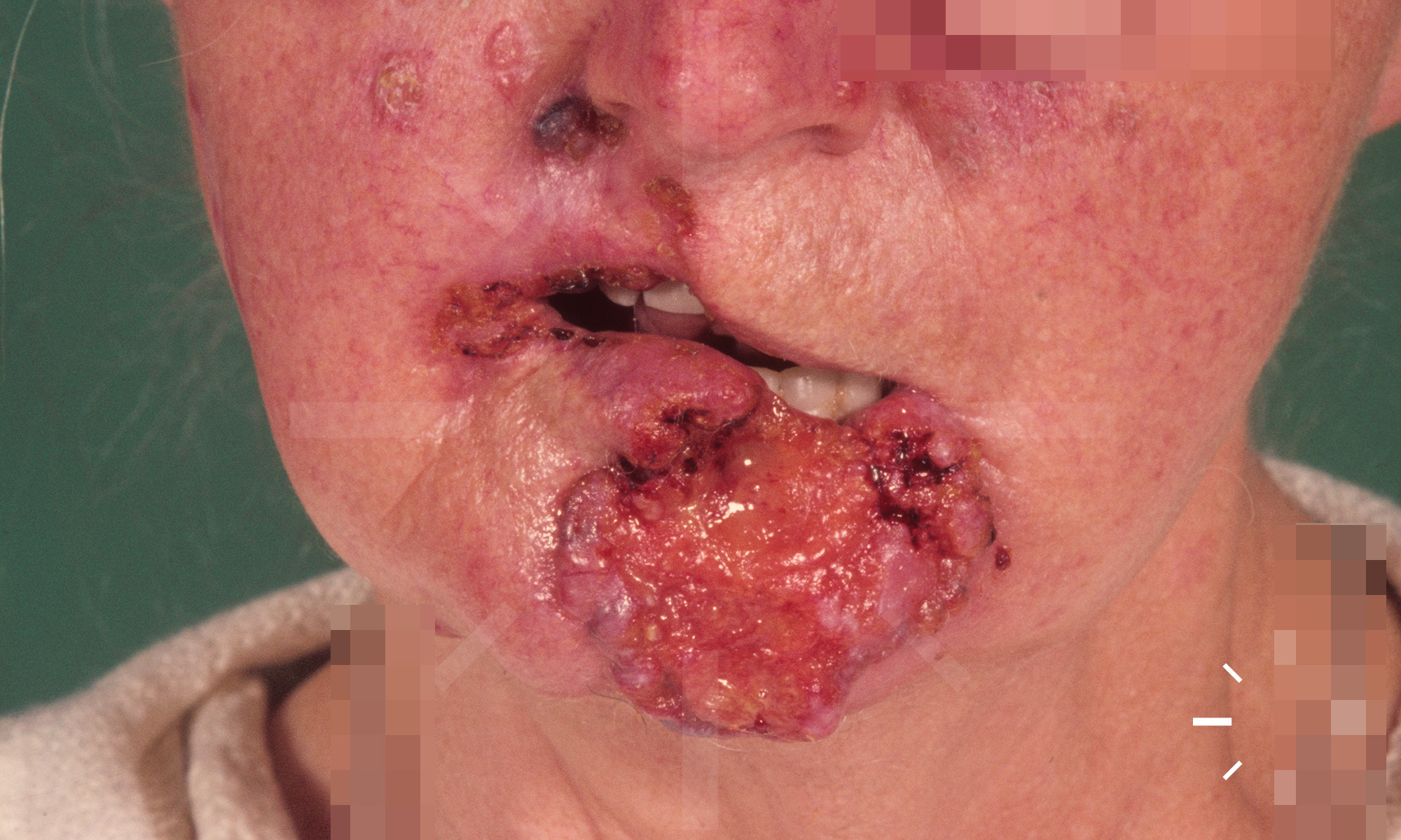
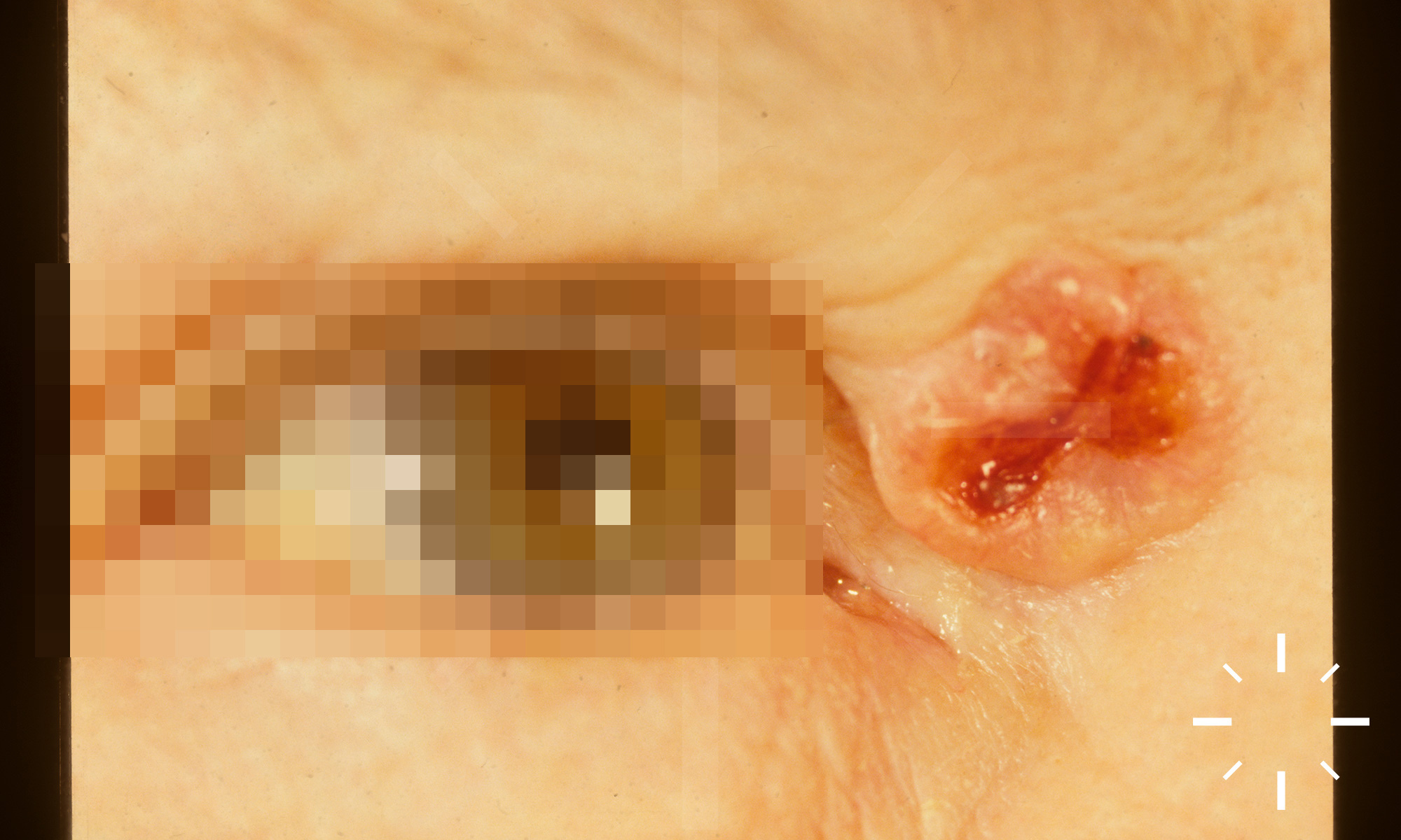
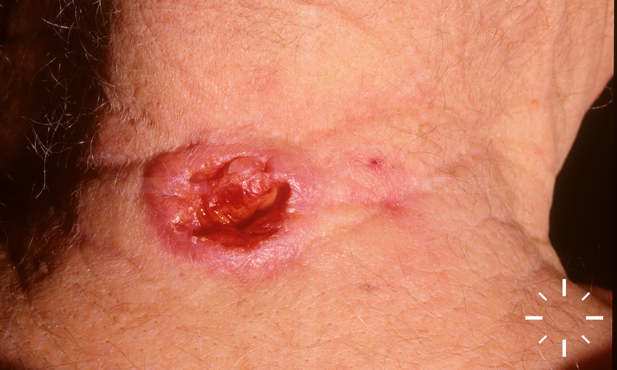
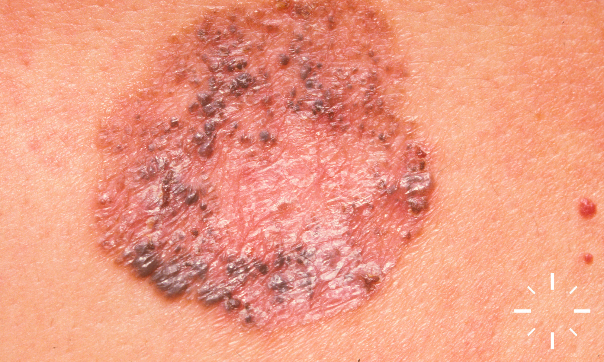
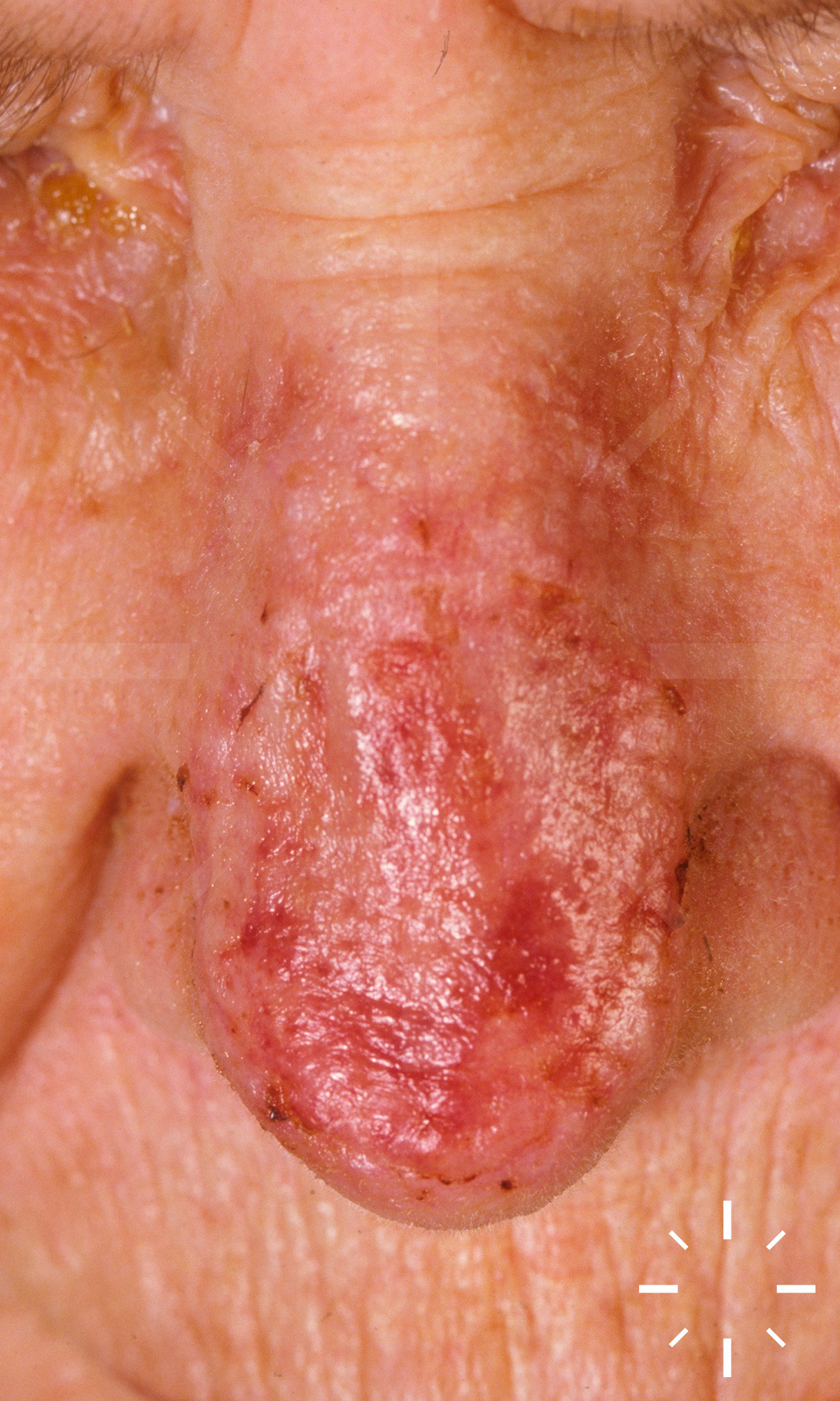
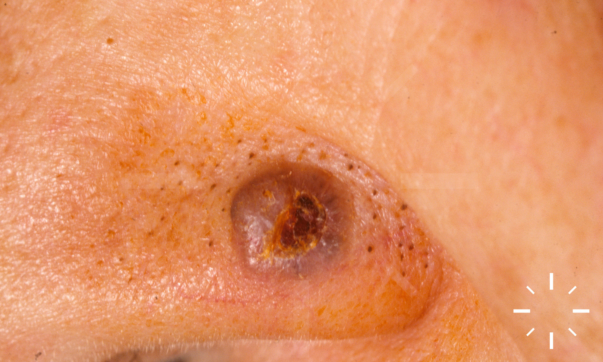
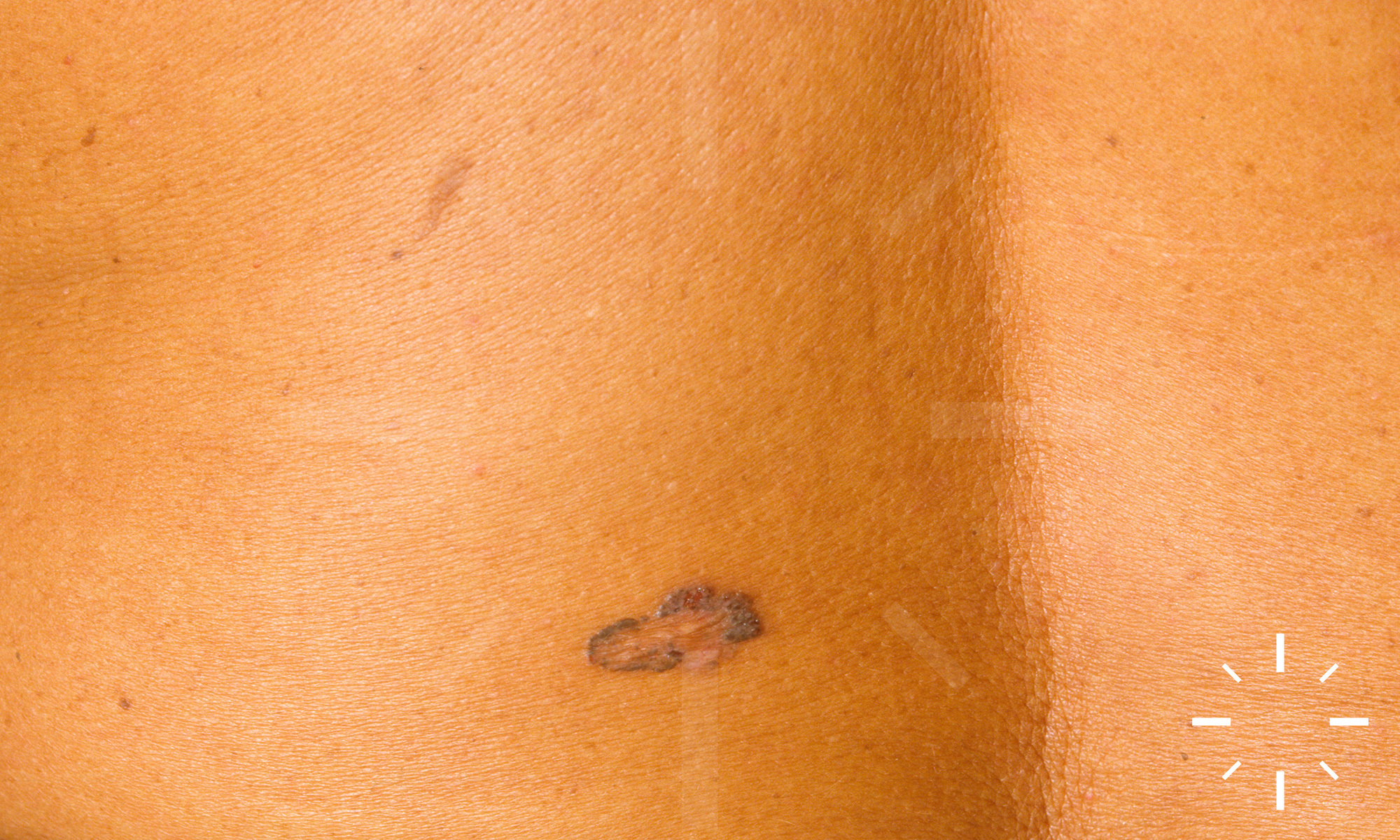
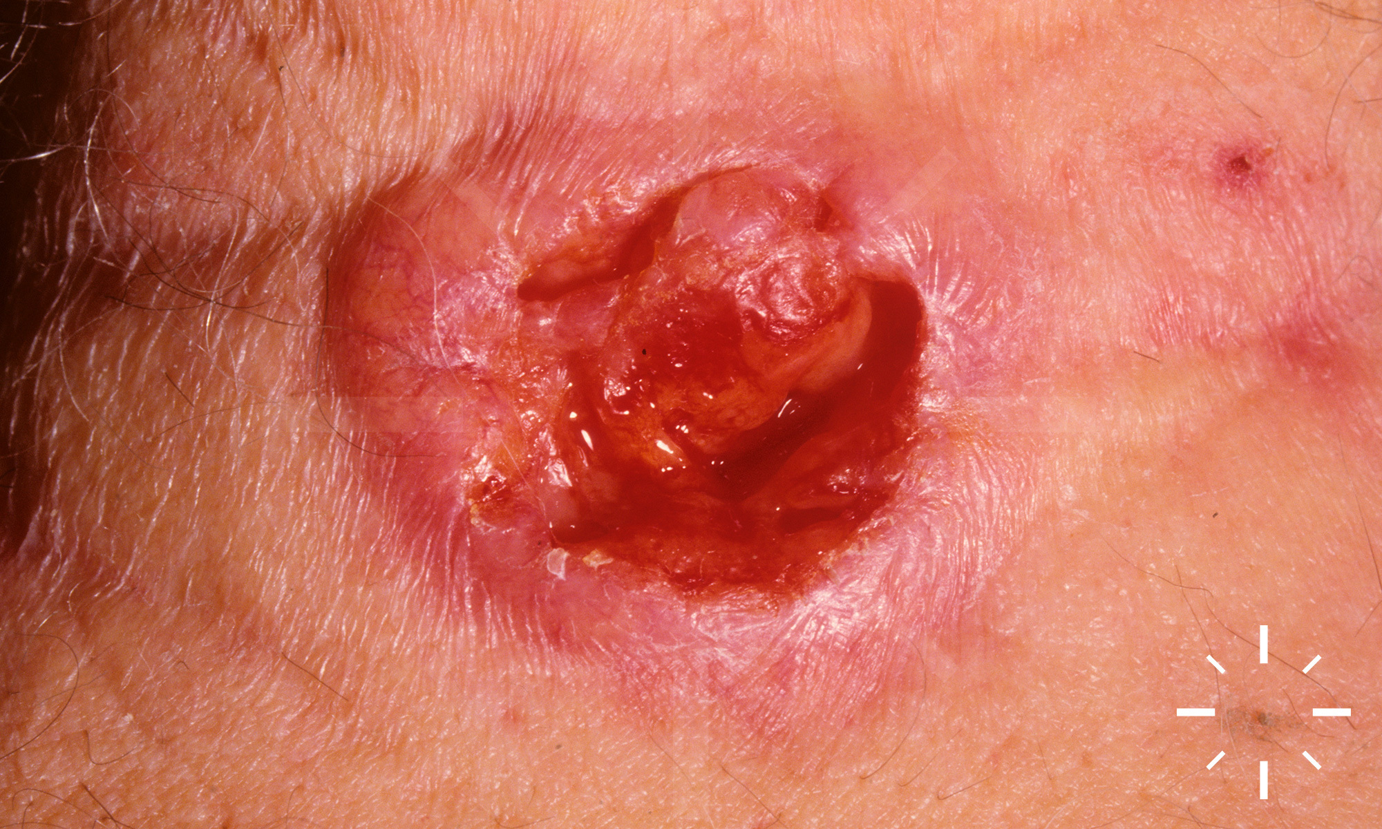
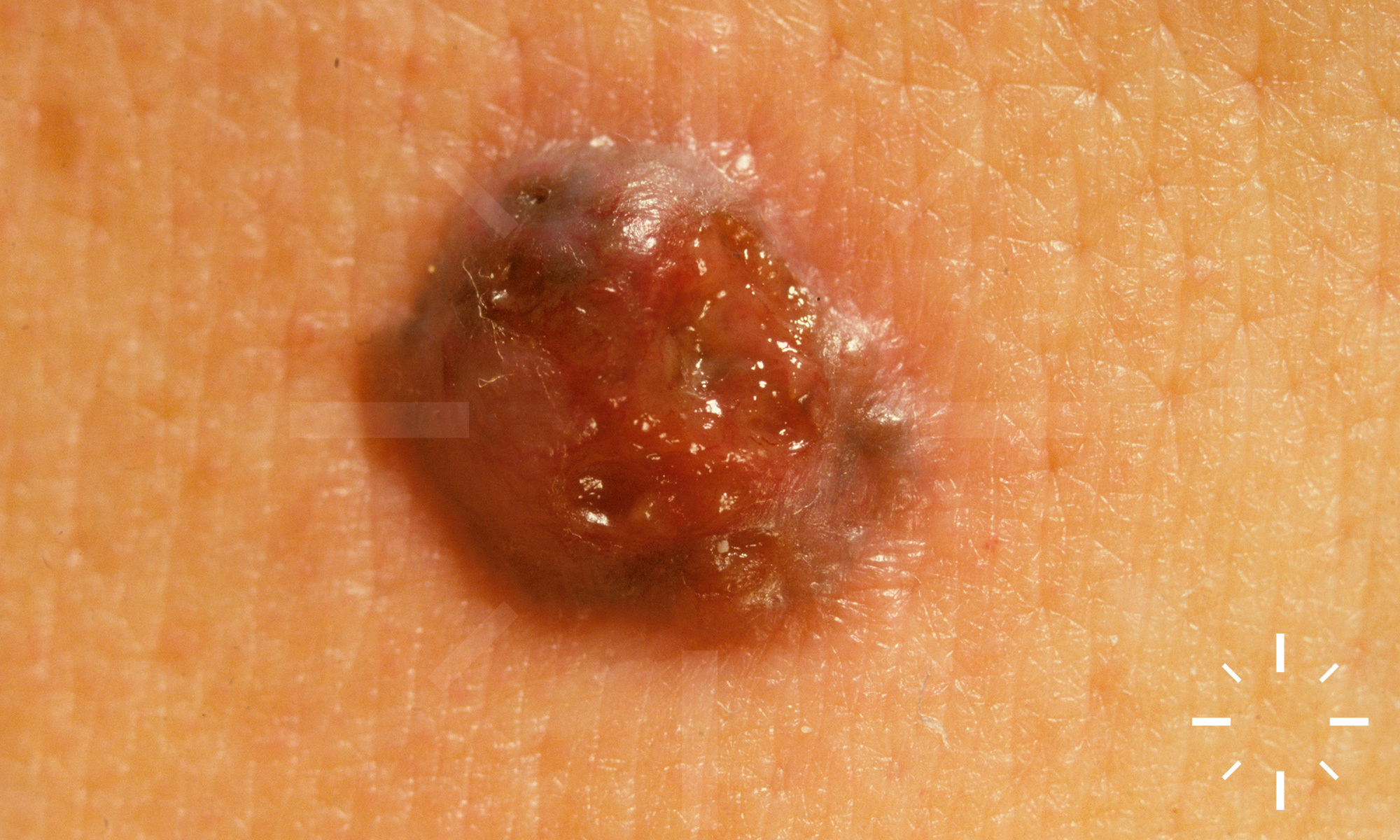
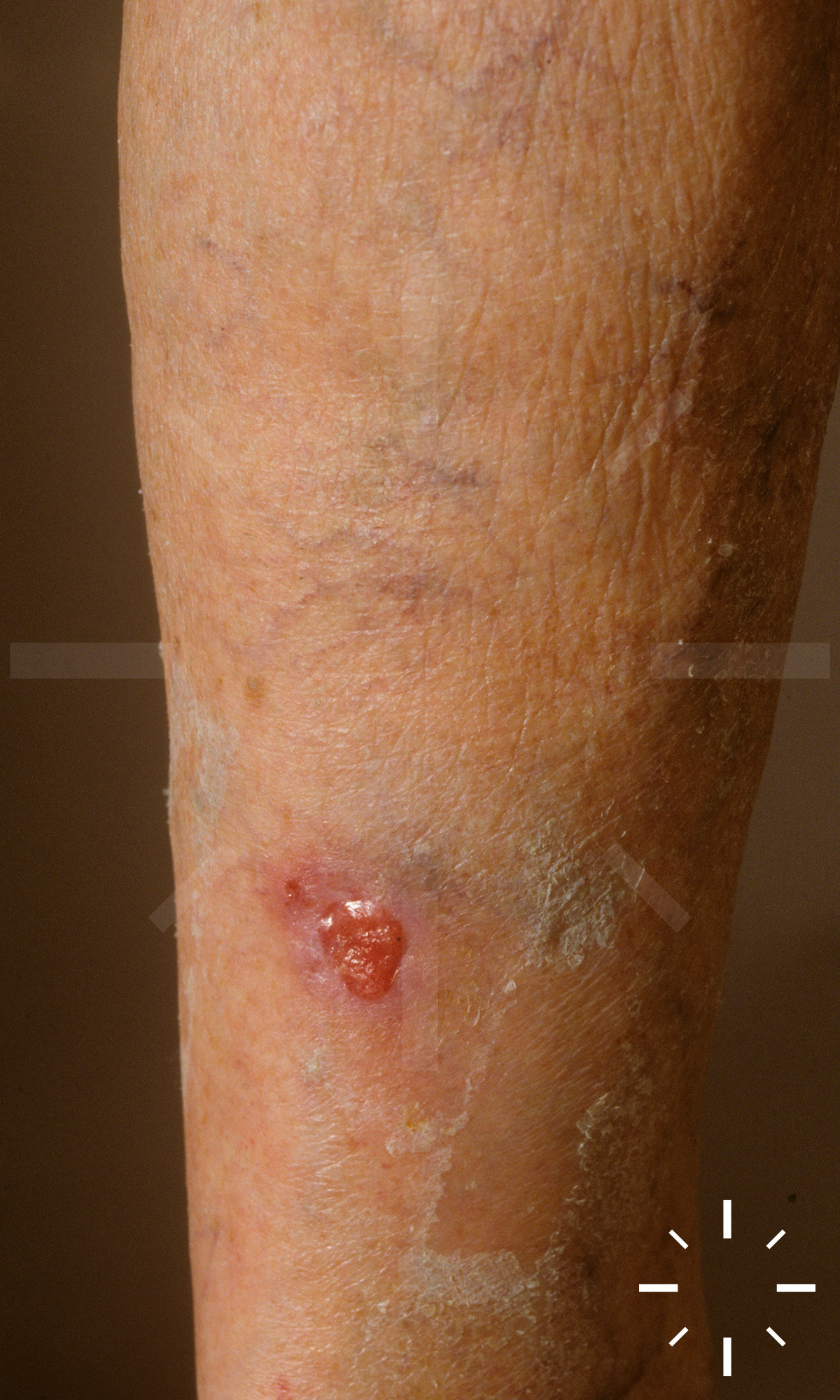
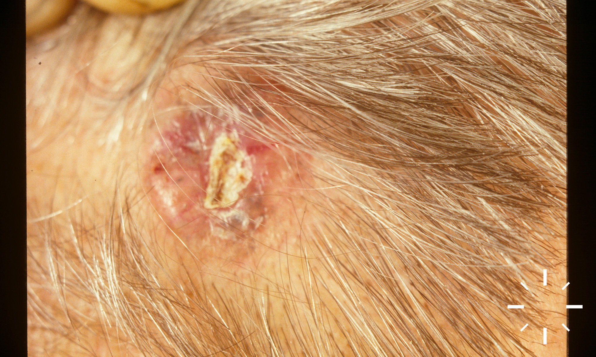
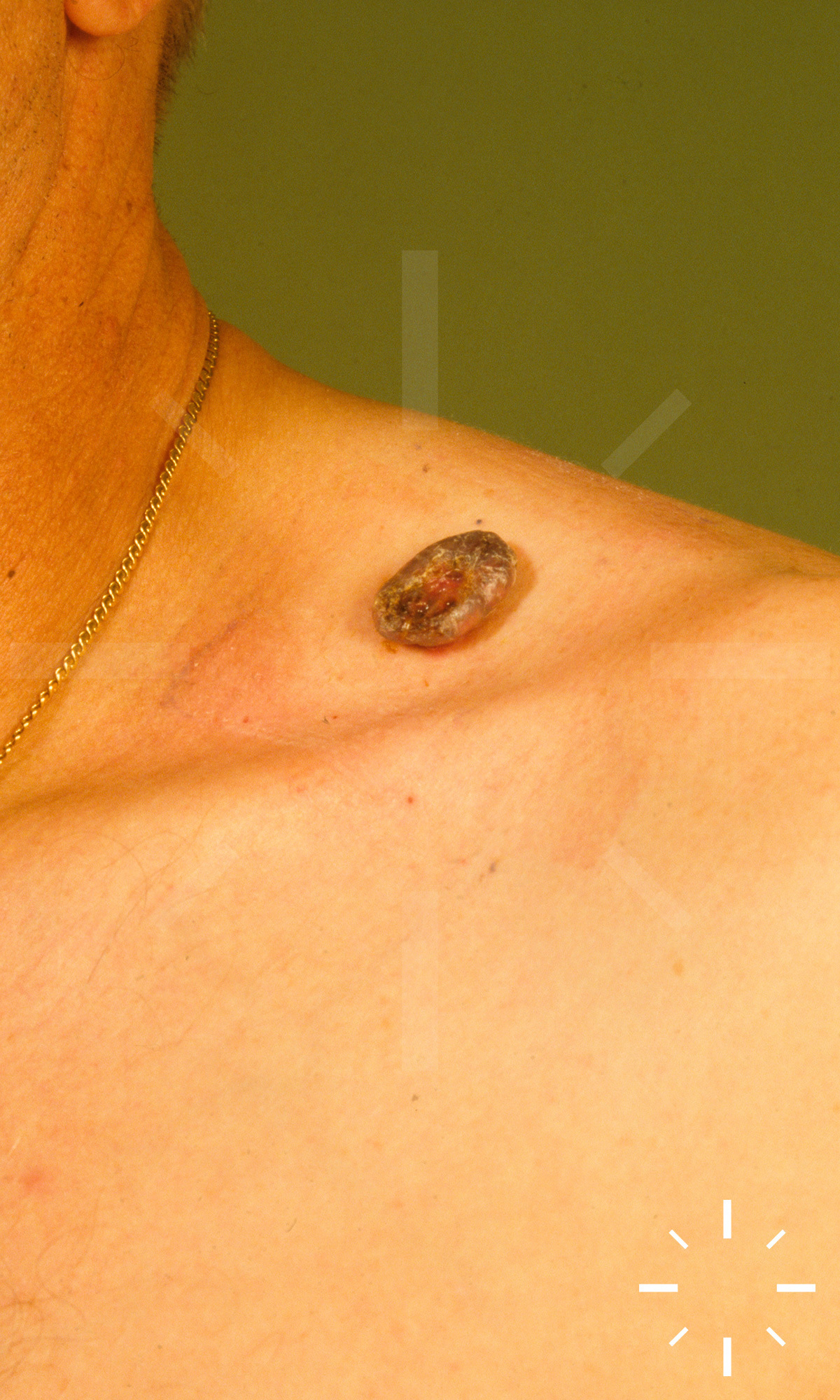
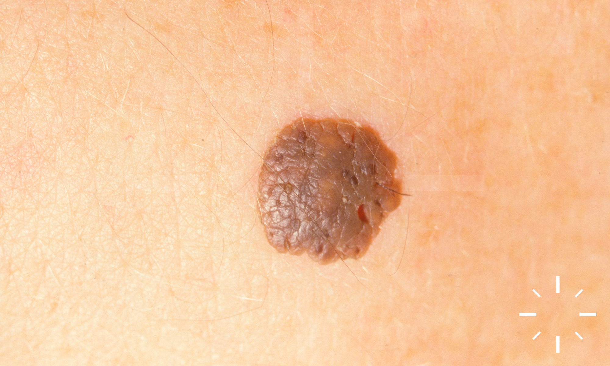
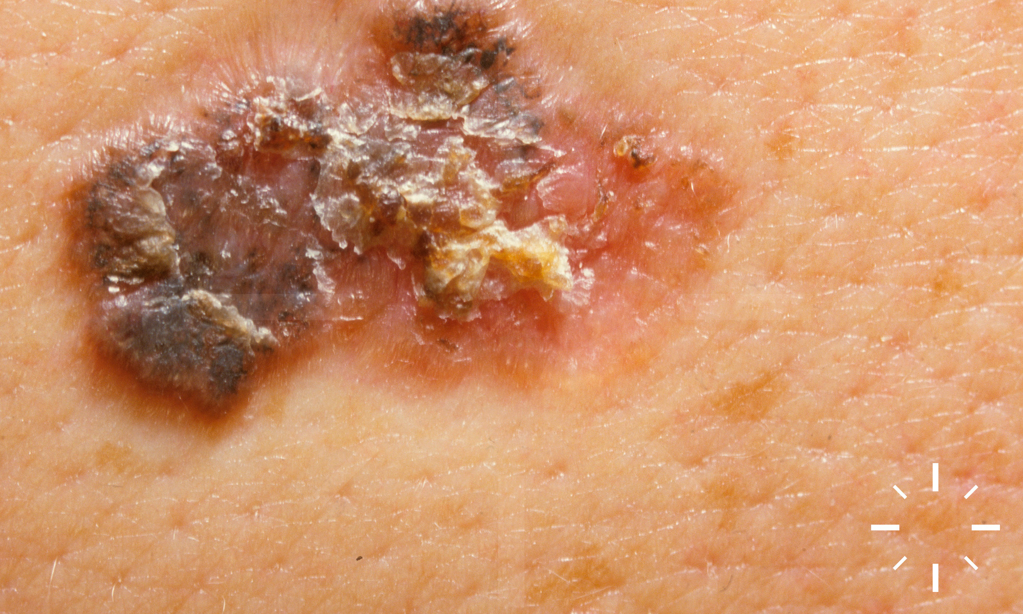
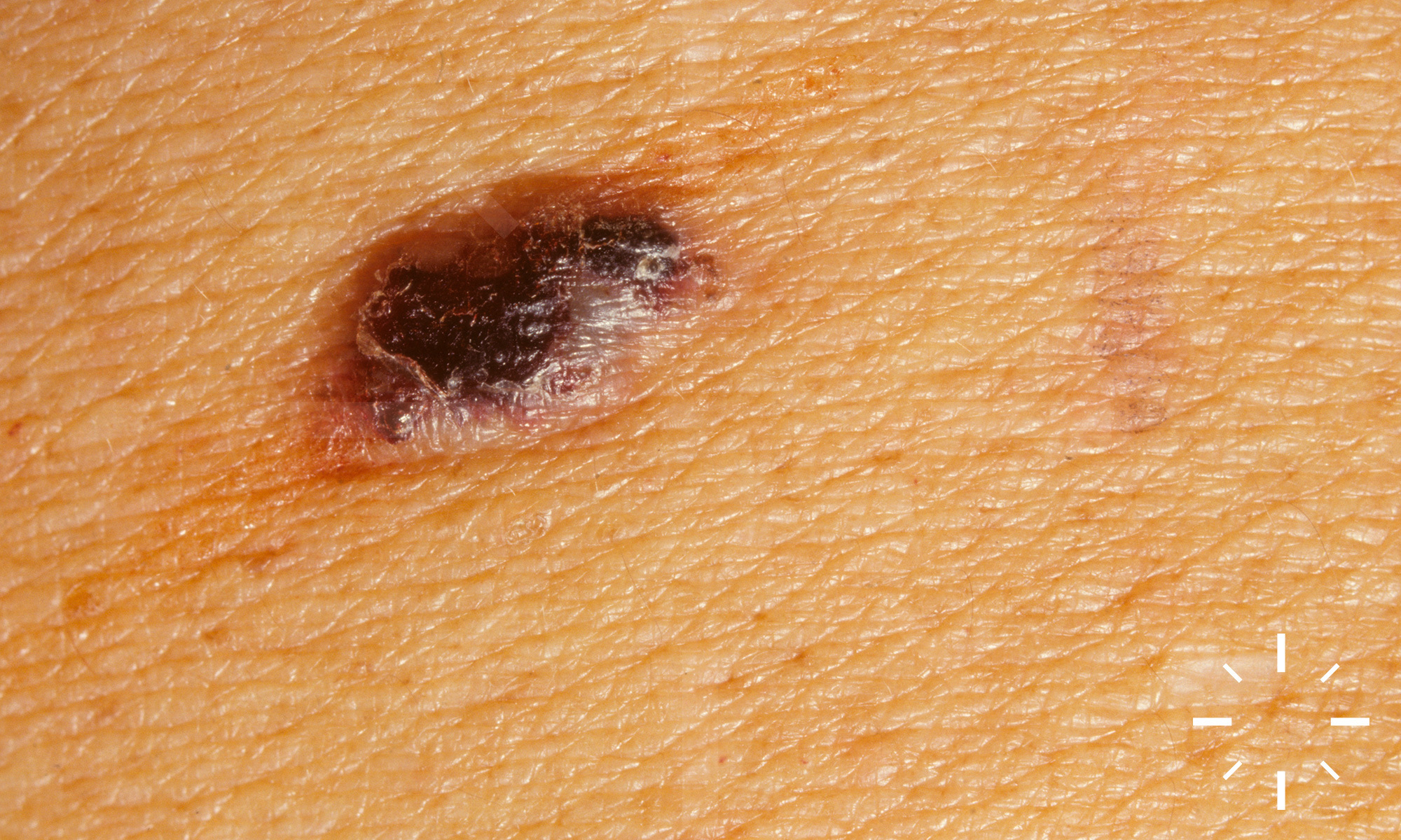
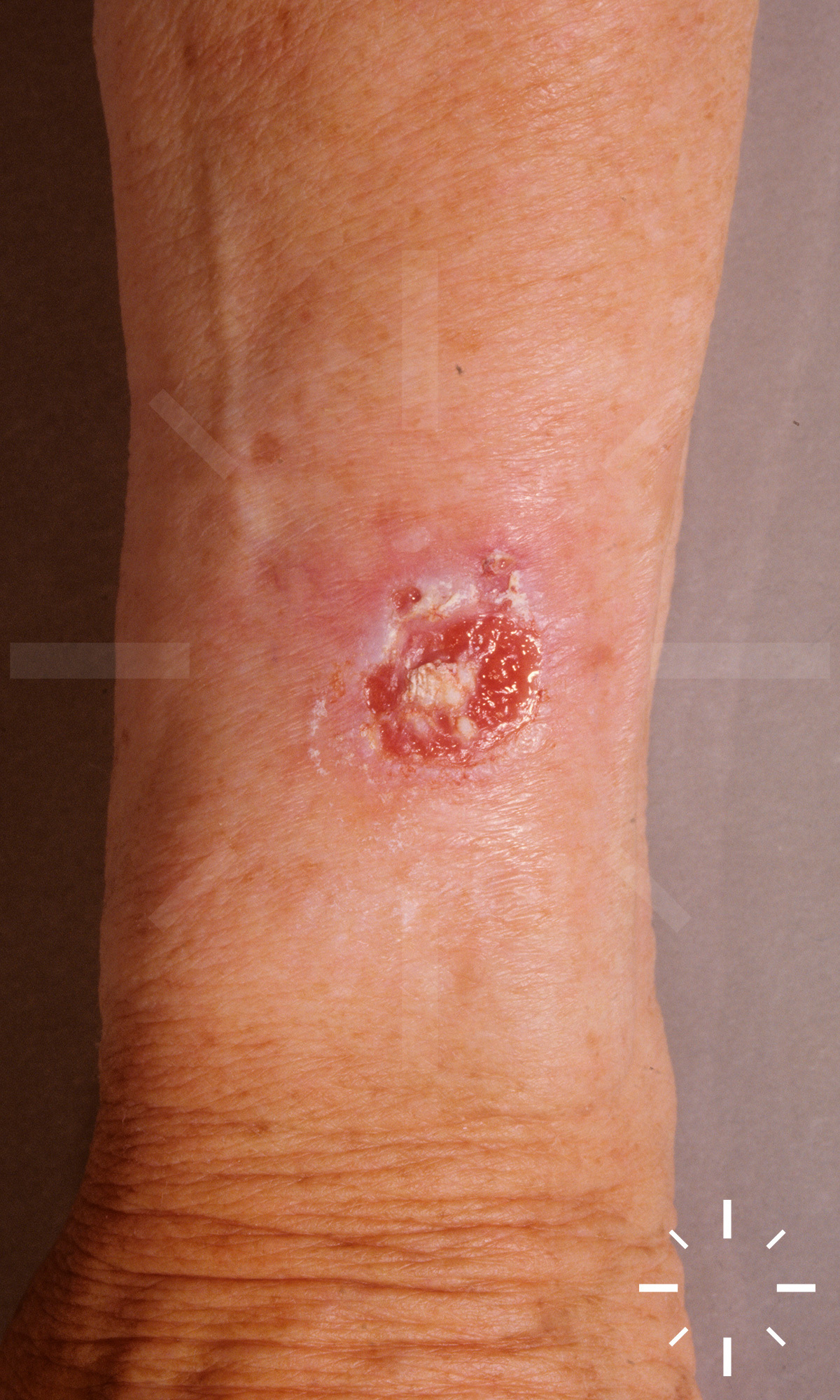
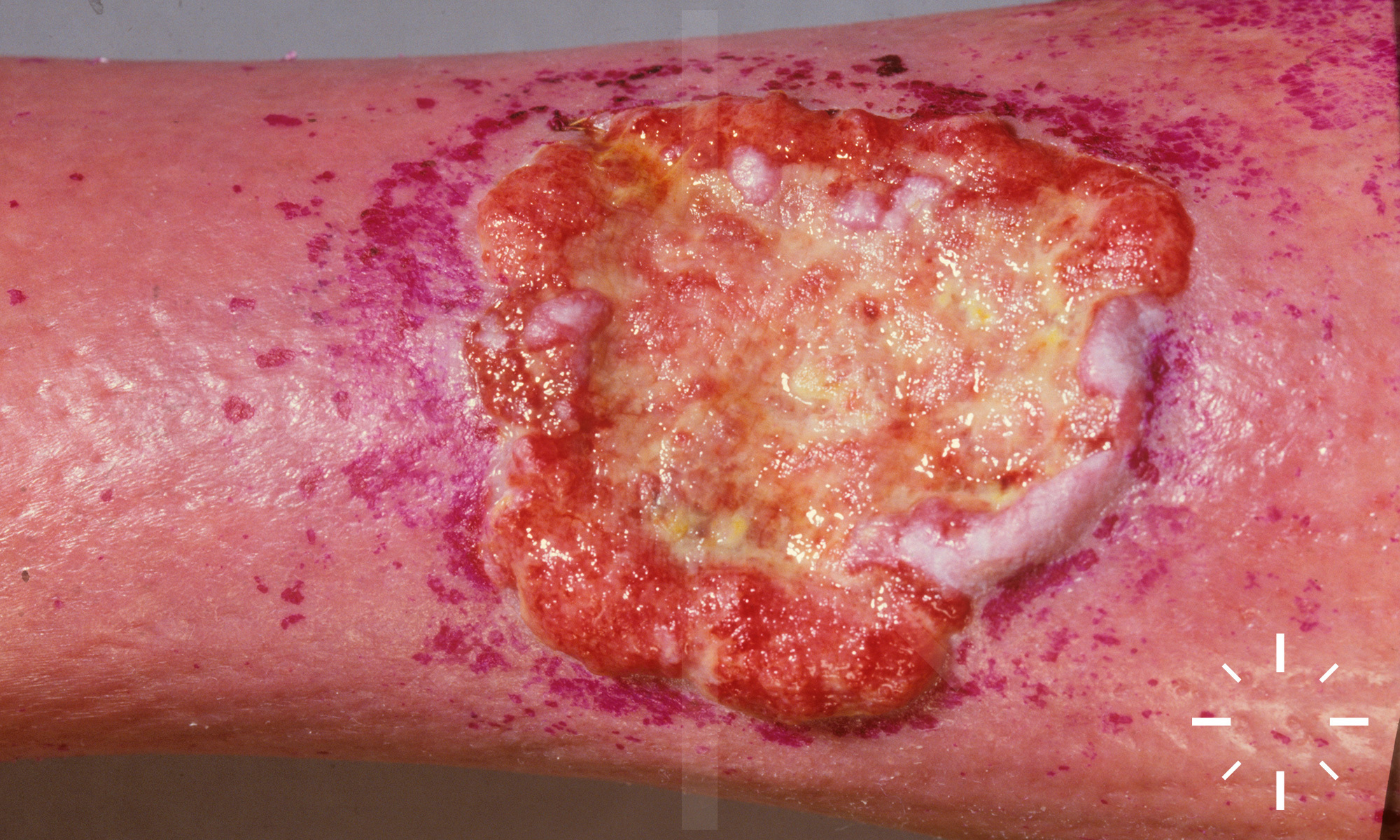
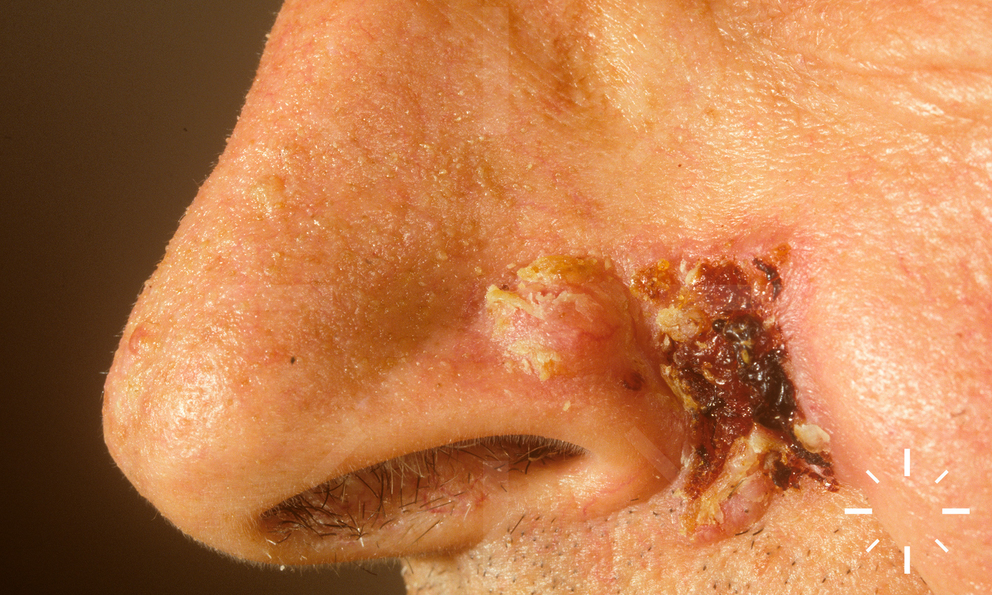
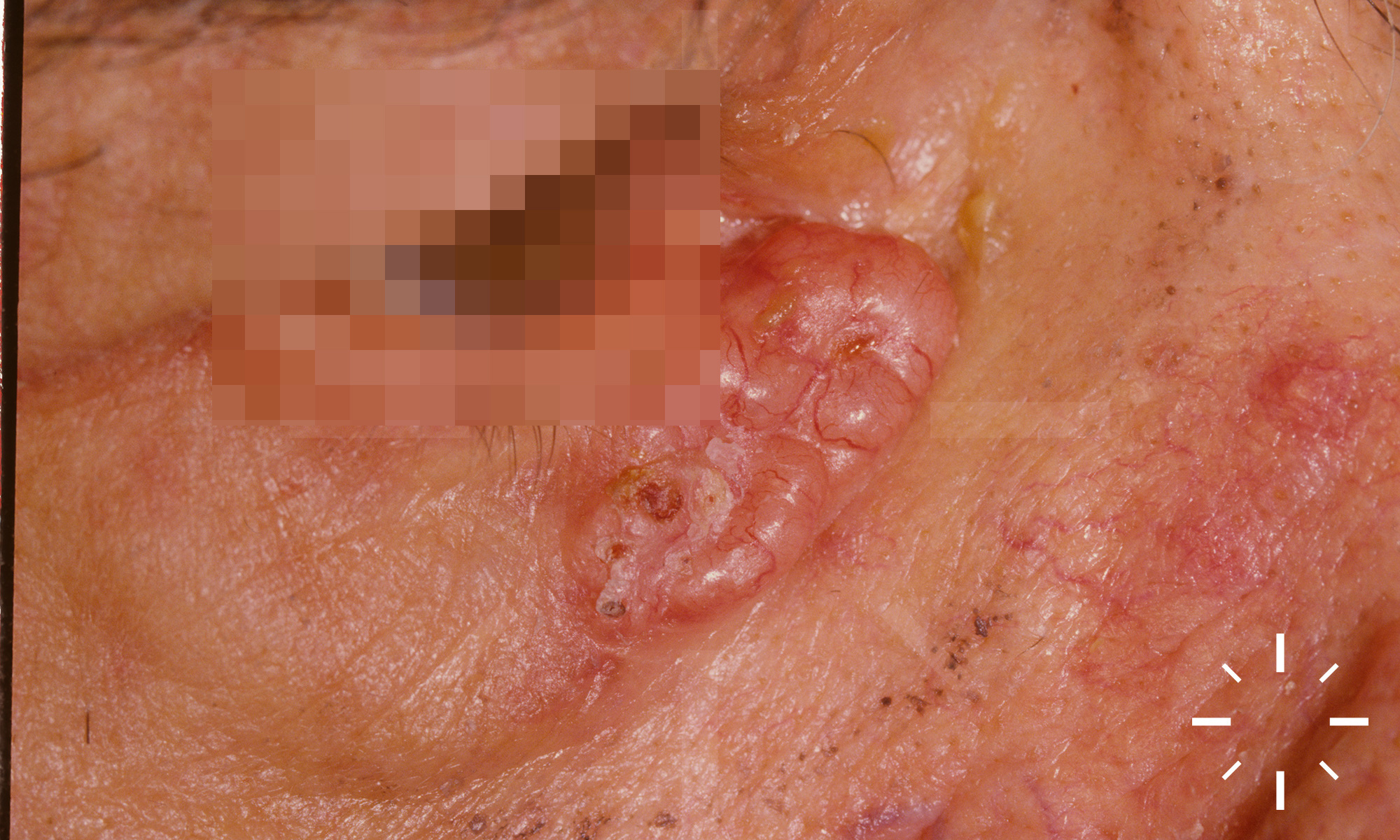
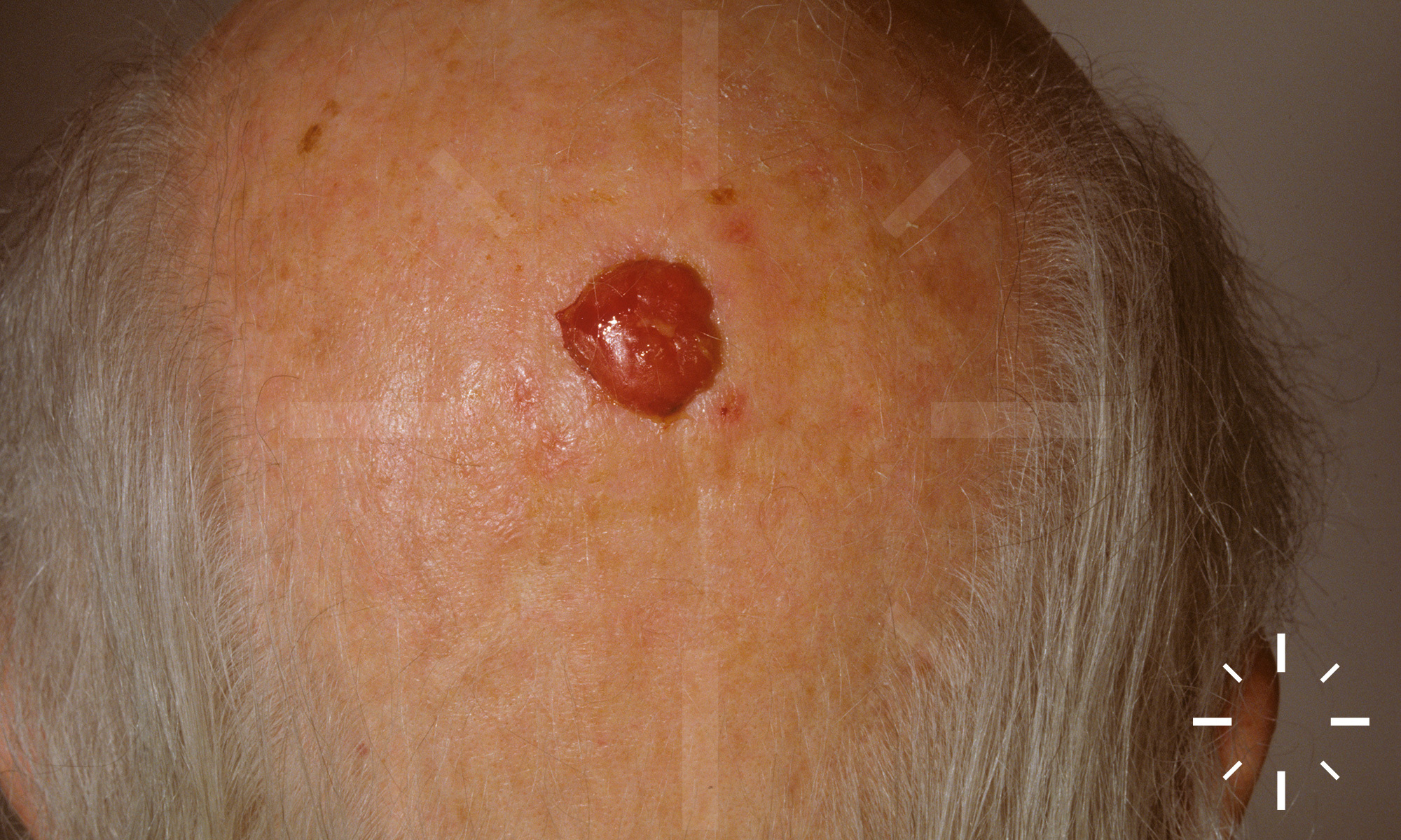
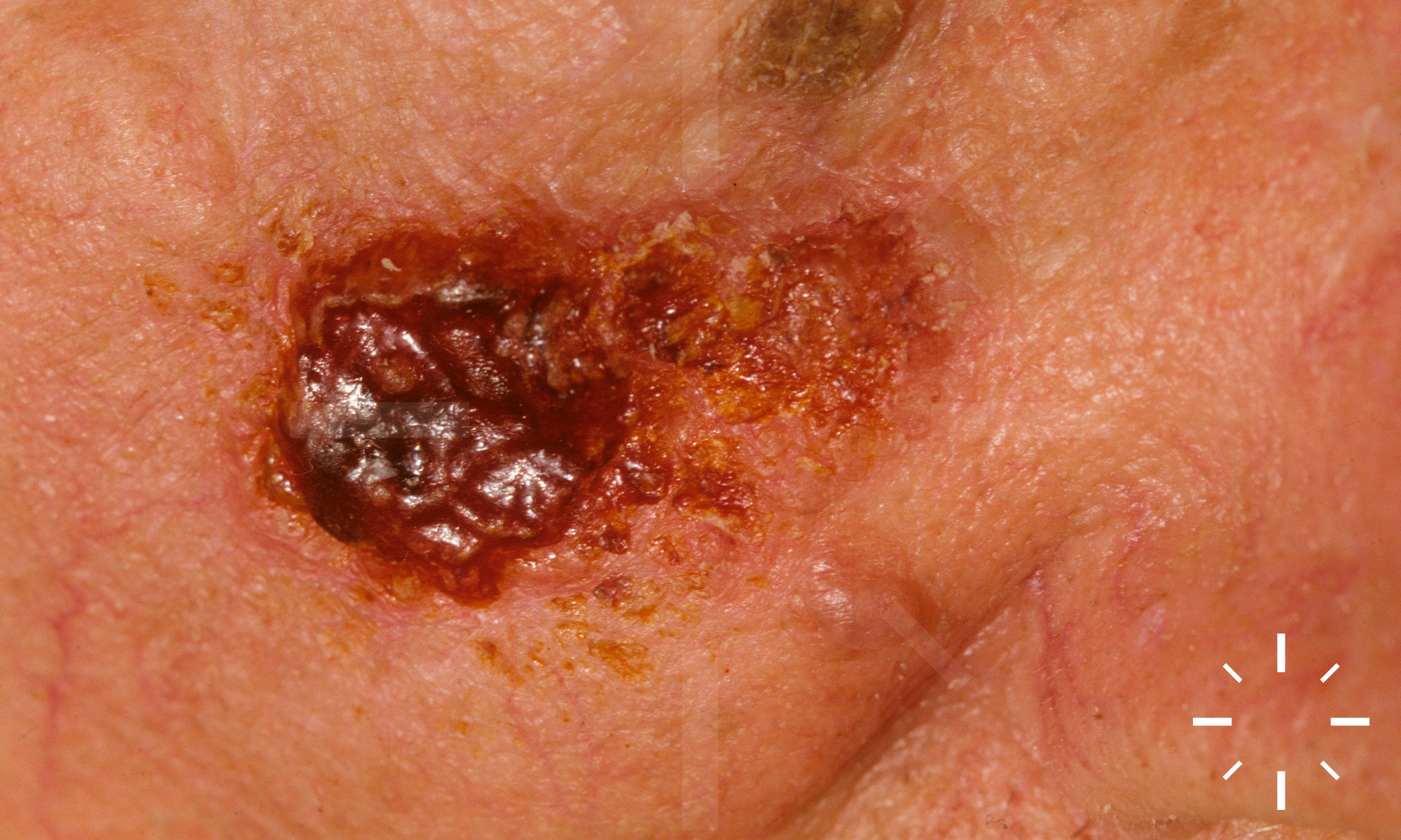
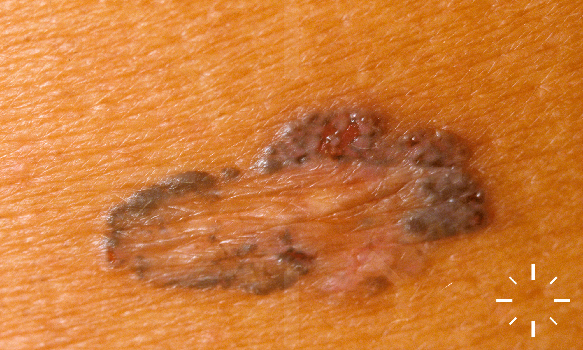
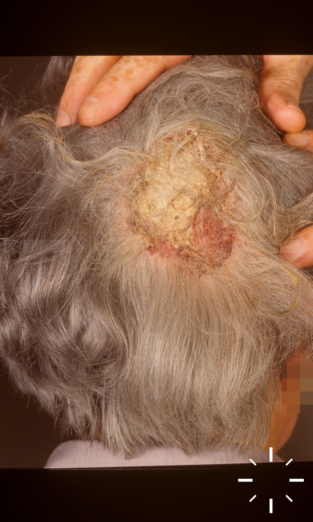
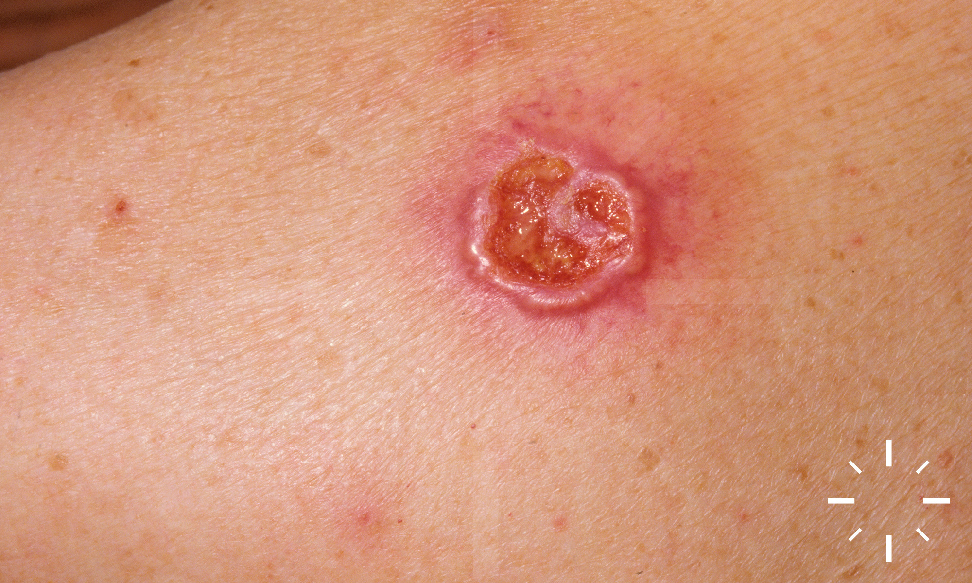
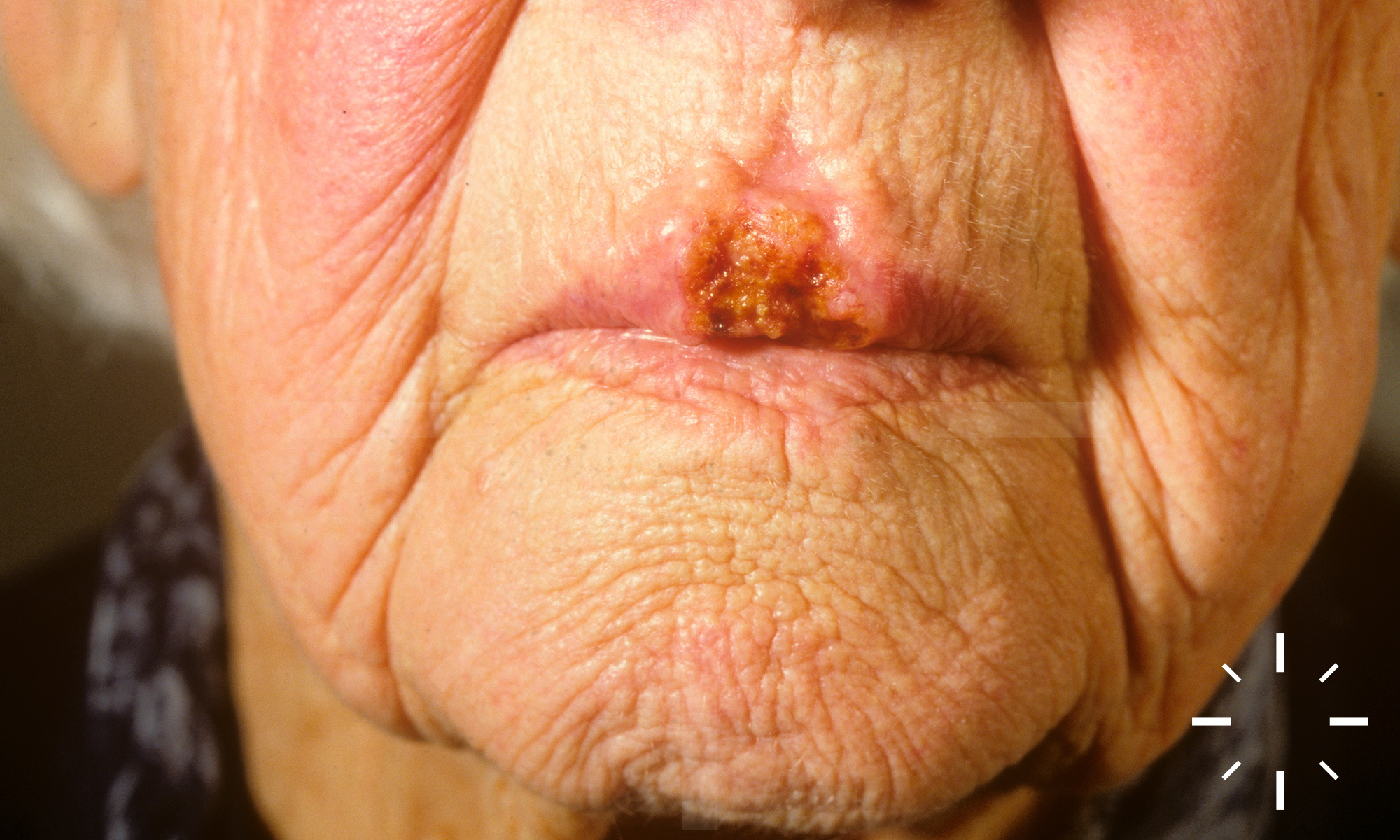
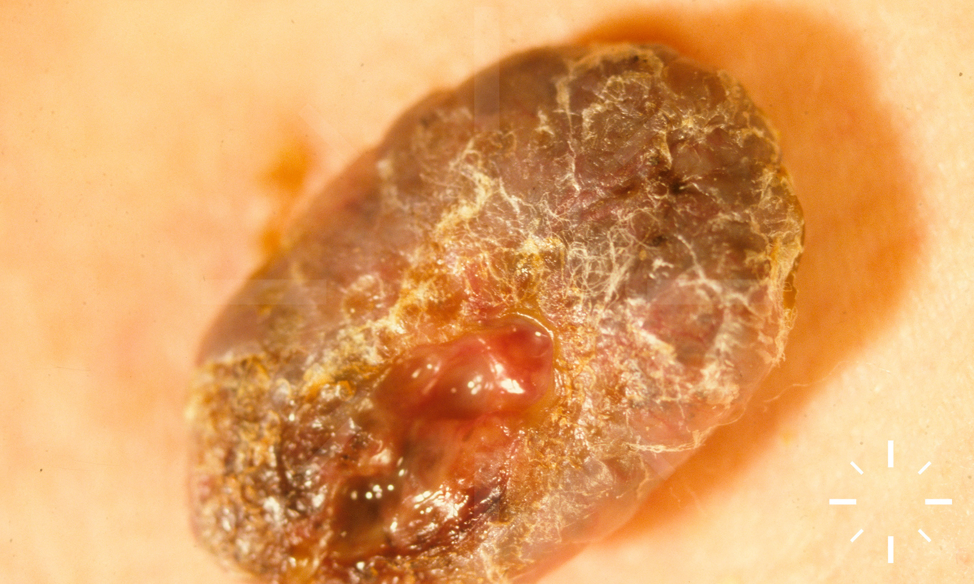
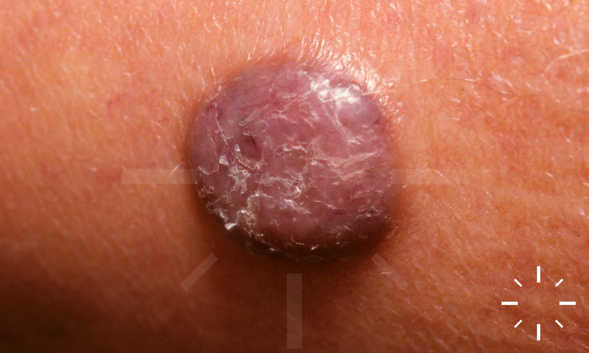
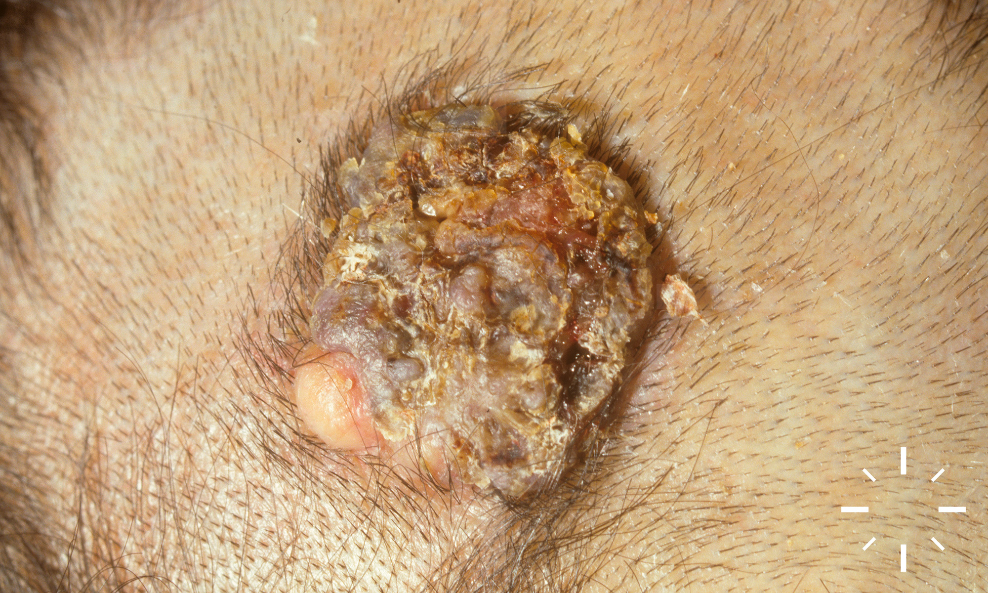
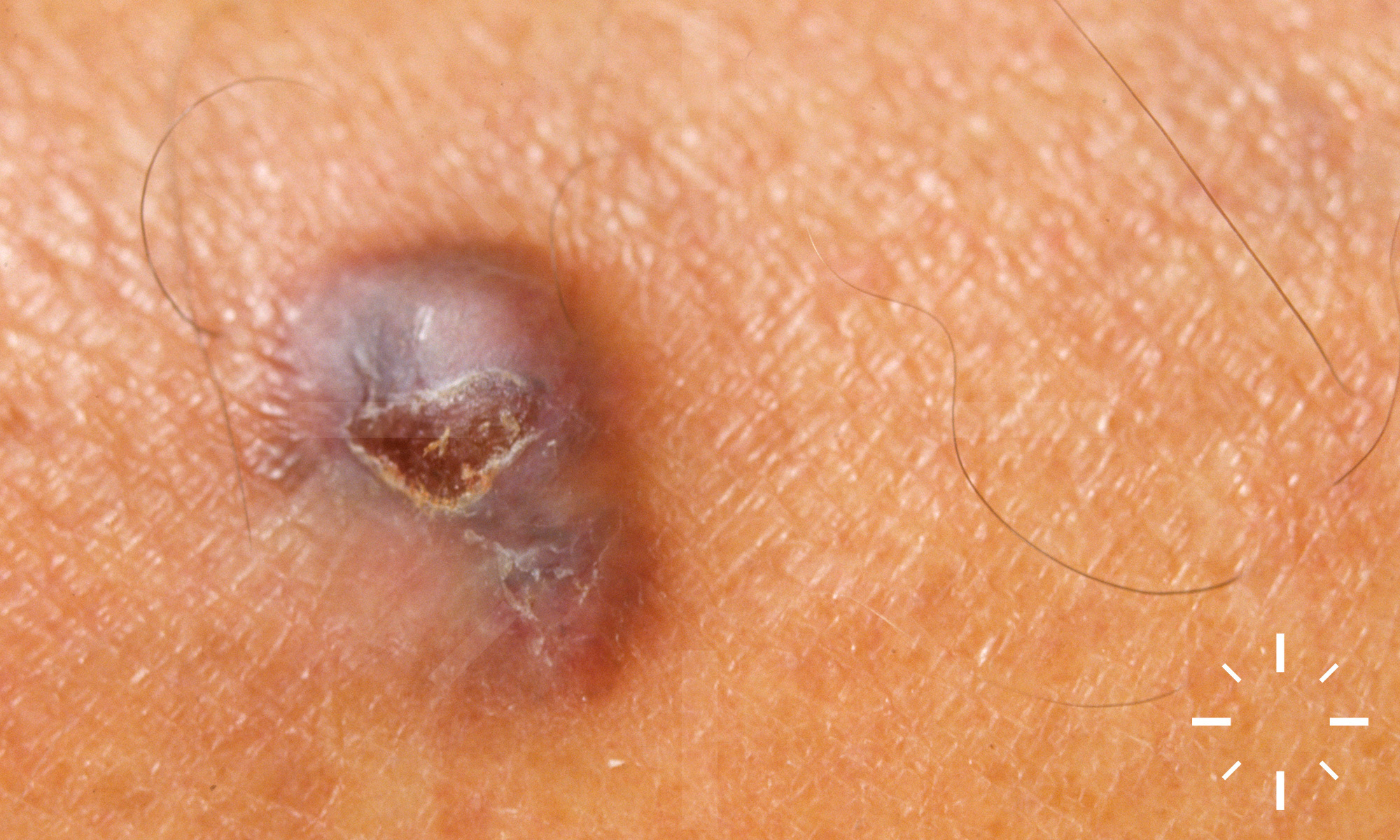
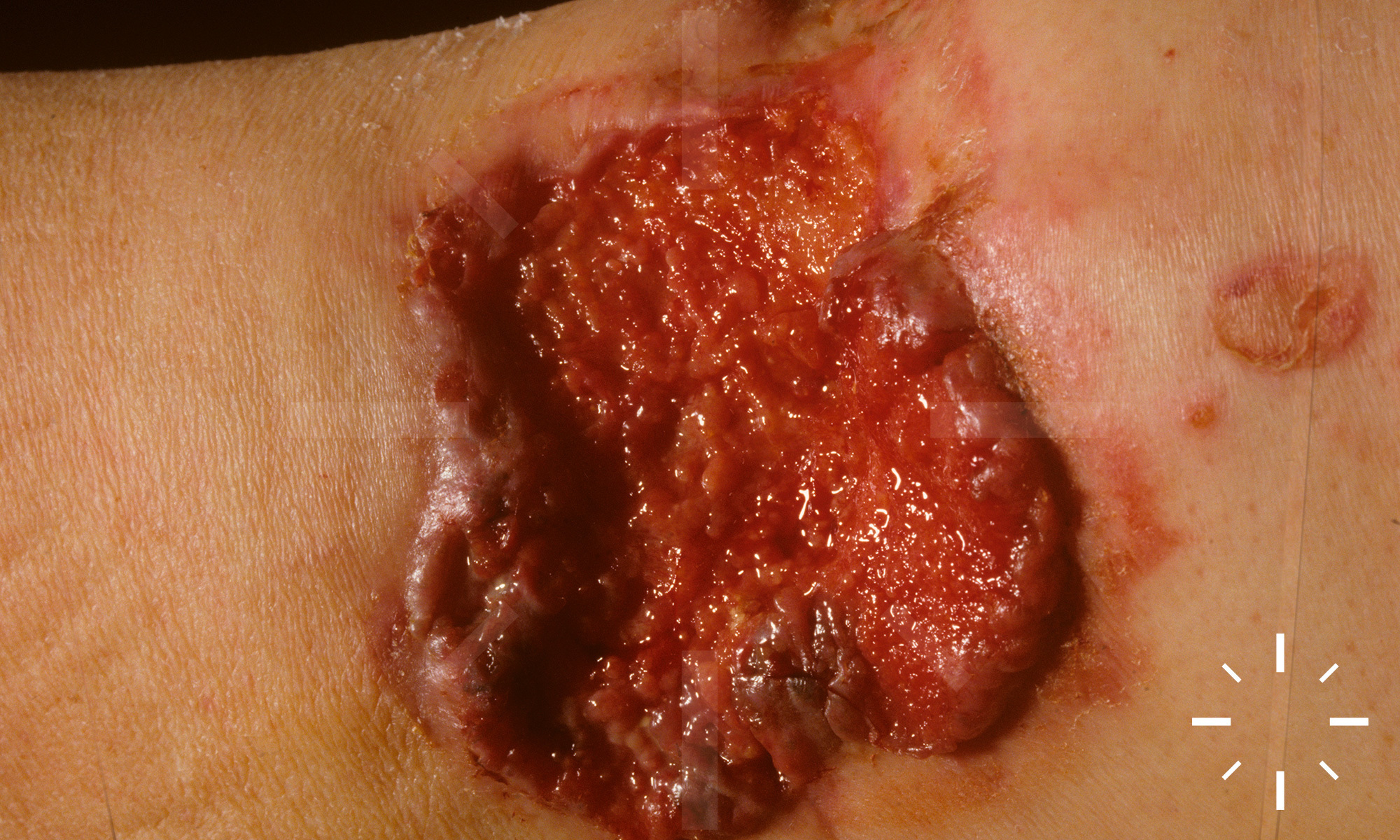
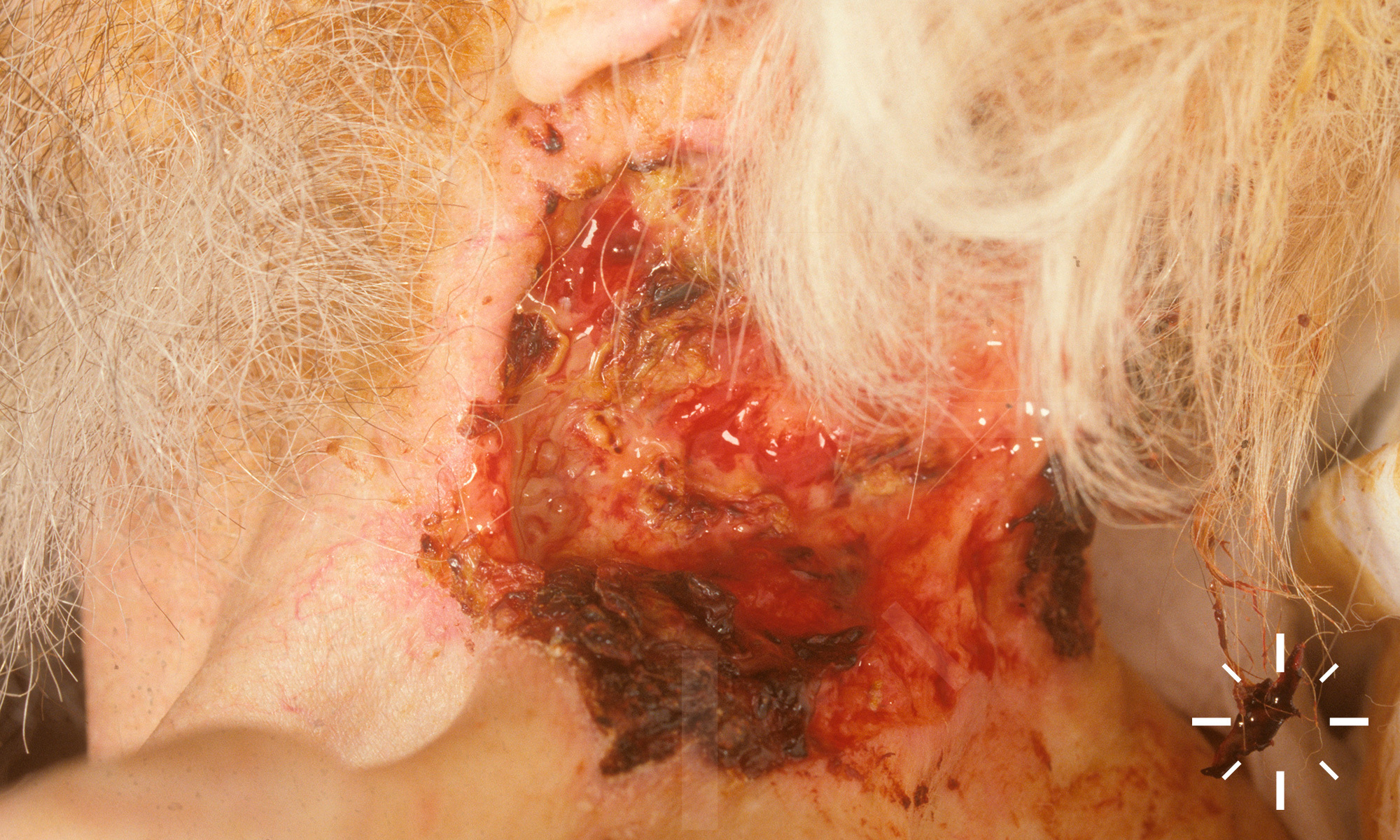
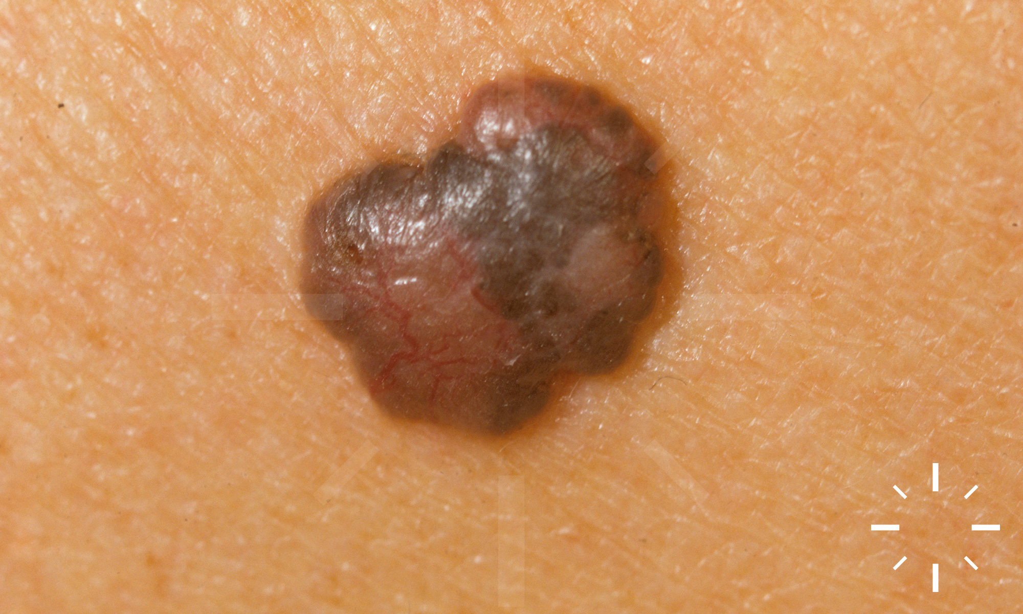
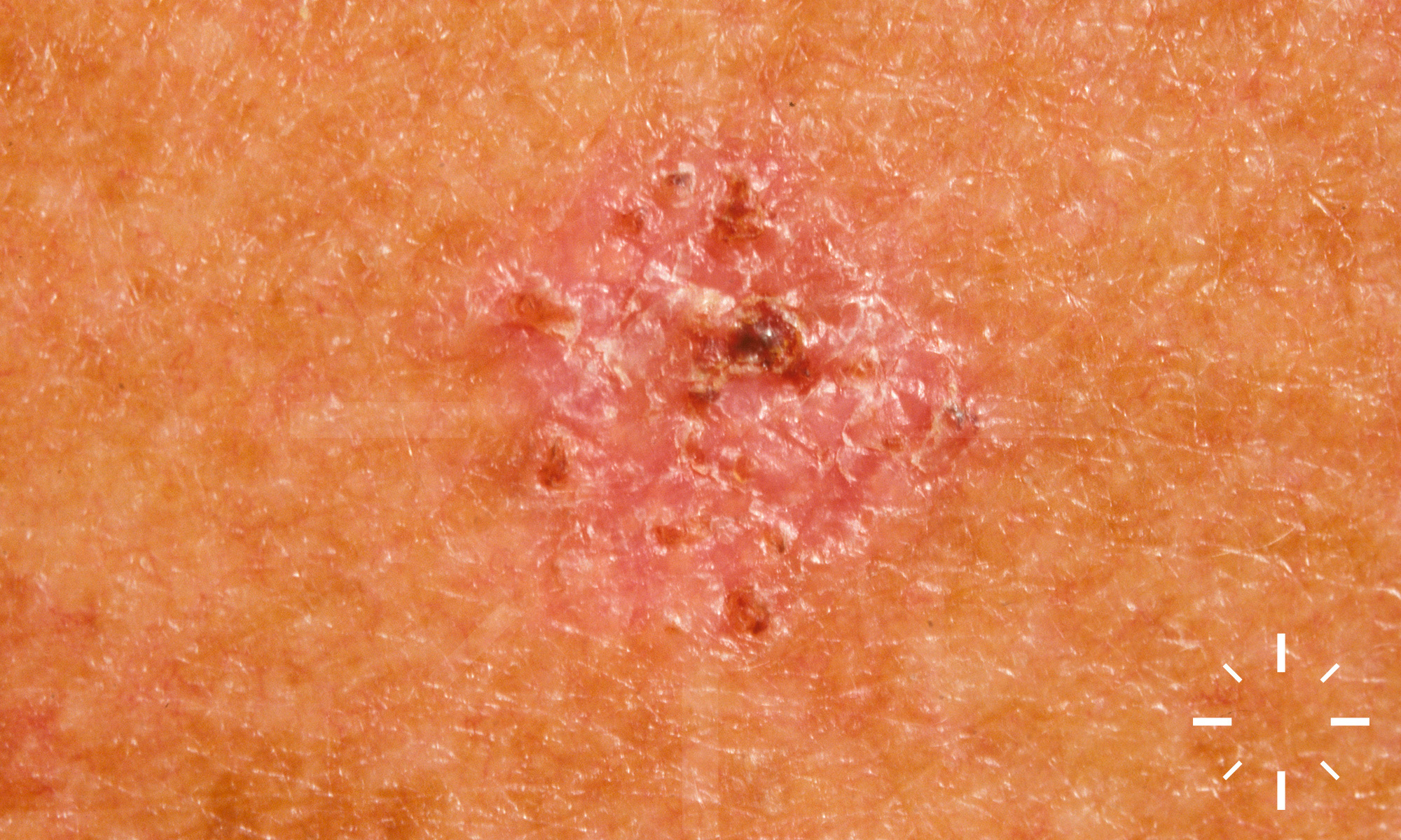
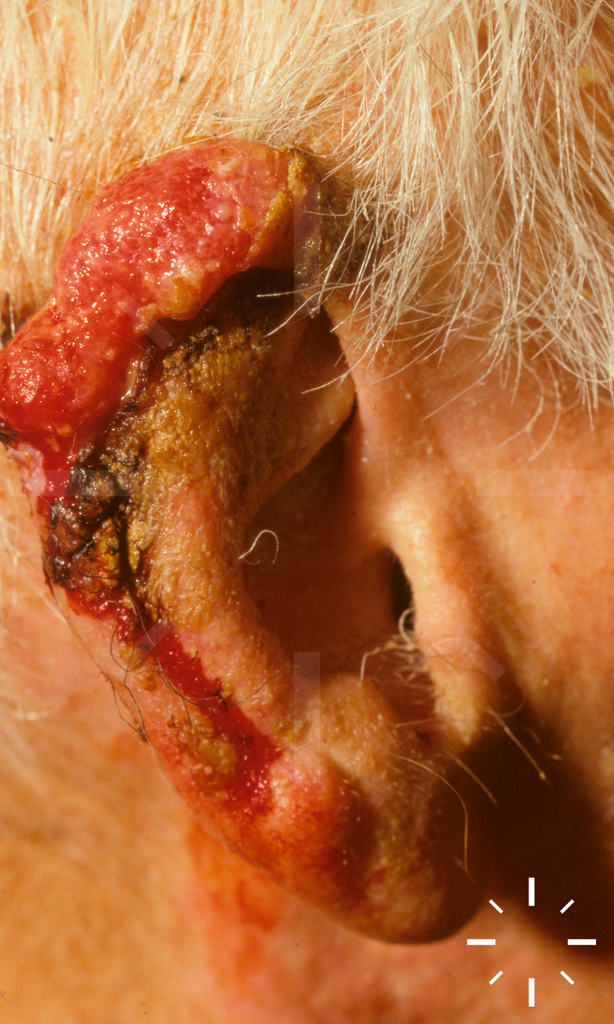
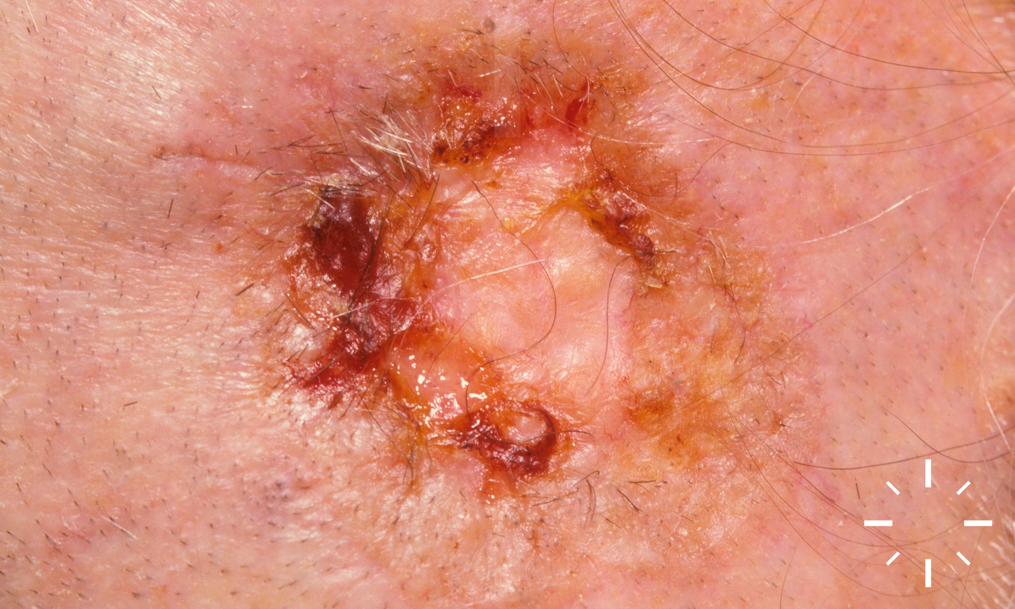
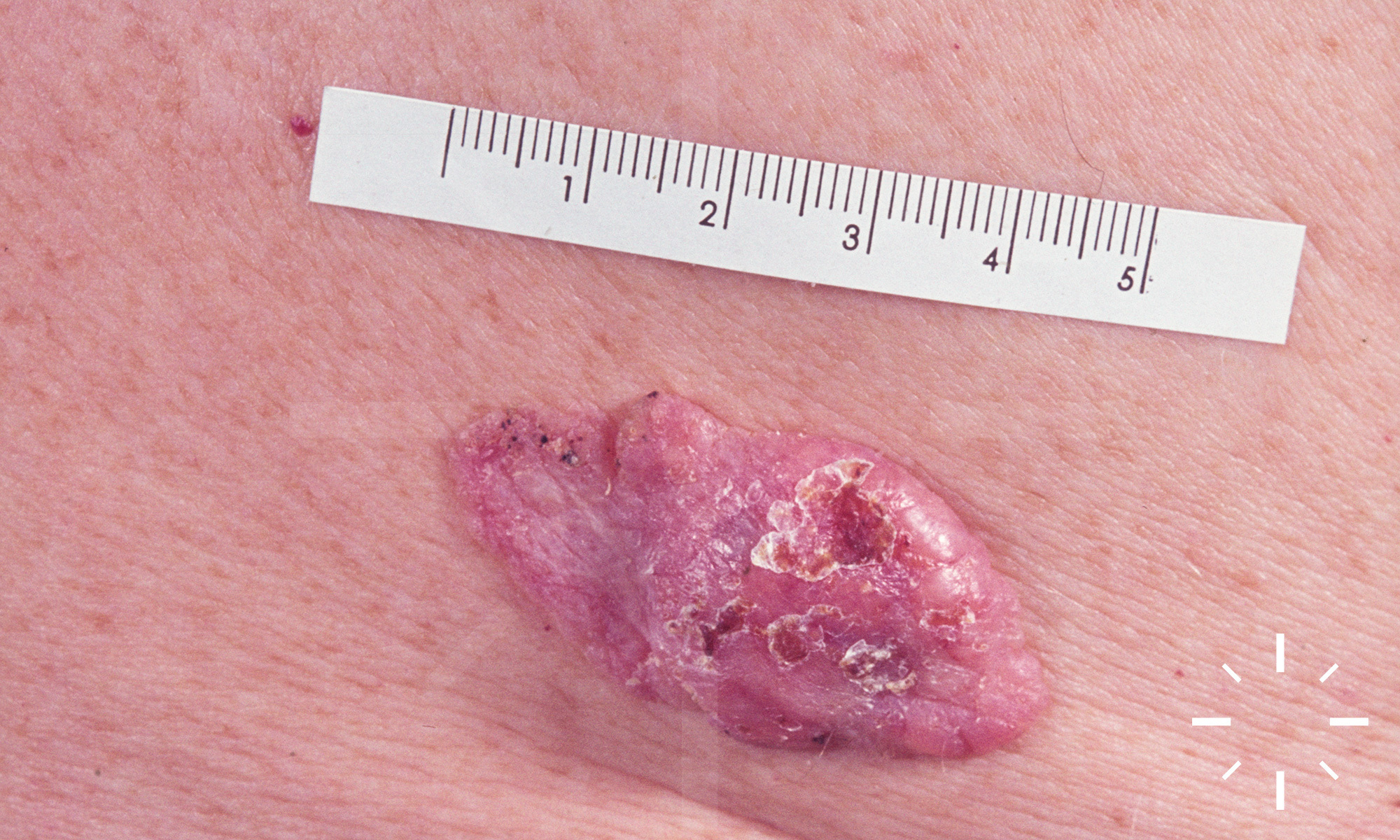
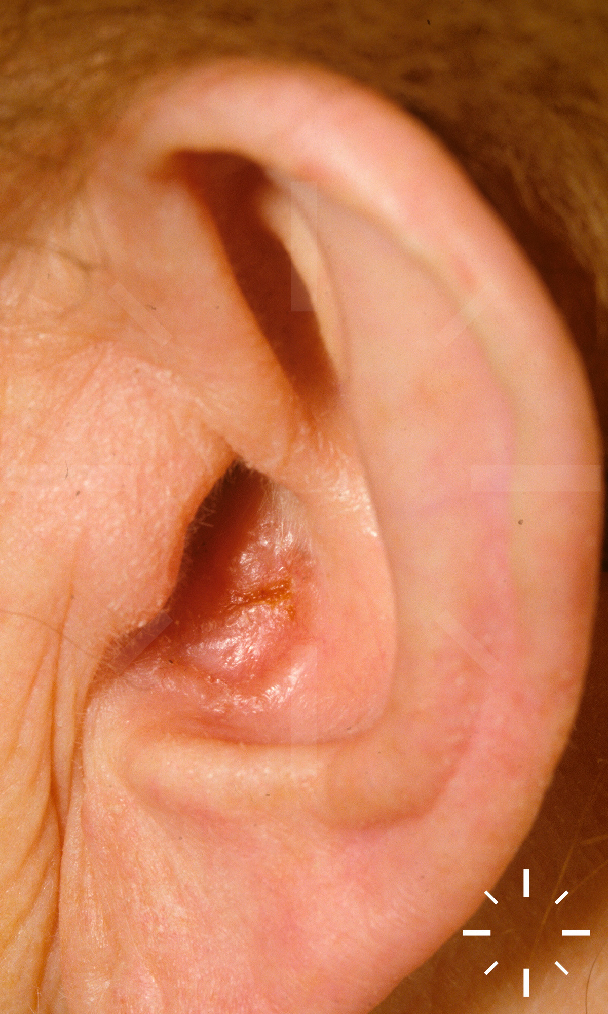
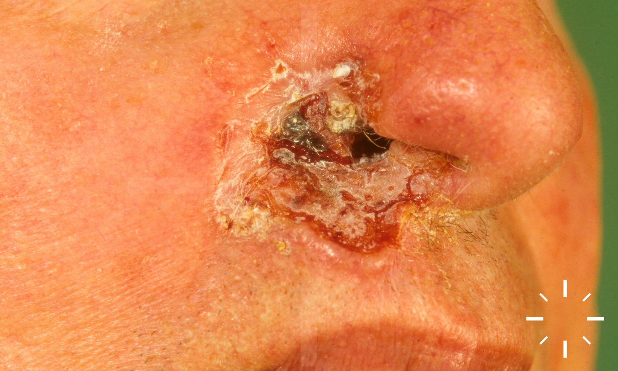
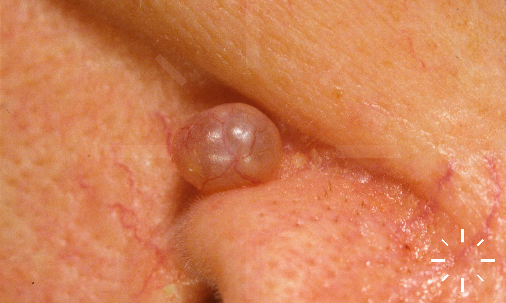
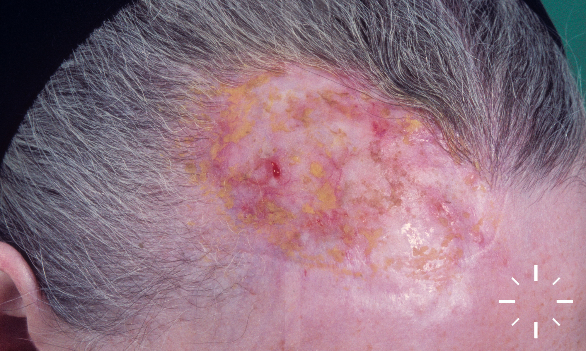
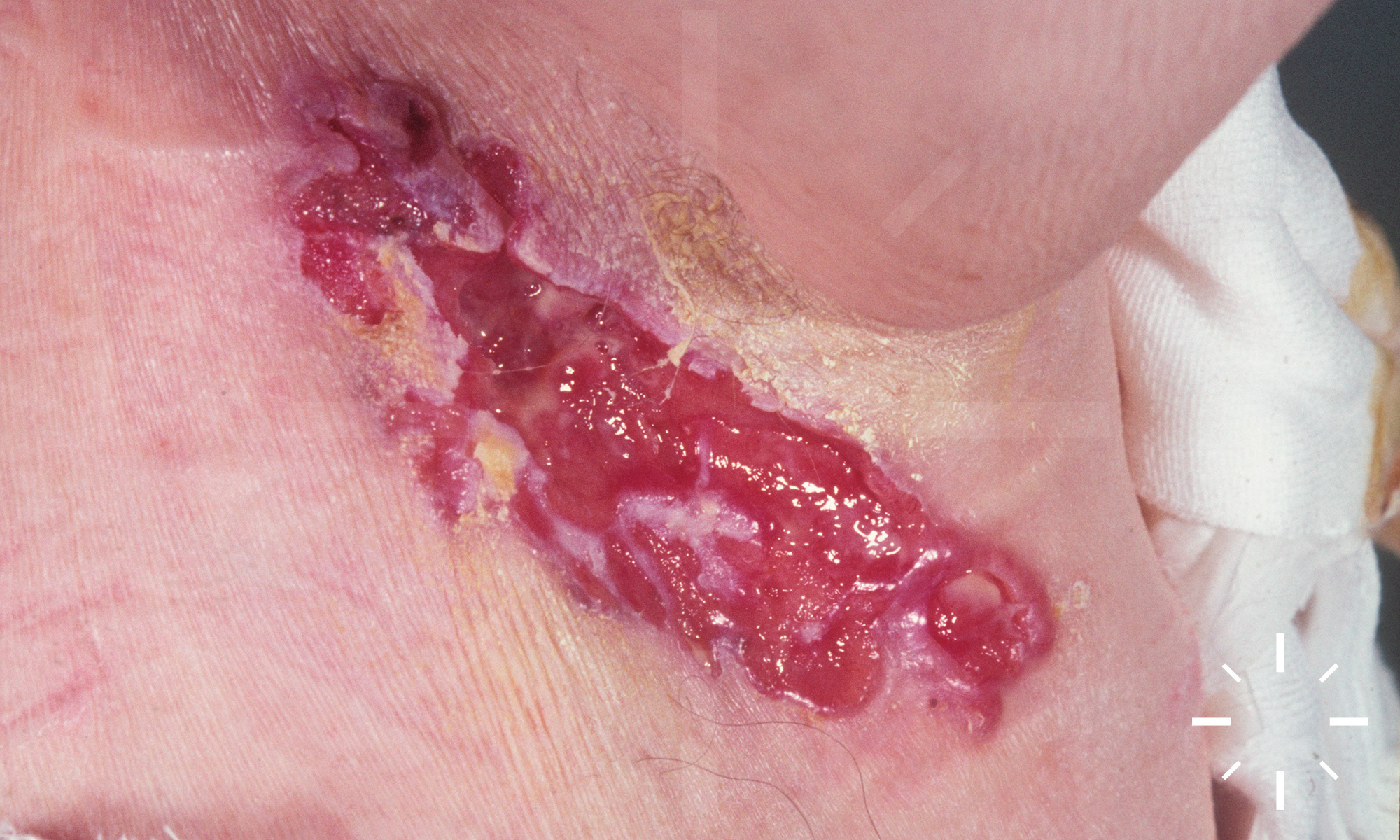
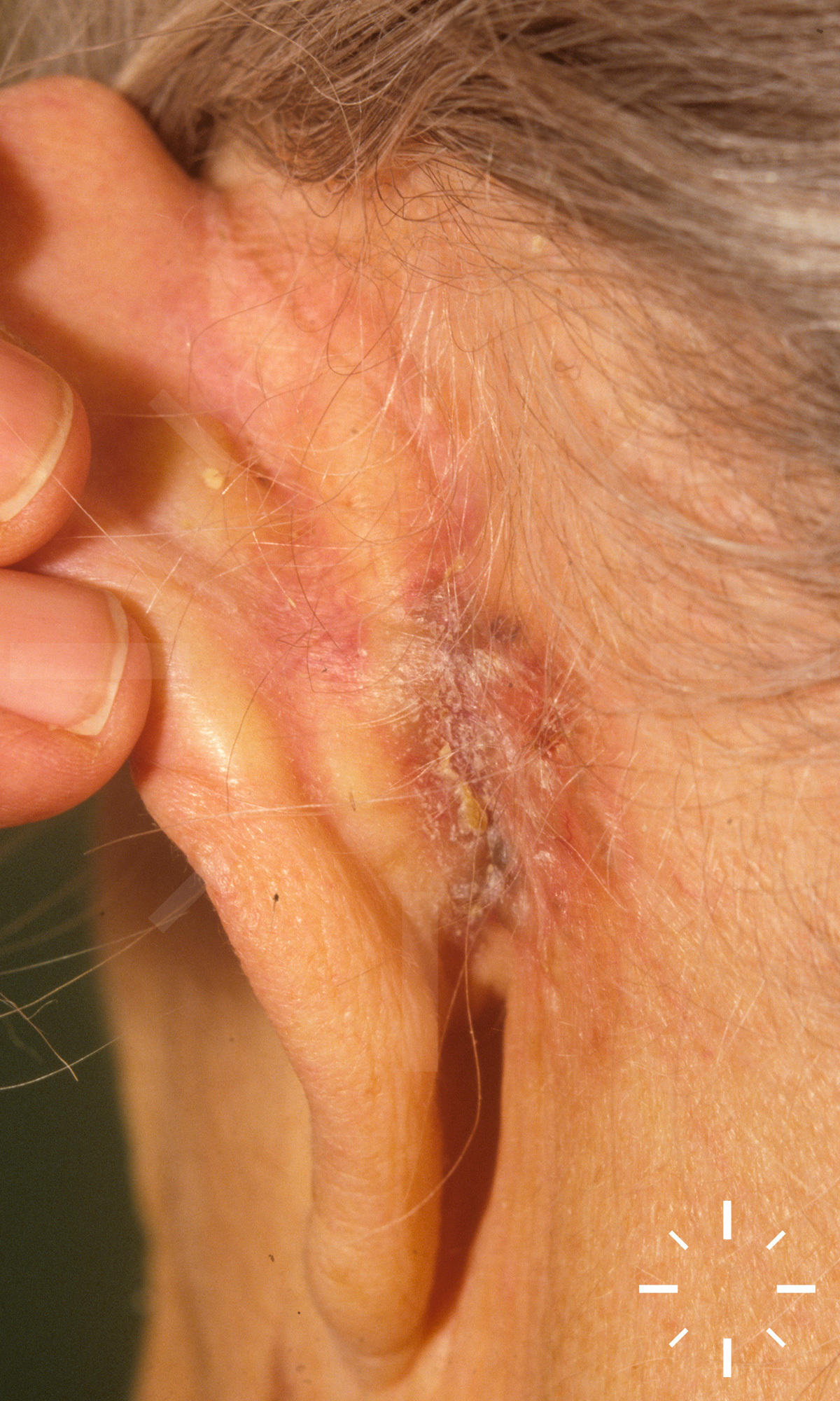
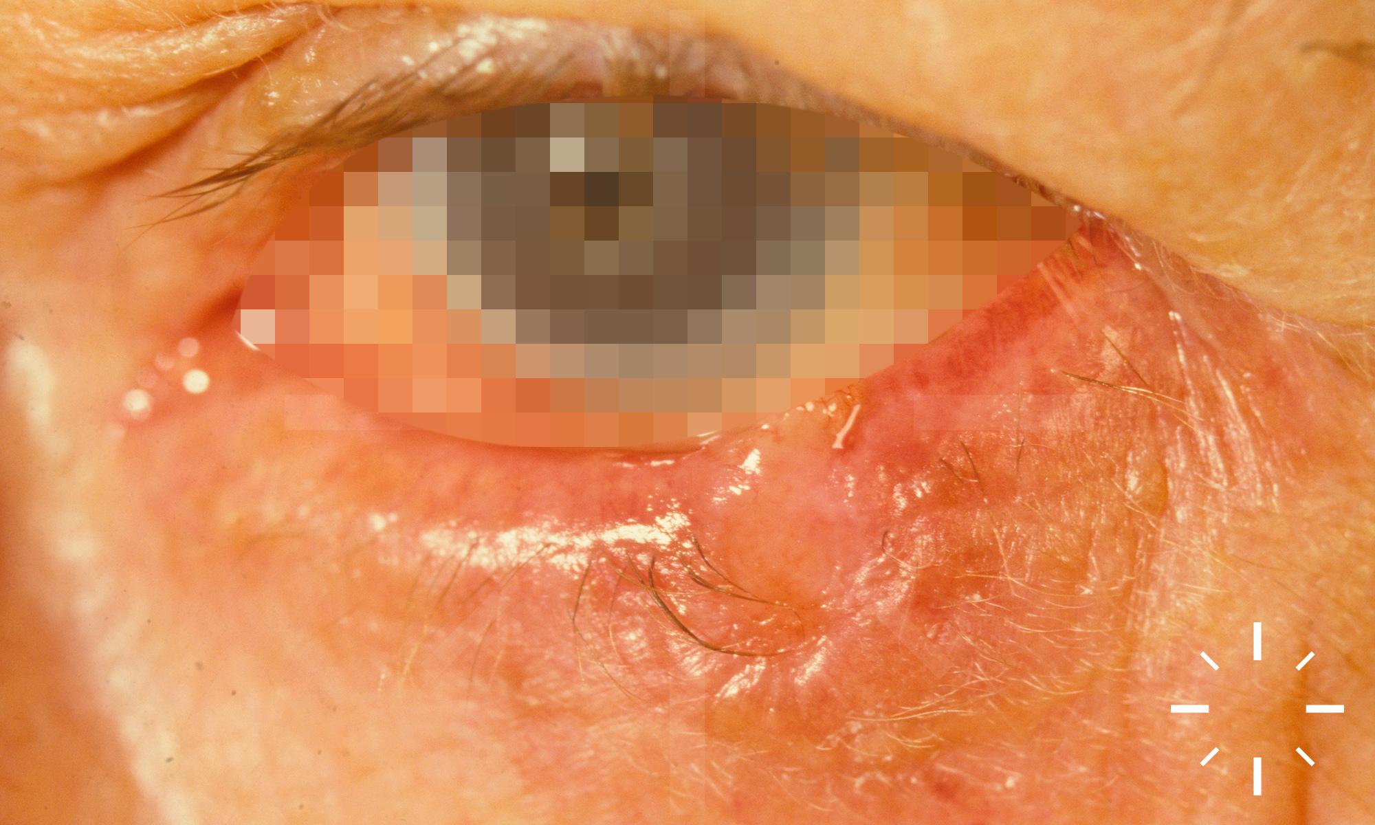
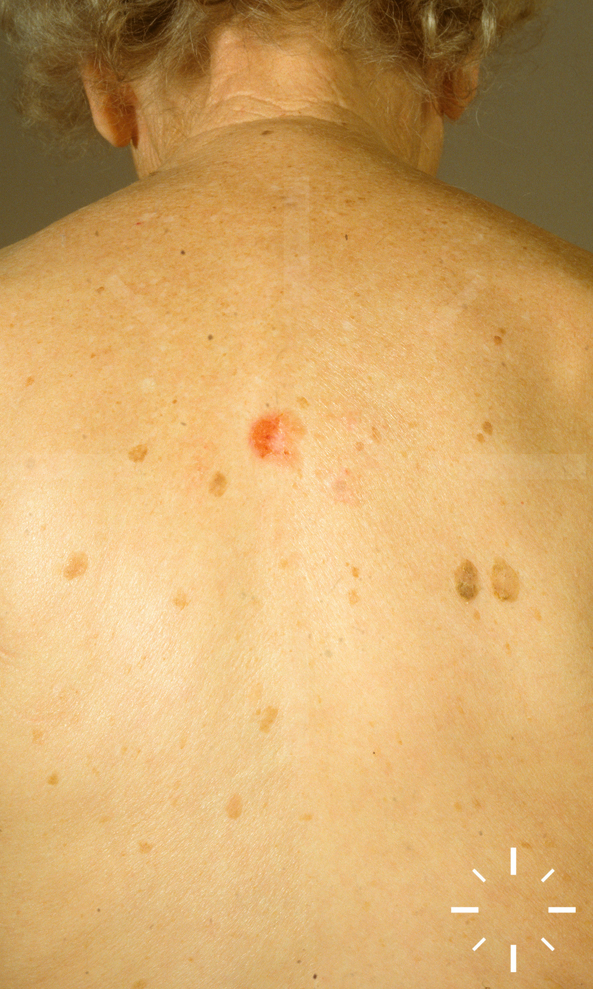
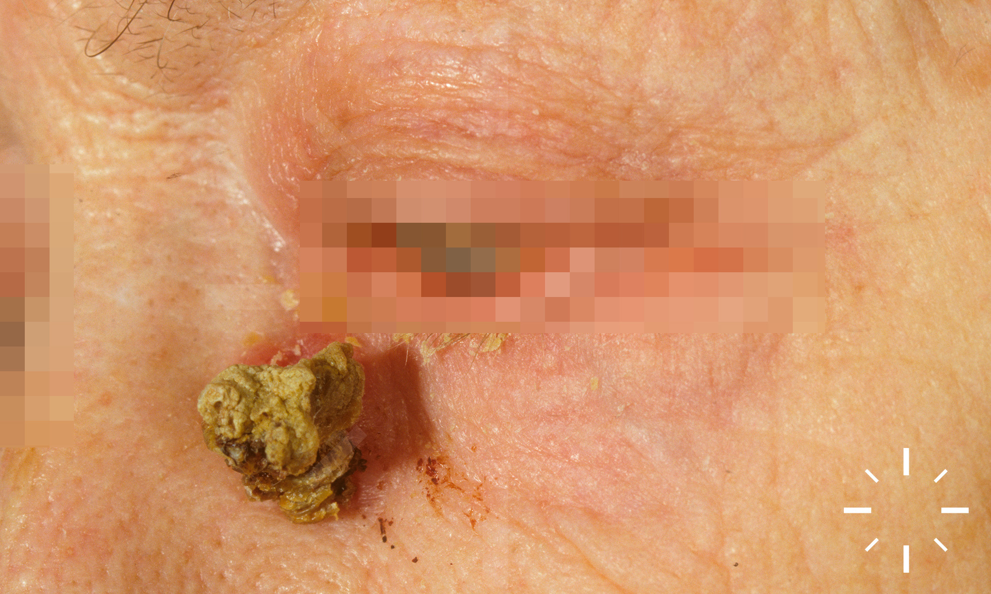
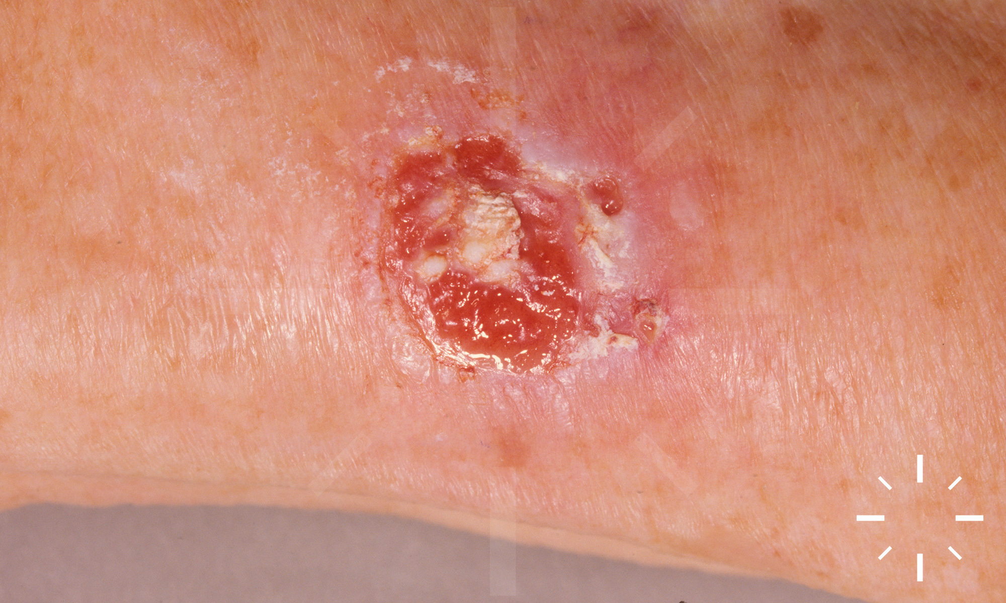
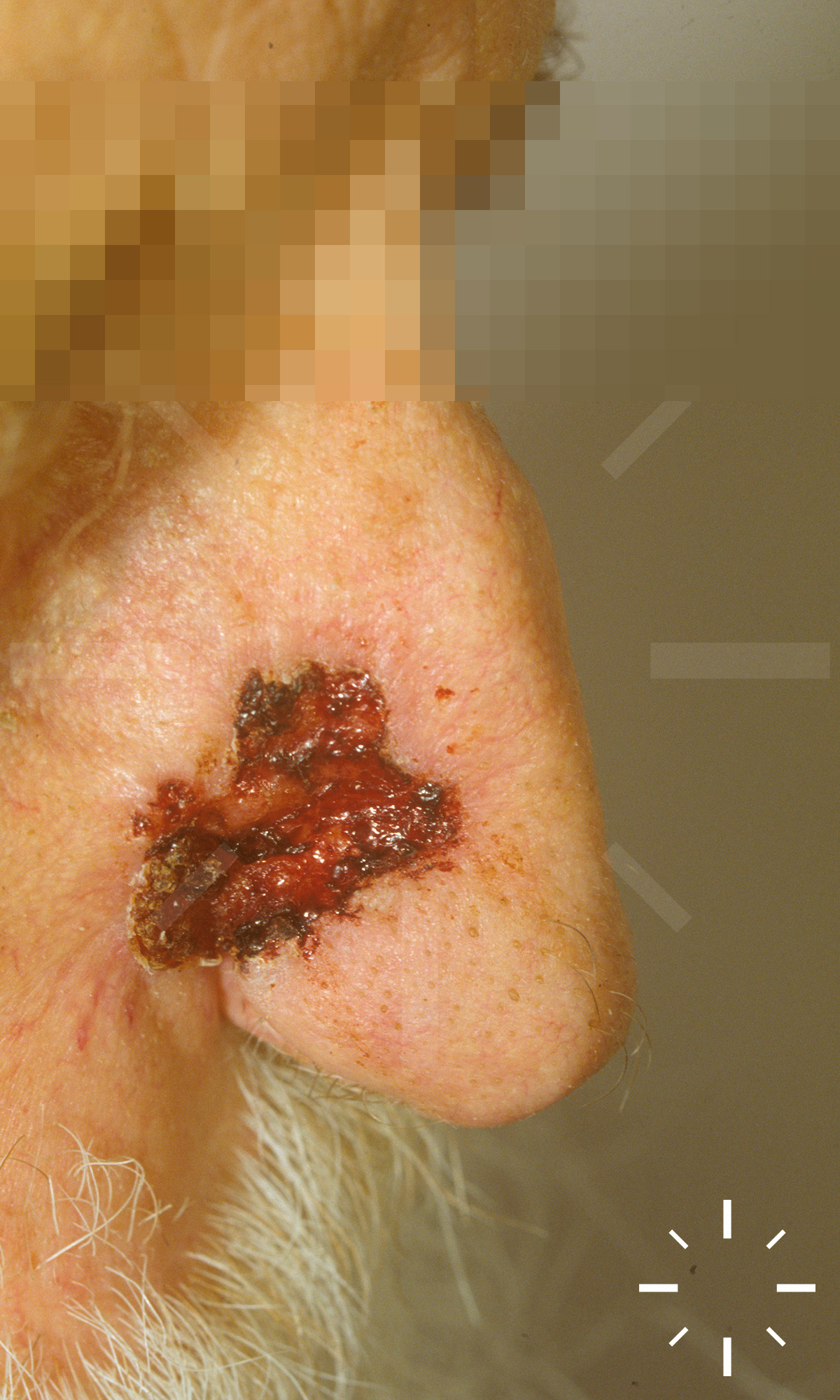
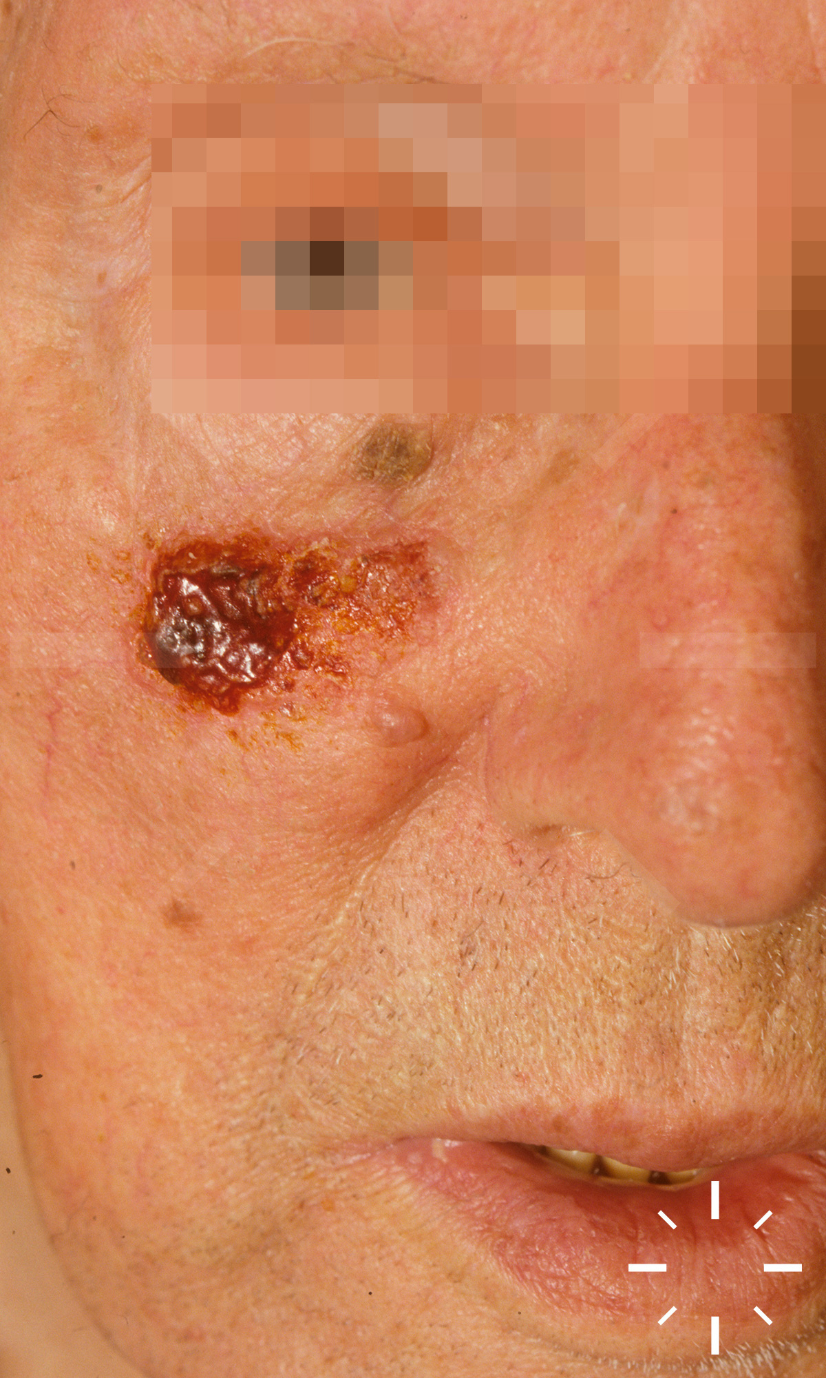
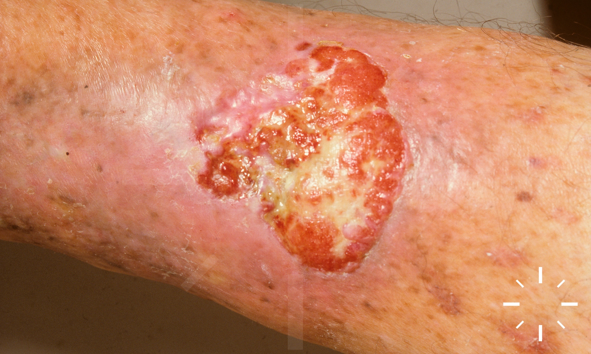
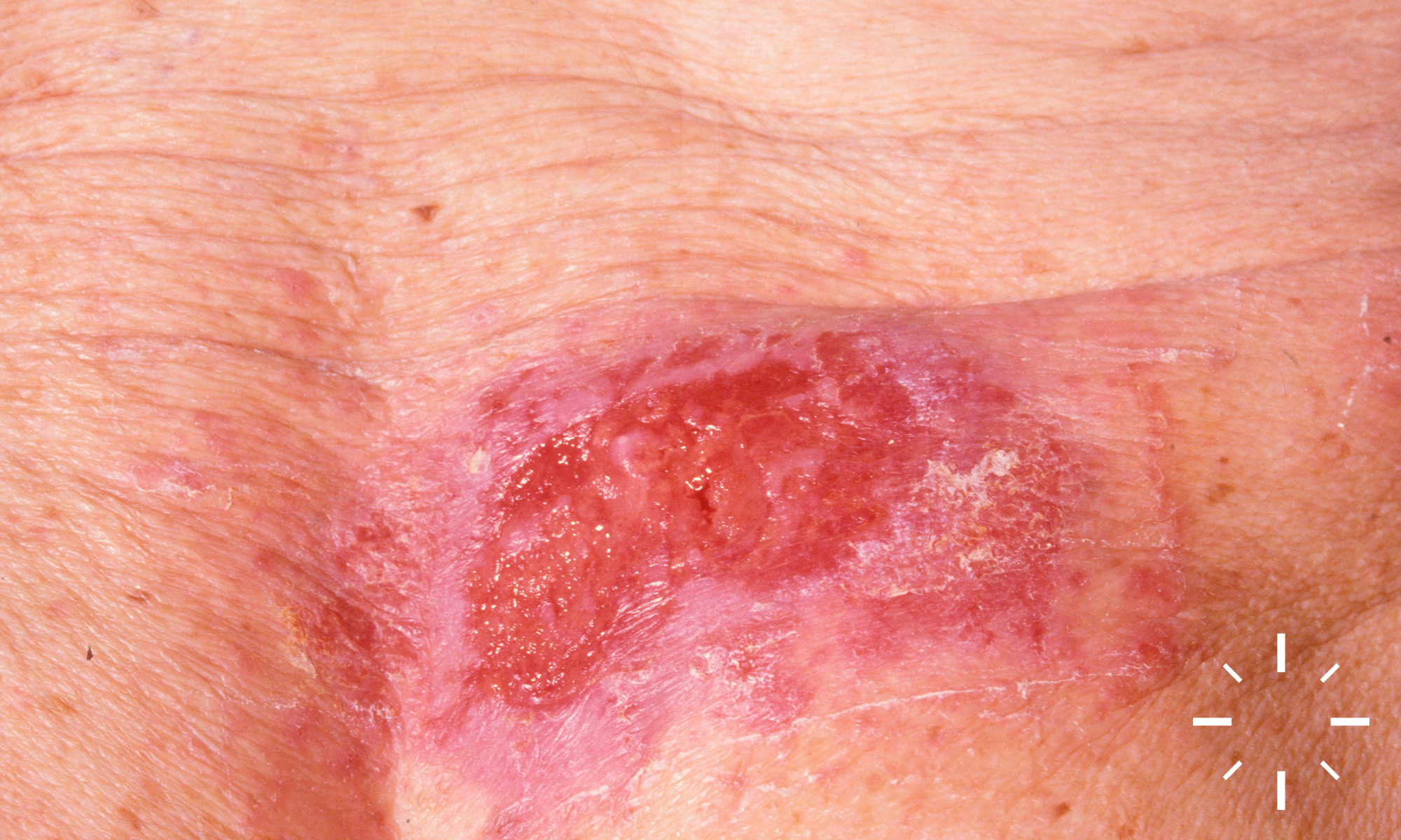
Basalzellkarzinom (incl. Subtypen)

Zuletzt aktualisiert: 2022-11-16

Autor(en): Anzengruber F., Navarini A.

ICD11: 2C32.Z

Krompecher 1900.

Basaliom, BCC, basalioma, basal cell carcinoma, basal cell epithelioma, Basalzellepitheliom.

Häufigste nicht gutartige, epitheliale Neoplasie, welche eine basaloide Differenzierung aufweist.

  • Inzidenz (Deutschland): 170/100000/Jahr
  • Häufigstes Hautkarzinom (Kaukasier)
  • Häufigster Krebs in den USA und Australien
  • Durchschnittsalter: ca. 60 Jahren
  • Inzidenz: Männer > Frauen (in den USA 30% höher)
  • In den USA ist in den letzten 10 Jahren die Inzidenz um 10% pro Jahr gestiegen
  • Lebenszeitrisiko: 30% (USA)
  • Im Alter von 55- 75 J. ist im Vergleich zu einer 20-jährigen Person das Erkrankungsrisiko 100-fach erhöht

Klinische/ätiologische Einteilung:

  • Noduläres Basalzellkarzinom
  • Superfizielles Basalzellkarzinom
  • Prämalignes Fibroepitheliom
  • Sklerodermiformes Basalzellkarzinom
  • Pigmentiertes Basalzellkarzinom
  • Destruierendes Basalzellkarzinom (Ulcus terebrans)
  • Narbenbasalzellkarzinom
  • Röntgen-Basalzellkarzinom
  • Polypoides Basalzellkarzinom

Histologische Einteilung:

  • Solides Basalzellkarzinom
  • Superfizielles Basalzellkarzinom
  • Sklerodermiformes Basalzellkarzinom
  • Fibroepitheliales Basazellkarzinom
  • Keratotisches Basalzellkarzinom
  • Zystisches Basalzellkarzinom
  • Metatypisches Basalzellkarzinom
  • Dimorphes Basalzellkarzinom

  • Das BCC entsteht aus epidermalen Stammzellen oder undifferenzierten Zellen des Haarfollikelwulst oder der äußeren Wurzelscheide
  • BCCs entstehen de novo, nicht über Präkanzerosen

Ätiologisch wichtige, prädispositionierende Faktoren:

  • Genetik (p53-Mutation)
  • Kumulative UVB-Belastung
  • Exposition zu Arsen
  • Strahlentherapie
  • Immunsuppression
  • Medikamentöse UV-Sensibilisierung
  • Heller Hauttyp.
  • Nävoides Basalzellkarzinom Syndrom (Gorlin-Goltz-Syndrom)
  • Rombo-Syndrom
  • Bazex-Dupré-Christol-Syndrom
  • Okulokutaner Albinismus
  • Xeroderma pigmentosum
  • Albinismus
  • Arsenexposition
  • Straffe Narben
  • Nävus sebaceus
  • Epidermolysis bullosa simplex, Dowling-Meara
  • Aktinische Keratosen (lässt auf ↑ UV-Exposition schließen)
  • Nikotinabusus
  • Kaffee?
  • Ekzeme?
  • NSAIDs?

Vitamin B3 dürfte protektiv wirken.

  • Anamnese (prädispositionierende Faktoren?)
  • Klinik.
  • Biopsie

  • Chronisch lichtexponierte Hautareale (Kapillitium, Gesicht, Hals, Dekolleté)
  • 80% der Basalzellkarzinome treten im Kopf-Hals-Bereich auf
  • 70% der Basalzellkarzinome treten im Gesicht auf
  • 15% der Basalzellkarzinome treten am Rumpf auf
  • Sehr selten treten Basalzellkarzinome am Penis, der Vulva oder perianal auf 

  • Exzellent, sofern keine Metastasierung stattgefunden hat
  • Rezidivwahrscheinlichkeit:
    • Verschiedene Autoren beziffern die Rezidivraten unterschiedlich.
Jahre nach Erstdiagnose eines BCCs Wahrscheinlichkeit des erneuten Auftretens eines BCC
1 Ca. 20%
2 Ca. 50%
3 Ca. 66%
5 Ca. 40-80% (je nach Datenlage)

Quelle: Hauschild, A. (2016). Langfassung der Leitlinie "Basalzellkarzinom der Haut". Awmf.org. Retrieved 30 May 2016, from http://www.awmf.org/leitlinien/detail/ll/032-021.html

  • Bei Patienten, welche bereits ein Basalzellkarzinom durchgemacht haben, ist die Wahrscheinlichkeit erneut an einem Basalzellkarzinom zu erkranken um das 4-fache erhöht.
  • Das Risiko sinkt, wenn das erste Basalzellkarzinom nach dem 75. Geburtstag auftritt.
  • Sofern eine obere Extremität die erste befallene Lokalisation darstellt, spricht dies für eine schlechtere Prognose.
  • 6-10% der Patienten mit einem BCC entwickeln ein SCC (3-fach höheres Risiko als in der Normalbevölkerung).
  • Das Risiko im Anschluss ein malignes Melanom zu entwickeln ist bei BCC Patienten 2-4-fach erhöht.  
  • Kontrolluntersuchungen sollten im 1. Jahre alle 6 Monate und danach 1x jährlich erfolgen.

  1. Leibovitch I, Huilgol SC, Selva D, Richards S, Paver R. Basal cell carcinoma treated with Mohs surgery in Australia II. Outcome at 5-year follow-up. Journal of the American Academy of Dermatology 2005;53:452-7.
  2. Peris K, Campione E, Micantonio T, Marulli GC, Fargnoli MC, Chimenti S. Imiquimod Treatment of Superficial and Nodular Basal Cell Carcinoma. Dermatologic Surgery 2005;31:318-23.
  3. Epstein EH. Basal cell carcinomas: attack of the hedgehog. Nature Reviews Cancer 2008;8:743-54.
  4. Geist DE, Garcia-Moliner M, Fitzek MM, Cho H, Rogers GS. Perineural Invasion of Cutaneous Squamous Cell Carcinoma and Basal Cell Carcinoma: Raising Awareness and Optimizing Management. Dermatologic Surgery 2008;34:1642-51.
  5. Gudbjartsson DF, Sulem P, Stacey SN, et al. ASIP and TYR pigmentation variants associate with cutaneous melanoma and basal cell carcinoma. Nature Genetics 2008;40:886-91.
  6. Pincus LB, McCalmont TH, Neuhaus IM, Kasper R, Oh DH. Basal cell carcinomas arising within multiple trichoepitheliomas. Journal of Cutaneous Pathology 2008;35:59-64.
  7. Redondo P, Marquina M, Pretel M, Aguado L, Iglesias ME. Methyl-ALA–Induced Fluorescence in Photodynamic Diagnosis of Basal Cell Carcinoma Prior to Mohs Micrographic Surgery. Arch Dermatol 2008;144.
  8. Szeimies RM, Ibbotson S, Murrell DF, et al. A clinical study comparing methyl aminolevulinate photodynamic therapy and surgery in small superficial basal cell carcinoma (8-20 mm), with a 12-month follow-up. Journal of the European Academy of Dermatology and Venereology 2008;22:1302-11.
  9. Telfer NR, Colver GB, Morton CA. Guidelines for the management of basal cell carcinoma. Br J Dermatol 2008;159:35-48.
  10. Love WE, Bernhard JD, Bordeaux JS. Topical Imiquimod or Fluorouracil Therapy for Basal and Squamous Cell Carcinoma. Arch Dermatol 2009;145.
  11. Muller FM, Dawe RS, Moseley H, Fleming CJ. Randomized Comparison of Mohs Micrographic Surgery and Surgical Excision for Small Nodular Basal Cell Carcinoma. Dermatologic Surgery 2009;35:1349-54.
  12. Swanson EL, Amdur RJ, Mendenhall WM, Morris CG, Kirwan JM, Flowers F. Radiotherapy for basal cell carcinoma of the medial canthus region. The Laryngoscope 2009;119:2366-8.
  13. Basset-Seguin N. Topical photodynamic therapy for superficial and nodular basal cell carcinoma. Expert Review of Dermatology 2010;5:61-6.
  14. Saran A. Basal cell carcinoma and the carcinogenic role of aberrant Hedgehog signaling. Future Oncology 2010;6:1003-14.
  15. Youssef KK, Van Keymeulen A, Lapouge G, et al. Identification of the cell lineage at the origin of basal cell carcinoma. Nature Cell Biology 2010.
  16. Sekulic A, Migden MR, Oro AE, et al. Efficacy and Safety of Vismodegib in Advanced Basal-Cell Carcinoma. New England Journal of Medicine 2012;366:2171-9.
  17. Sezer E, Erkek E, Sahin S. A Case Report of Syringocystadenoma Papilliferum Mimicking Basal Cell Carcinoma on the Face. Turkish Journal of Dermatology / Türk Dermatoloji Dergisi 2012;6:21-3.
  18. Dreier J, Felderer L, Barysch M, Rozati S, Dummer R. Basal cell carcinoma: a paradigm for targeted therapies. Expert Opinion on Pharmacotherapy 2013;14:1307-18.
  19. Harms KL, Dlugosz AA. Harnessing Hedgehog for the Treatment of Basal Cell Carcinoma. JAMA Dermatol 2013;149:607.
  20. Iarrobino A, Messina JL, Kudchadkar R, Sondak VK. Emergence of a squamous cell carcinoma phenotype following treatment of metastatic basal cell carcinoma with vismodegib. Journal of the American Academy of Dermatology 2013;69:e33-e4.
  21. Sekulic A, Mangold AR, Northfelt DW, LoRusso PM. Advanced basal cell carcinoma of the skin. Current Opinion in Oncology 2013:1.
  22. McCusker M, Basset-Seguin N, Dummer R, et al. Metastatic basal cell carcinoma: Prognosis dependent on anatomic site and spread of disease. European Journal of Cancer 2014;50:774-83.
  23. Majores M, Bierhoff E. [Actinic keratosis, Bowen's disease, keratoacanthoma and squamous cell carcinoma of the skin]. Pathologe 2015;36:16-29.
  24. Hauschild, A. (2016). Langfassung der Leitlinie "Basalzellkarzinom der Haut". Awmf.org. Retrieved 30 May 2016, from http://www.awmf.org/leitlinien/detail/ll/032-021.html
  25. Lebwohl, Mark. Treatment of Skin Disease: Comprehensive Therapeutic Strategies. Elsevier, 2014. Print.