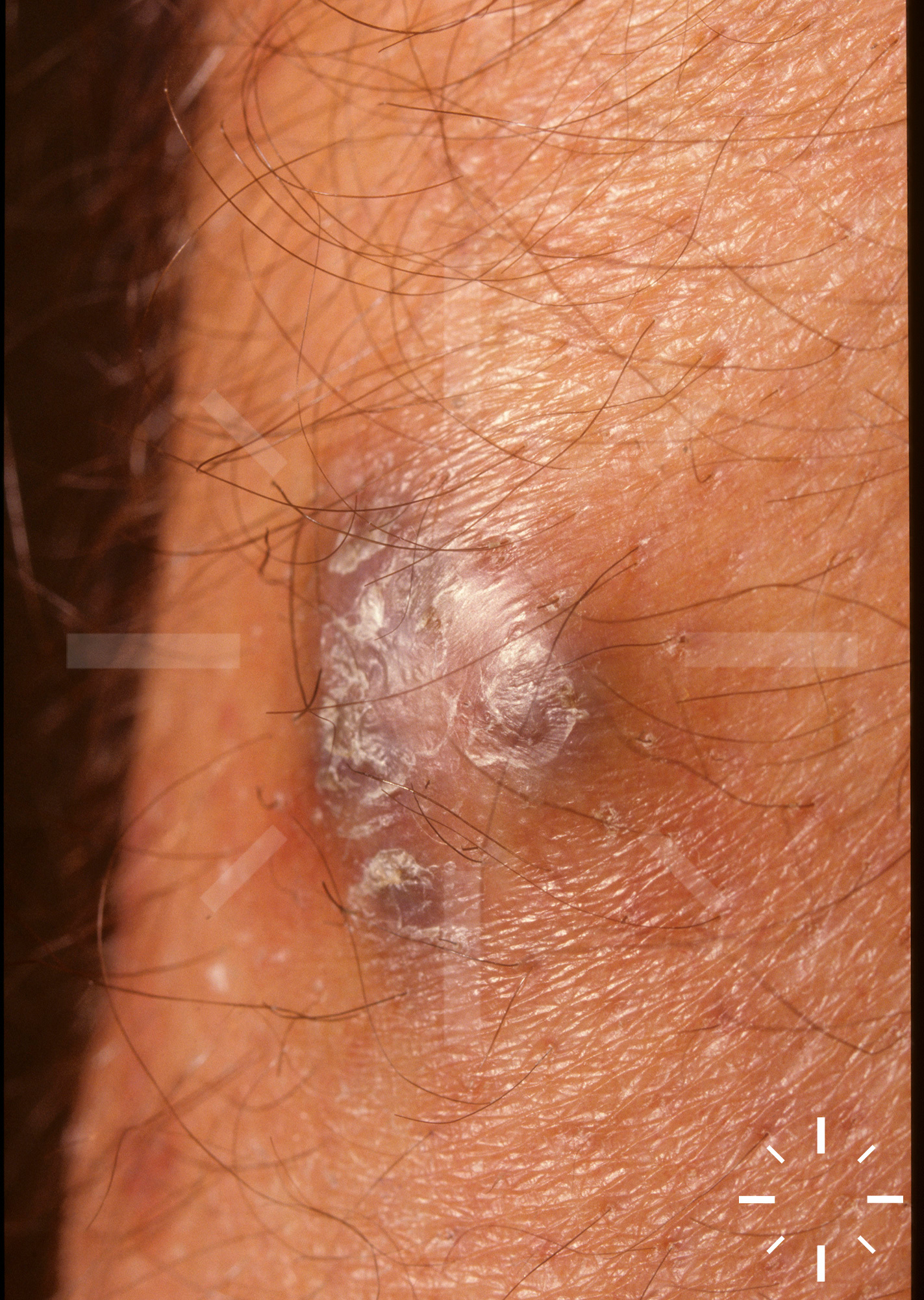
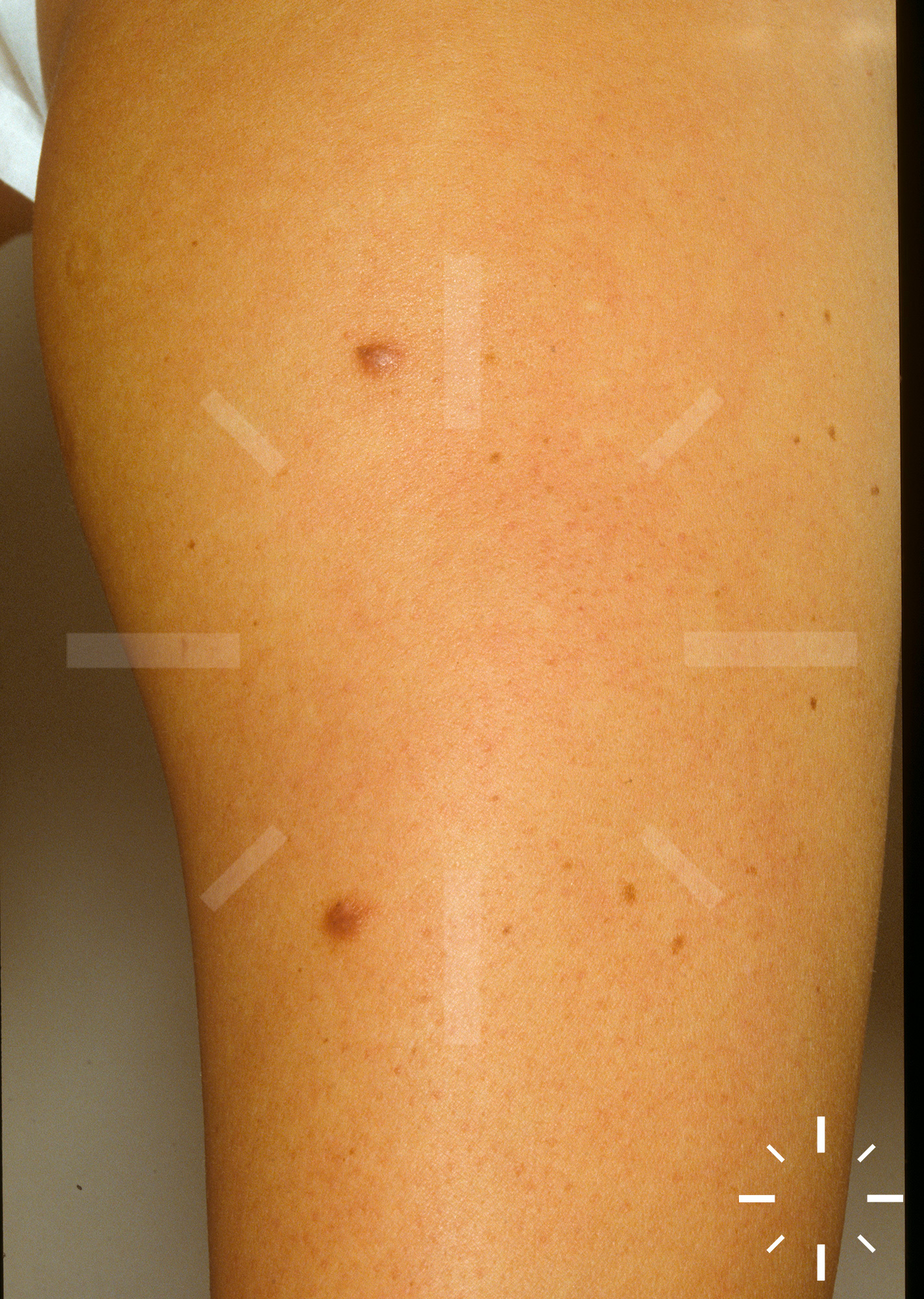
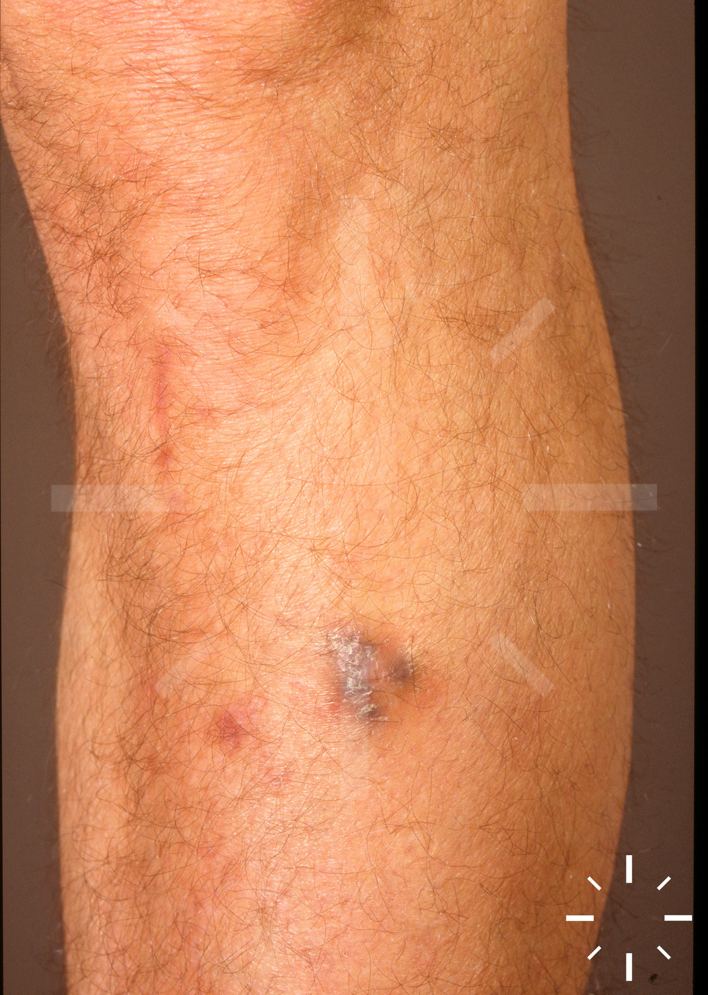
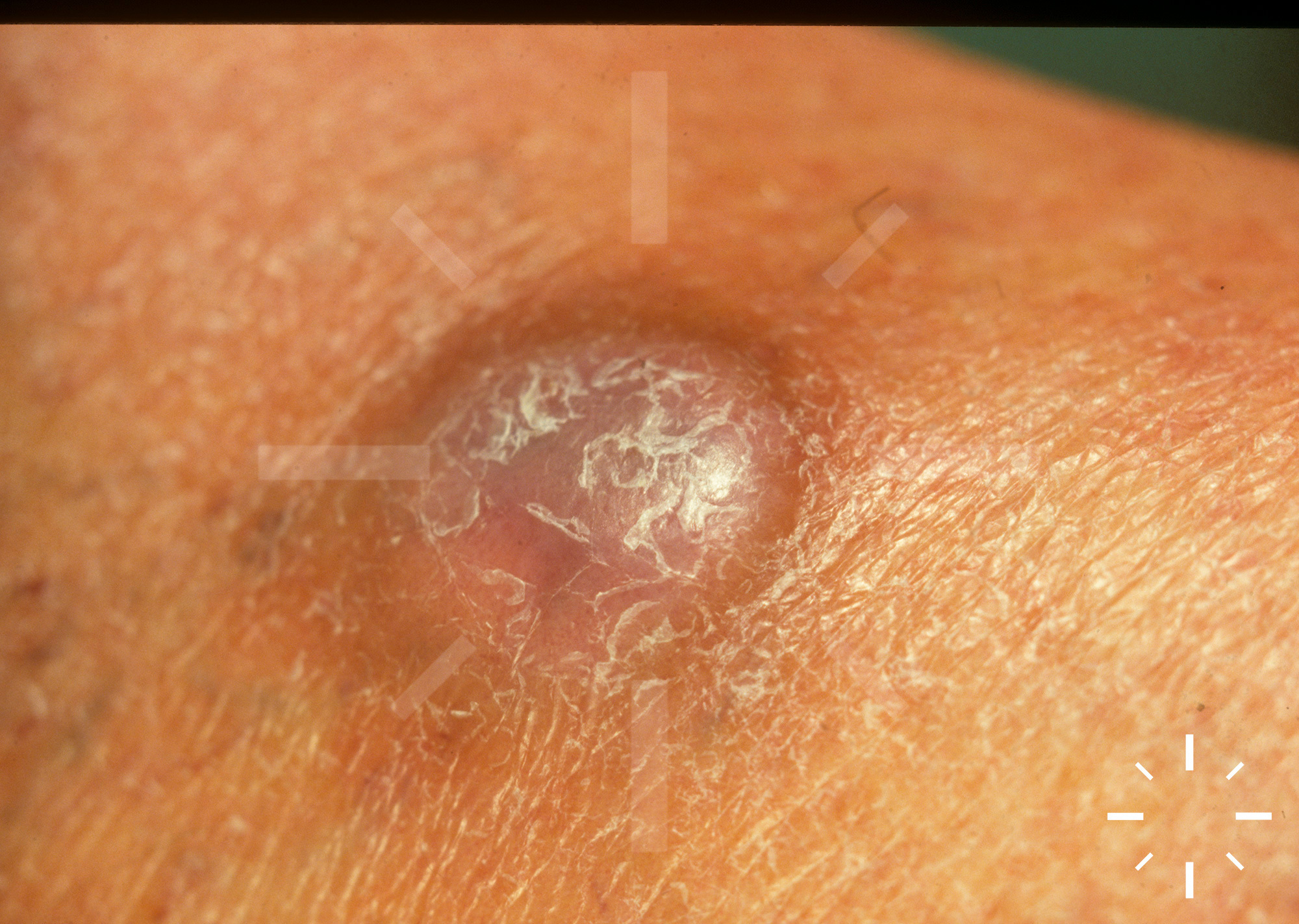
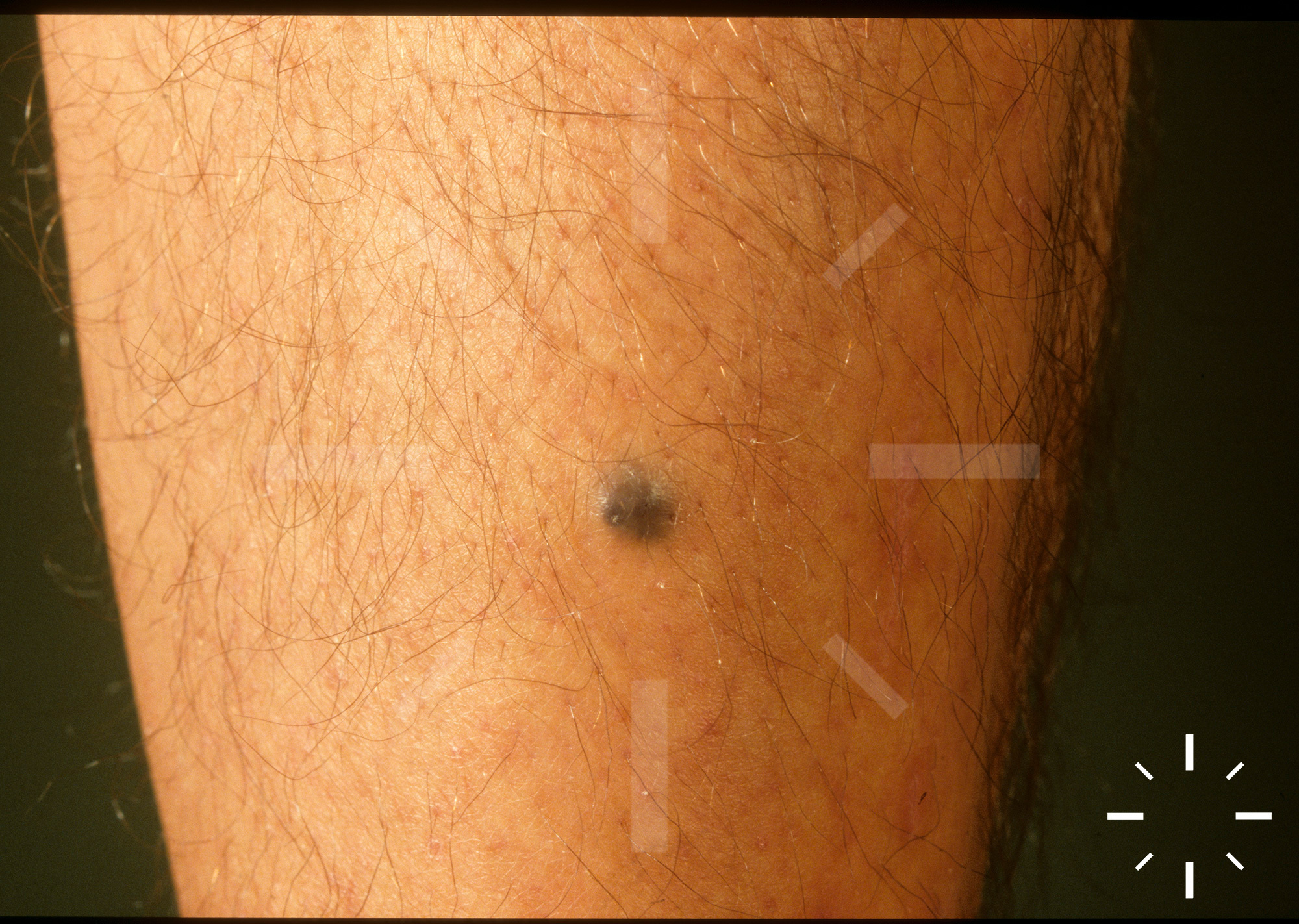
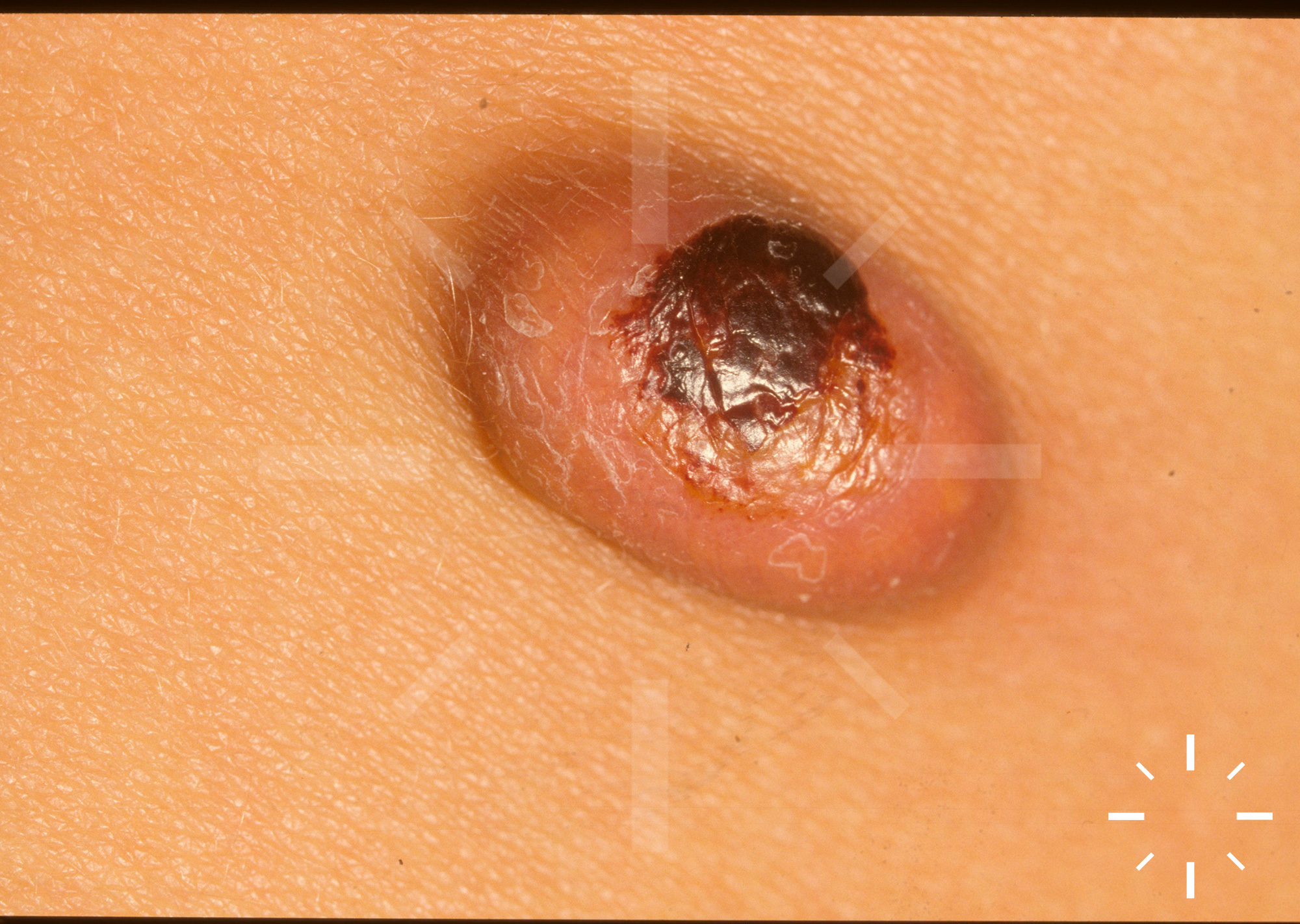
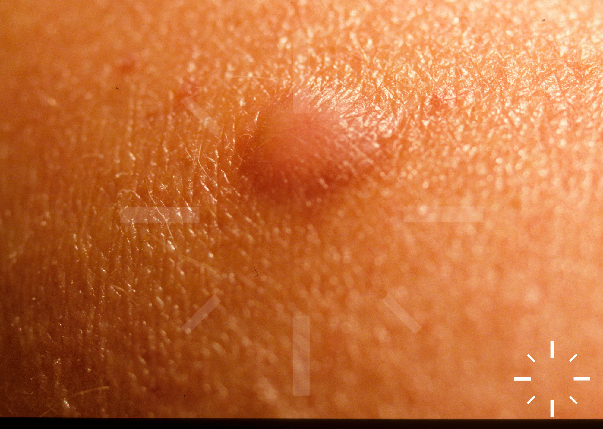
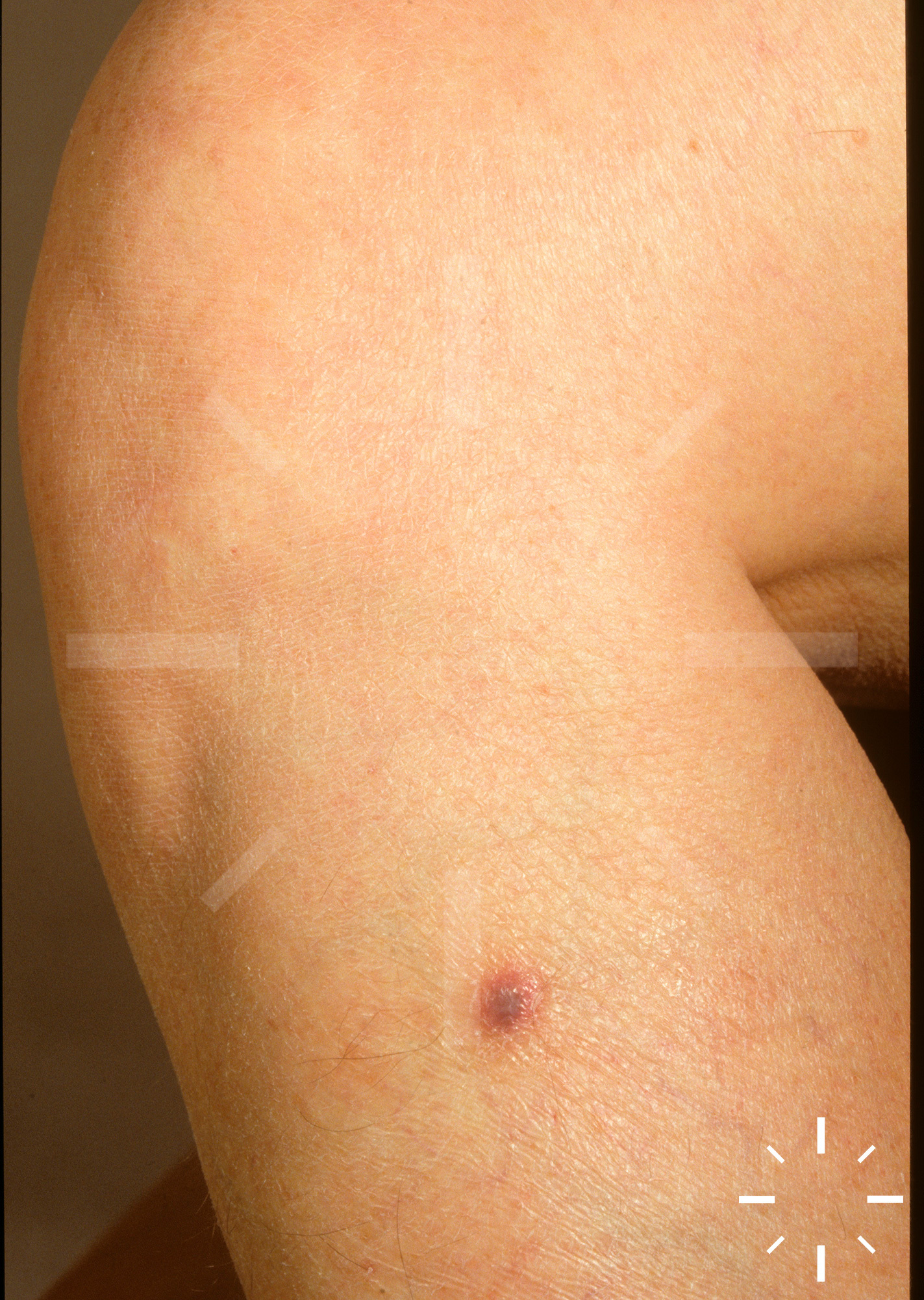
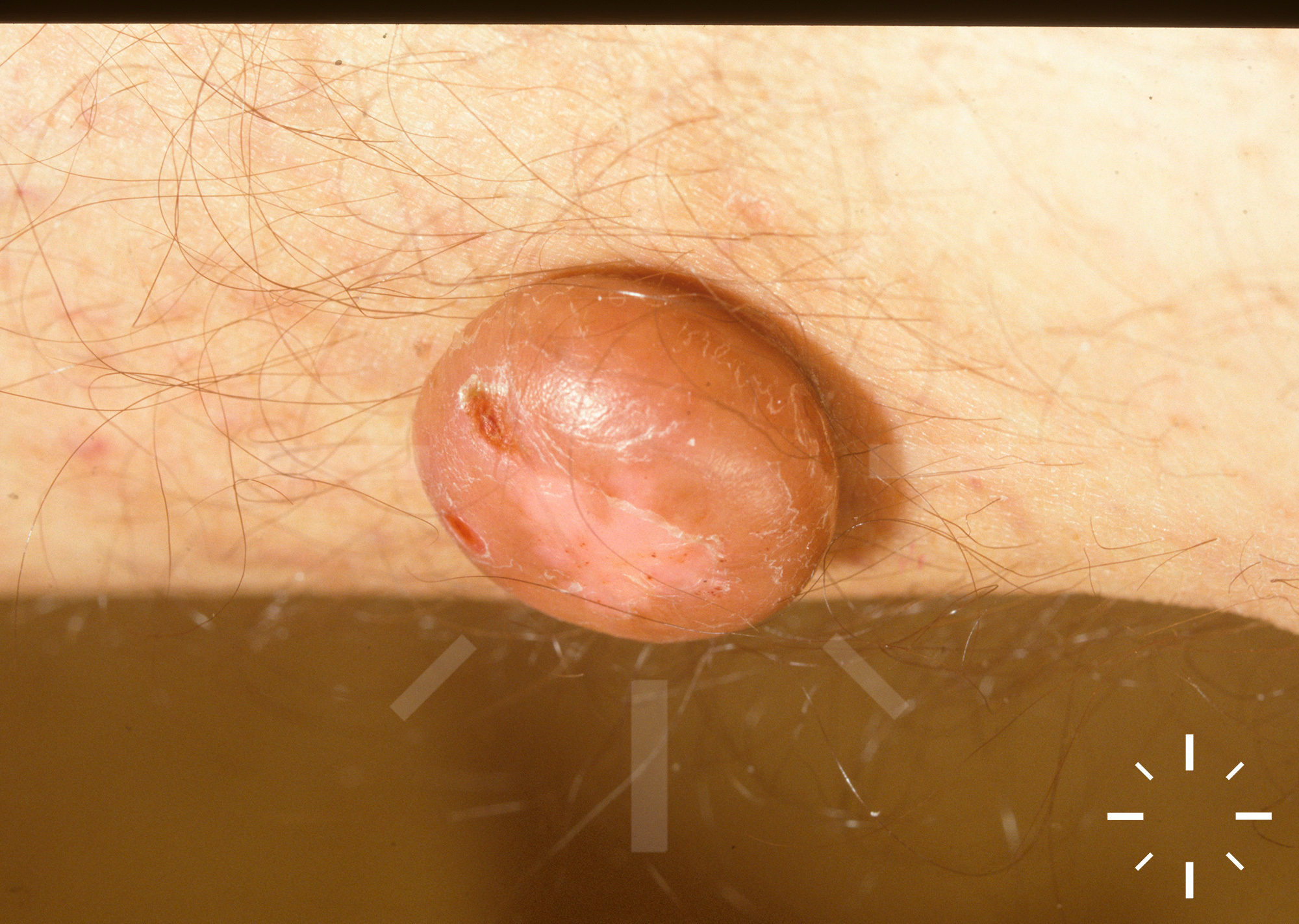
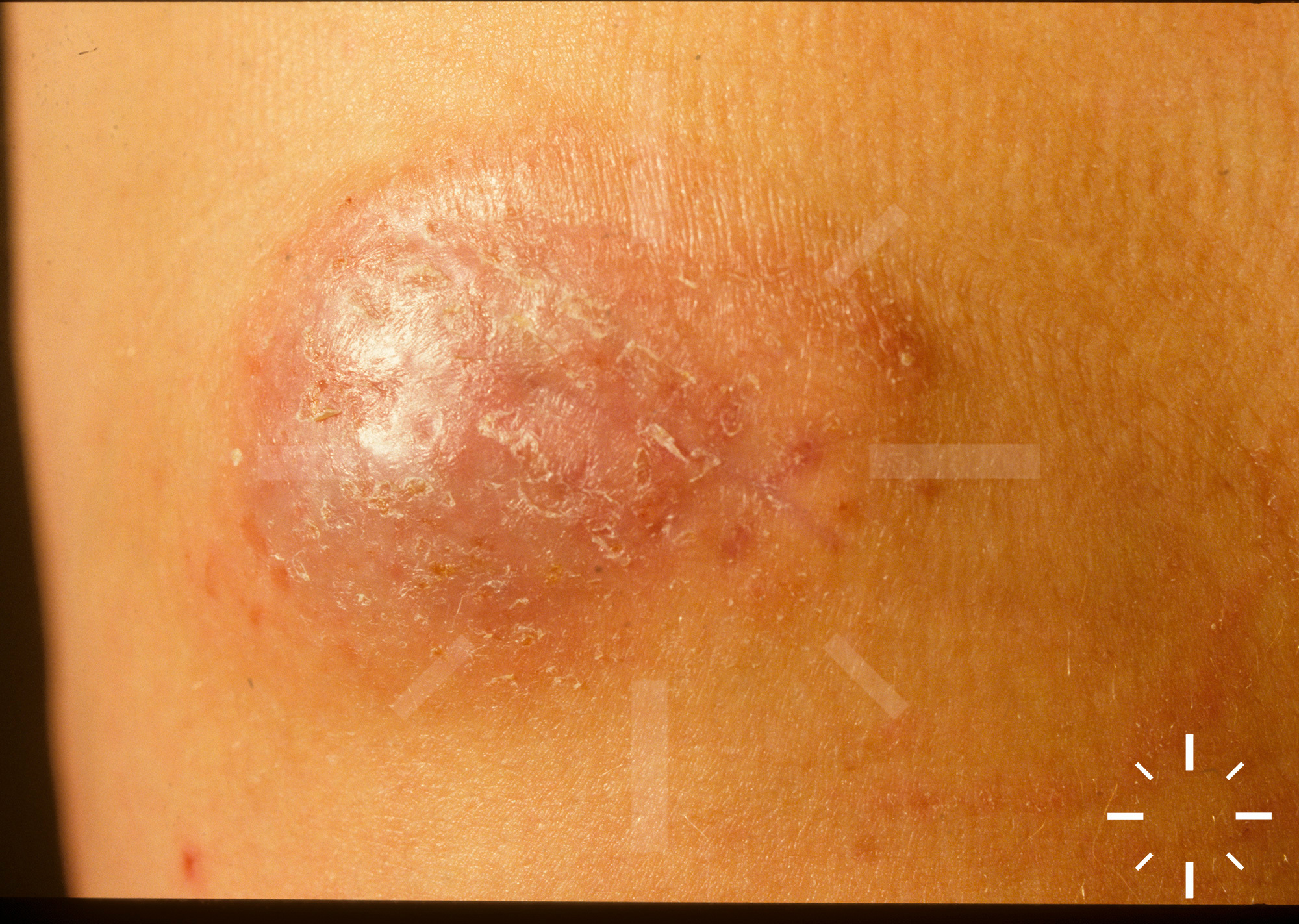
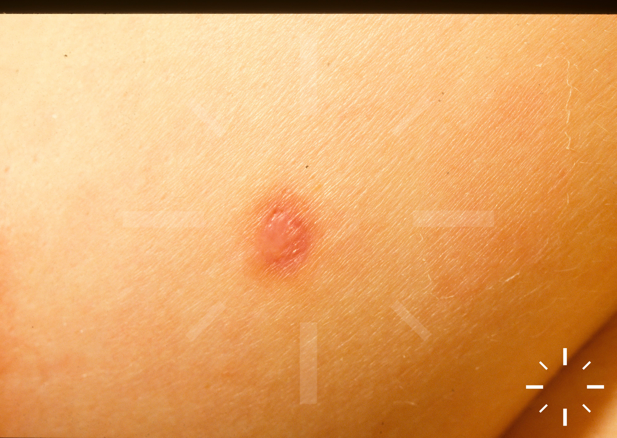
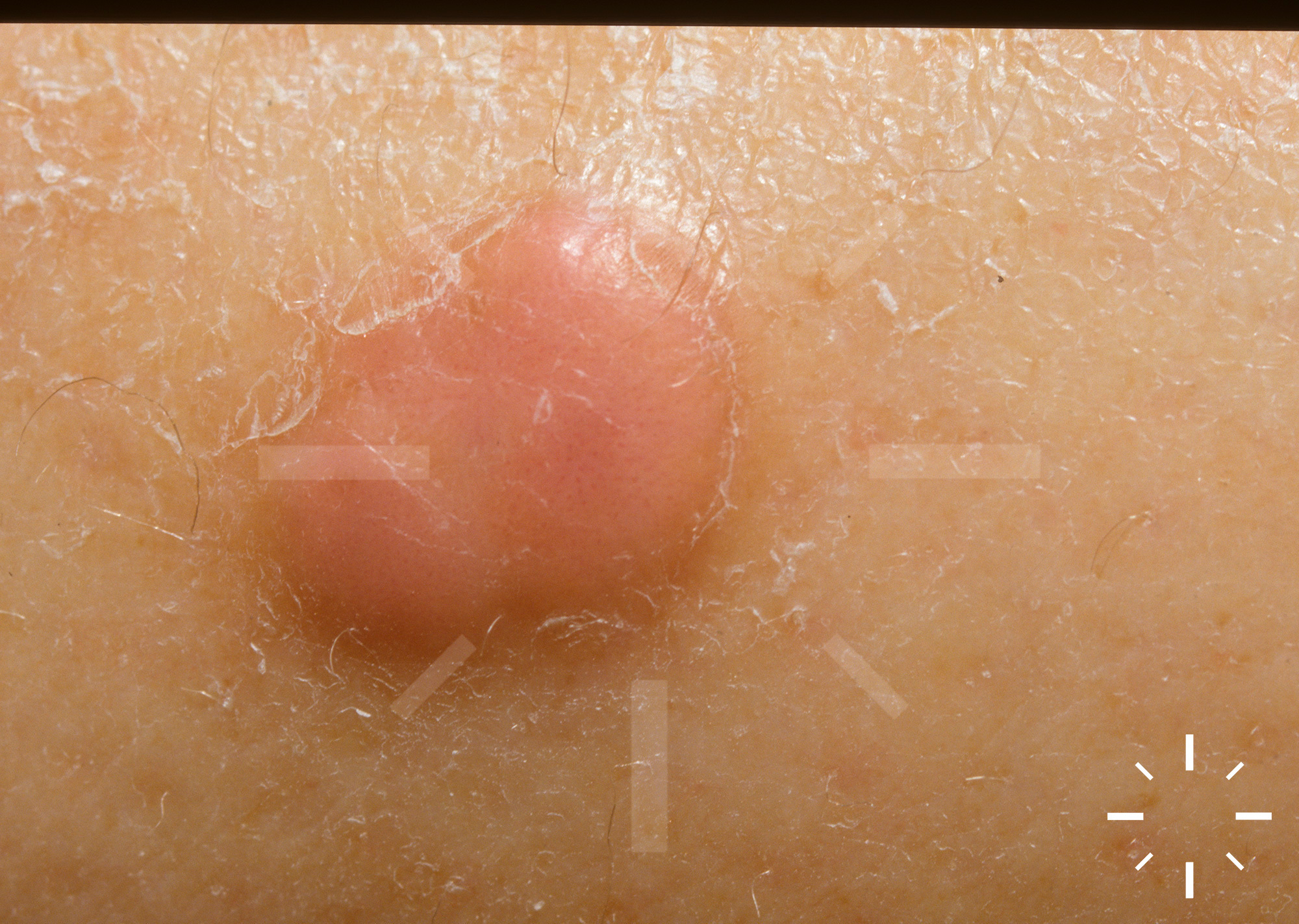
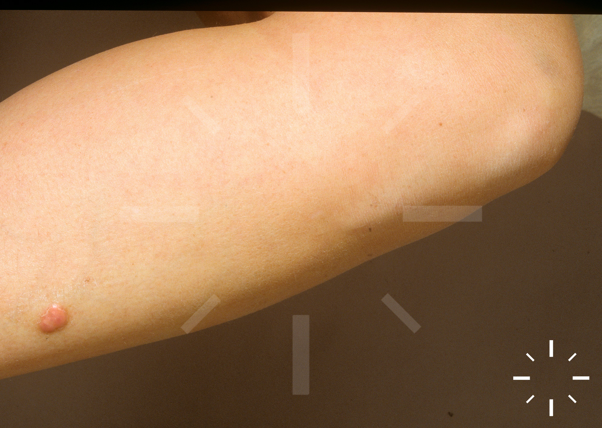
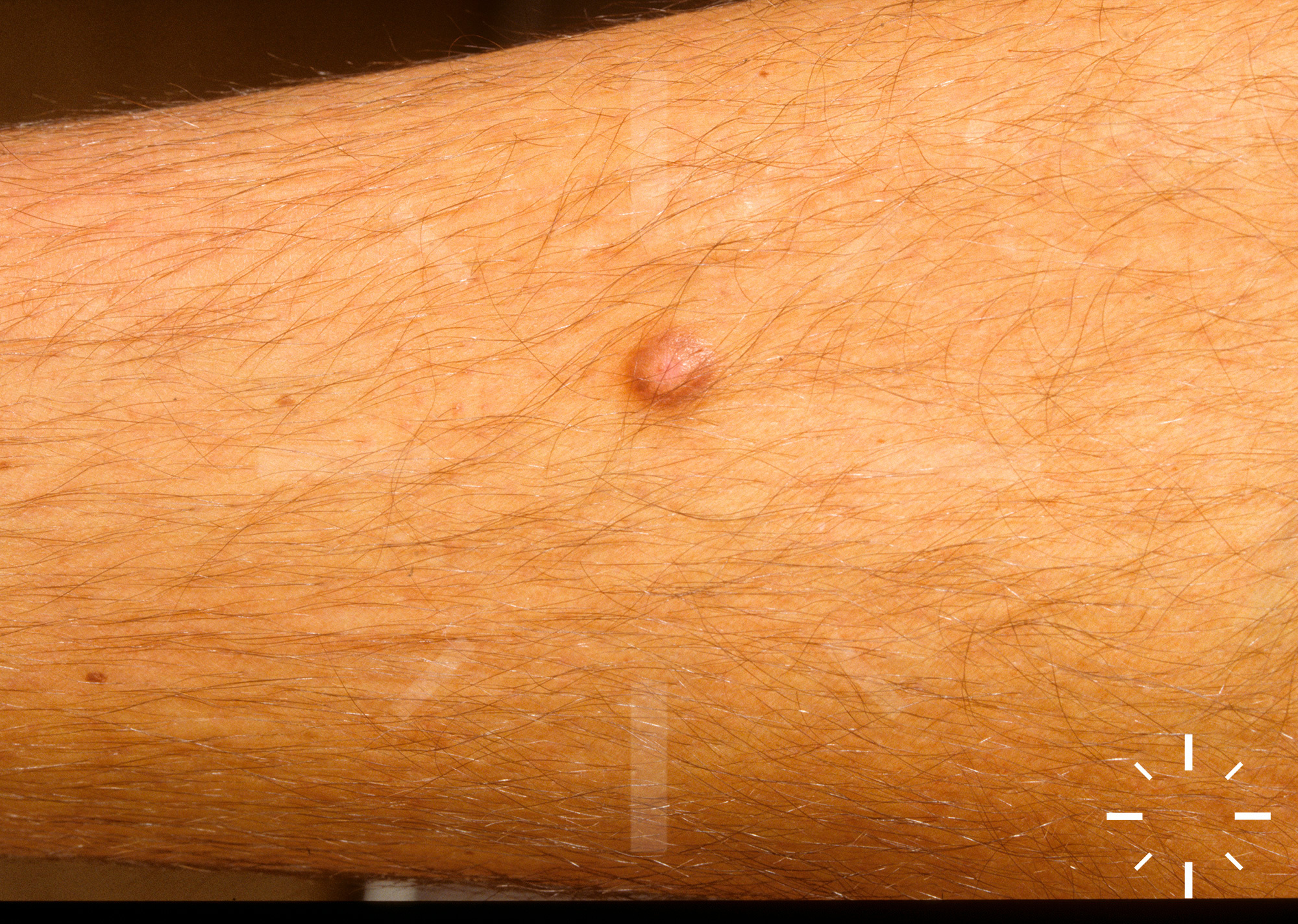
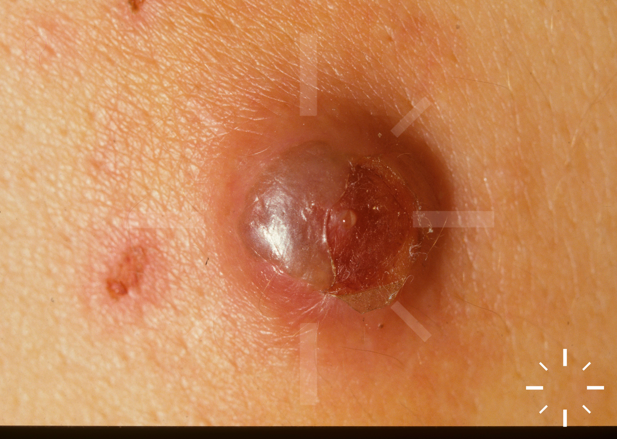
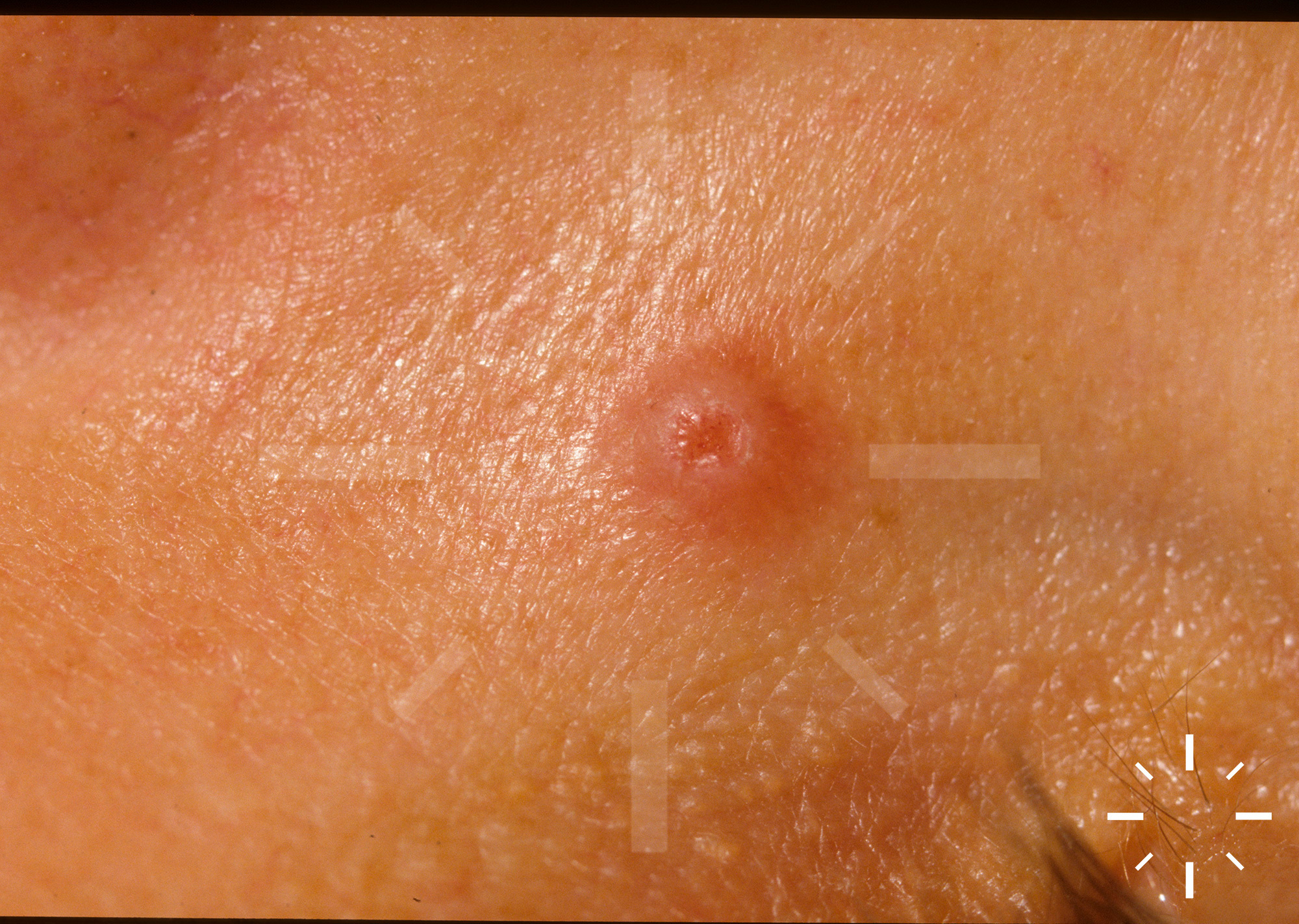
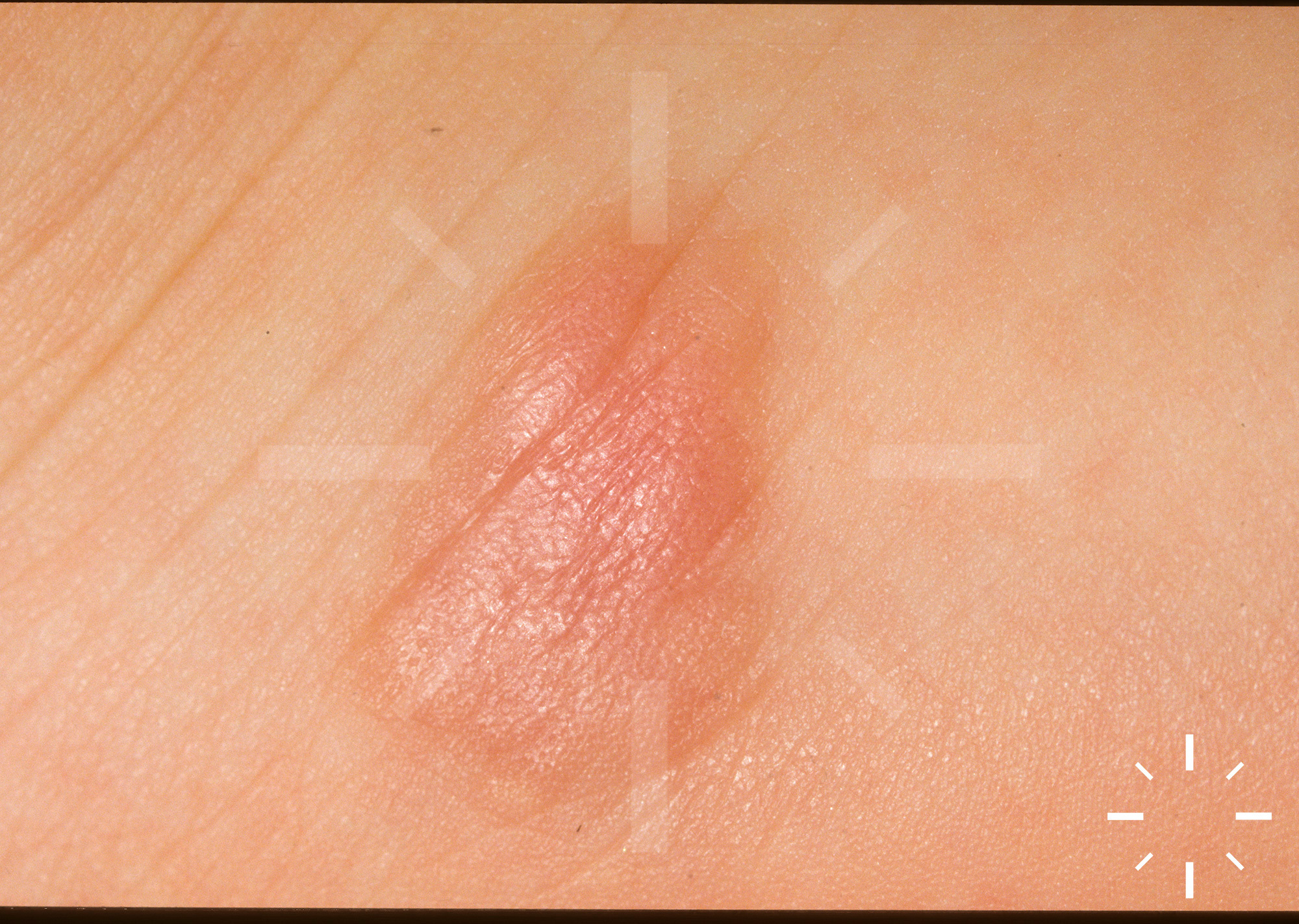
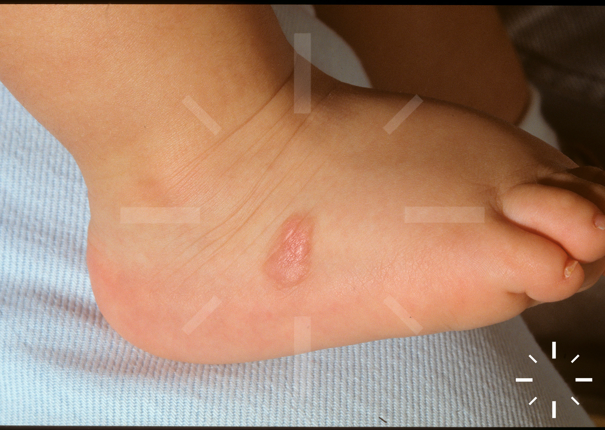
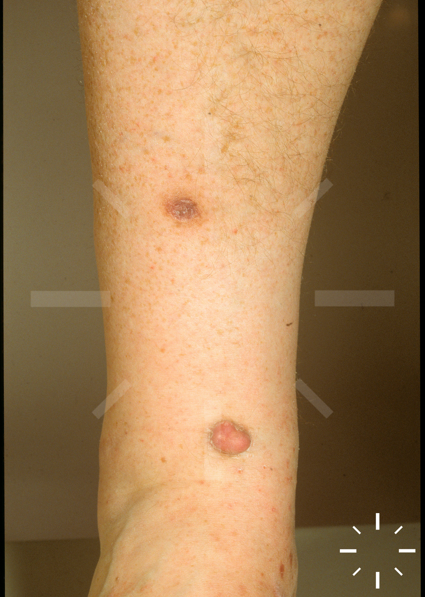
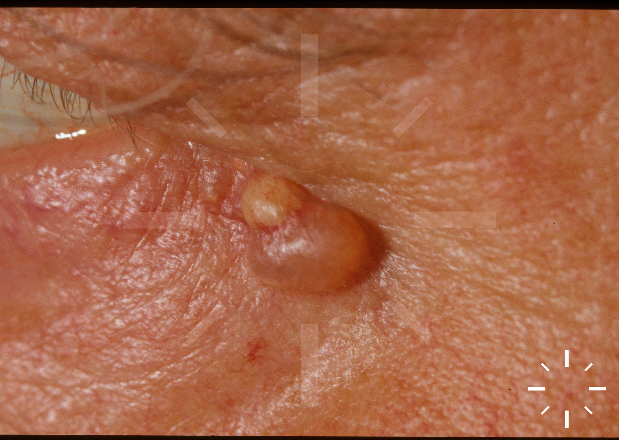
Dermatofibroma, histiocytoma, DF

Last Updated: 2023-07-07

Author(s): Anzengruber F., Navarini A.

ICD11: 2F23.0

  • Histiocytoma, dermatofibroma lenticulare, fibrome en pastille, hard fibroma.

Mostly women in young adulthood, but also older people. The prevalence is around 50%. Histiocytomas are often not immediately presented to dermatologists

In addition to dermatofibroma vulgaris, there are several subtypes that can be primarily differentiated dermatopathologically:

Histological structure:

Ossifying, sclerosing, desmoplastic, myxoid, xanthomatous, haemosiderotic

Anatomical:

Deep infiltrating, facial area

Cellular architecture / typology:

Cell-rich (high recurrence rate, in males on legs), granular cell-containing, neurothekeoma, DF with monster cells, epithelioid cellular, pseudosarcomatous, metastatic,

  • It is unclear whether this is a neoplasm or a skin reaction pattern. However, dendritic cell markers are indeed found, suggesting a relationship with the non-Langerhans cell histiocytoses. Since certain histiocytoma types tend to recur, this may also postulate the neoplasia.
  • Trigger
    • Ictus reaction
    • Folliculitis
    • Ruptured cyst
    • Microtrauma
  • There is a subsequent inflammatory connective tissue reaction

  • Mostly on the lower extremity, a red/brown, sharply demarcated, lenticular nodule is seen
  • If the skin lesion is fixed between the thumb and index finger, the nodule sinks (Fitzpatrick's sign)
  • Unusually large dermatofibromas are called giant dermatofibromas

  • Clinic
  • Dermatoscopic pattern:
  • Brown, rather light pigment network in the periphery of the lesion. No sharp demarcation against the surrounding skin. A scar is found in the centre of the lesion, which may be bizarre and irregularly configured. With polarisation, "shiny white lines" are found.
  • Papular changes with pigment network should be excised if uncertain, no prolonged observation period recommended. Unfortunately, dermatofibromas have already turned out to be melanomas several times - deep-threshold excisions are useful and should also always be covered by the insurance company.

Extremities, especially legs, are the most frequent localisation. Dermatofibromas are rare on the face (separate subtype).

  • Thickened (acanthotic) epidermis
  • No sharp border to side and base
  • No capsule
  • Diffuse collections of spindle cells, some containing lipid or haemosiderin
  • Subtypes: Cellular, Aneurismal, Epithelioid, Atypical, Lipid-containing, Cholesterol-containing, Palisade DF (see also at Classification).

  • May bleed in trauma
  • Differential diagnosis of amelanotic melanoma may be missed for a long time, there are known legal cases on this

Does not start, but persists for a long time.

  1. Goette DK. Basal Cell Carcinomas and Basal Cell Carcinoma-like Changes Overlying Dermatofibromas. Arch Dermatol 1975;111:589.
  2. Tamada S, Ackerman AB. Dermatofibroma with Monster Cells. The American Journal of Dermatopathology 1987;9:380-7.
  3. Kamino H, Jacobson M. Dermatofibroma Extending into the Subcutaneous Fat. The American Journal of Dermatopathology 1990;12:315.
  4. Abenoza P, Lillemoe T. CD34 and Factor XIII a in the Differential Diagnosis of Dermatofibroma and Dermatofibrosarcoma Protuberans. The American Journal of Dermatopathology 1993;15:429-34.
  5. Kuo T-t, Hu S, Chan H-L. Keloidal Dermatofibroma. The American Journal of Surgical Pathology 1998;22:564-8.
  6. Rudolph P, Schubert C, Zelger BG, Zelger B, Parwaresch R. Differential Expression of CD34 and Ki-M1p in Pleomorphic Fibroma and Dermatofibroma With Monster Cells. The American Journal of Dermatopathology 1999;21:414.
  7. Chen T-C, Kuo T-t, Chan H-L. Dermatofibroma is a clonal proliferative disease. J Cutan Pathol 2000;27:36-9.
  8. Yazici AC, Baz K, Ikizoglu G, Koca A, Kokturk A, Apa DD. Familial eruptive dermatofibromas in atopic dermatitis. Journal of the European Academy of Dermatology and Venereology 2006;20:90-2.
  9. Kim HJ, Lee JY, Kim SH, et al. Stromelysin-3 expression in the differential diagnosis of dermatofibroma and dermatofibrosarcoma protuberans: comparison with factor XIIIa and CD34. Br J Dermatol 2007;157:319-24.