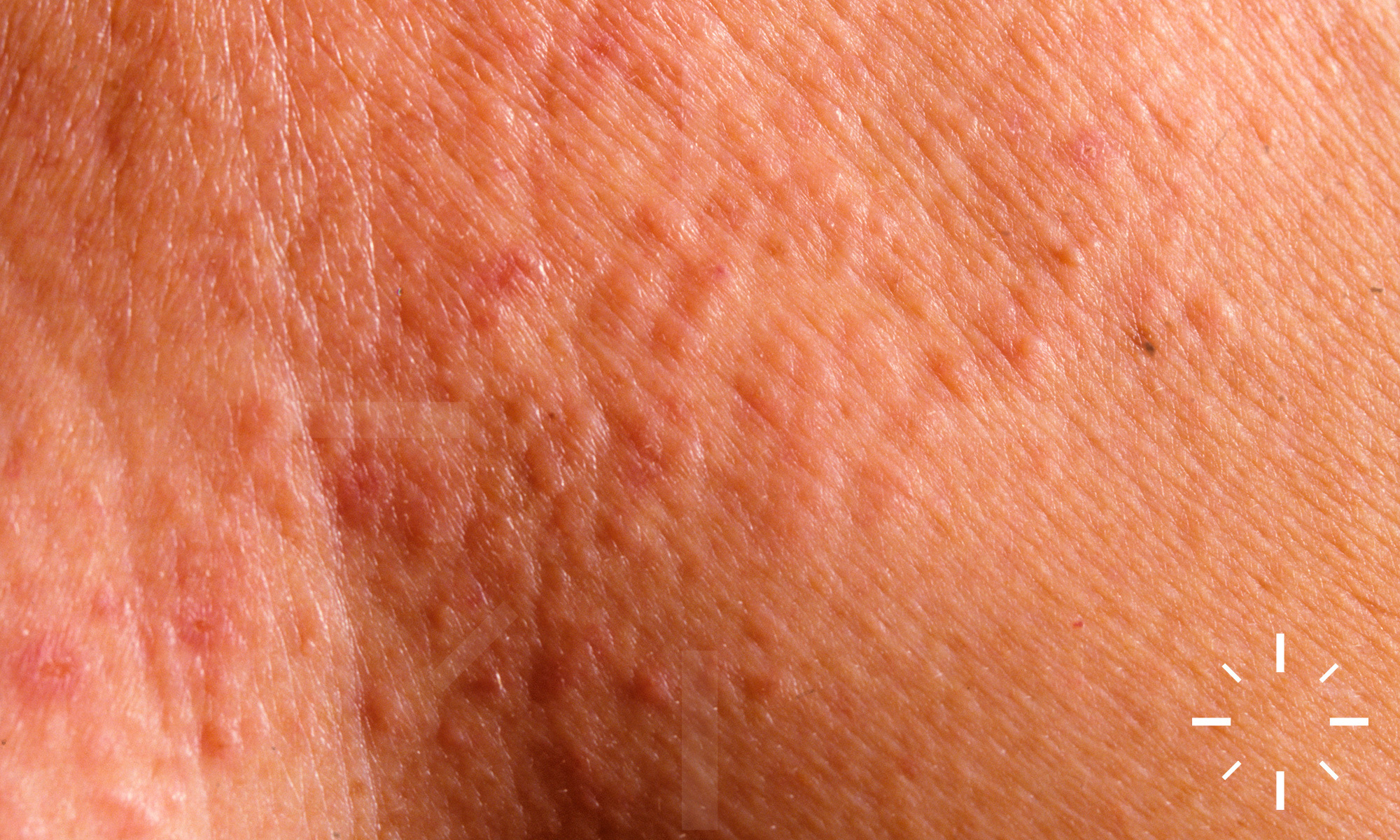
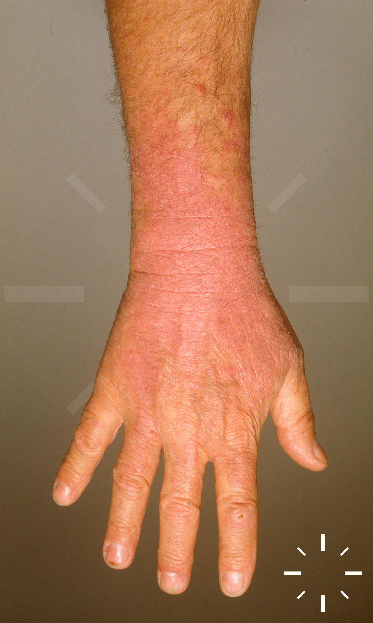
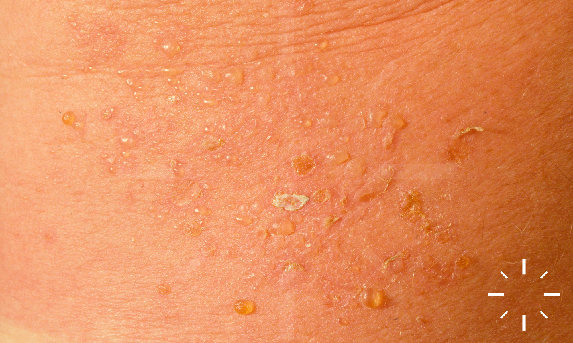
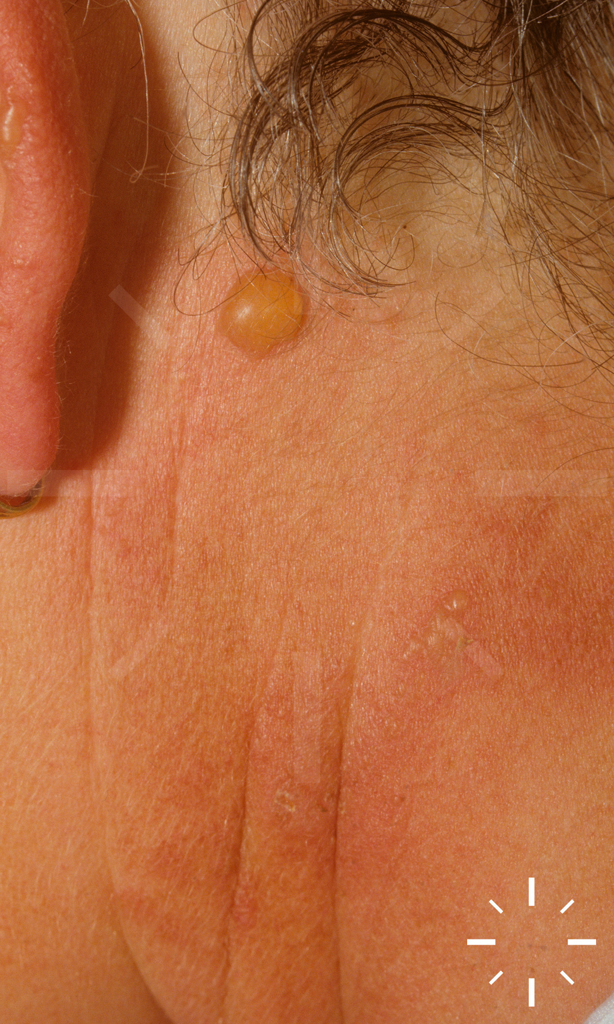
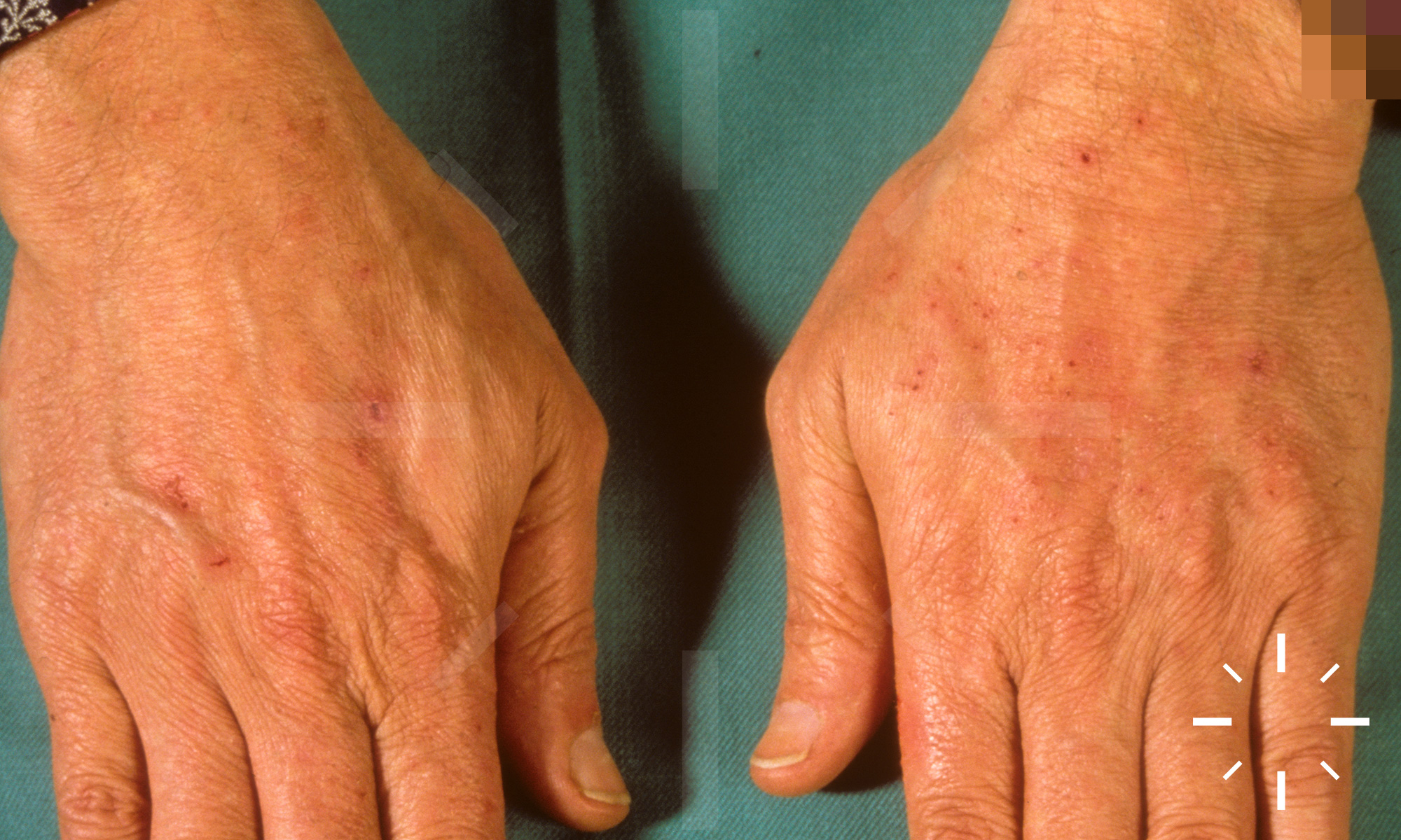
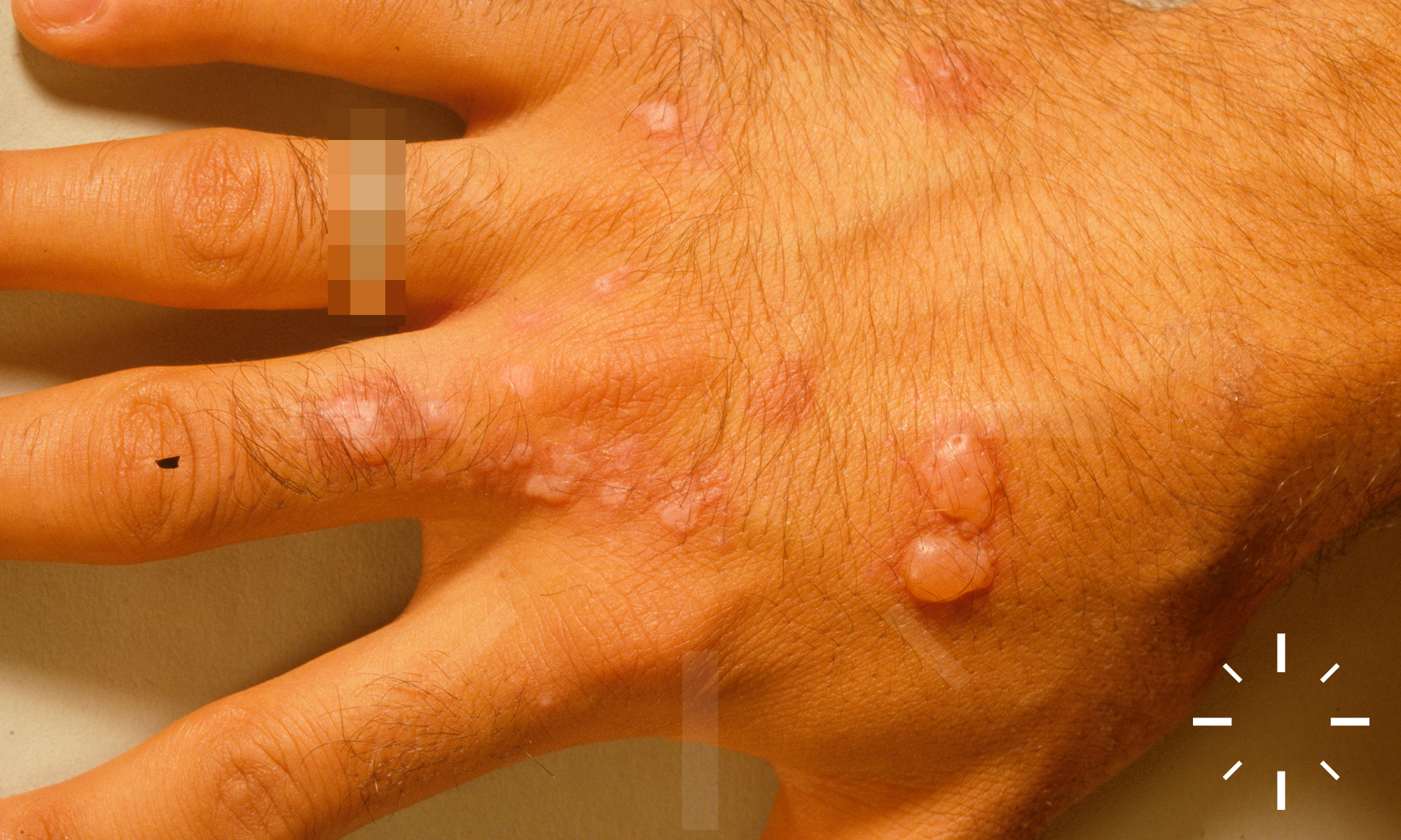
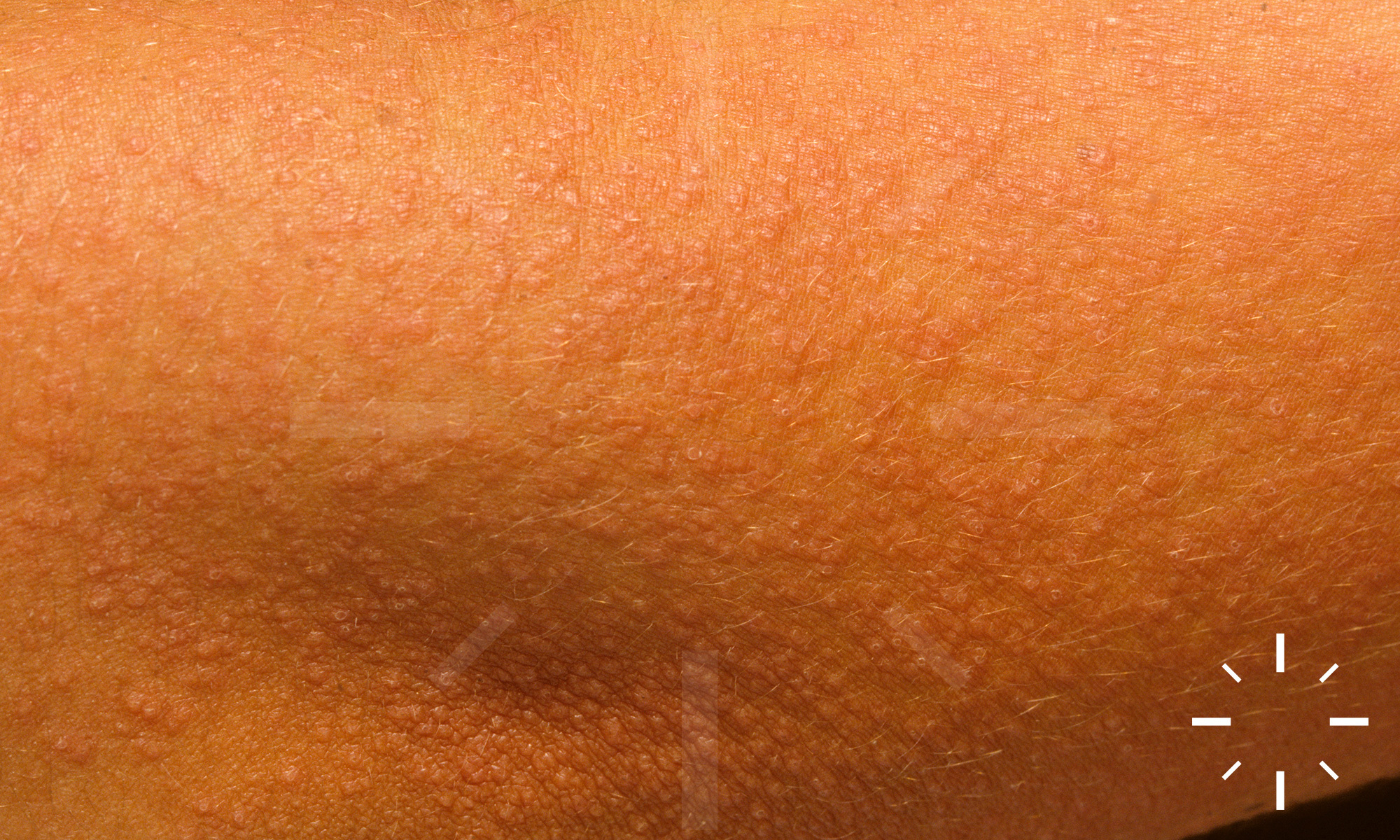
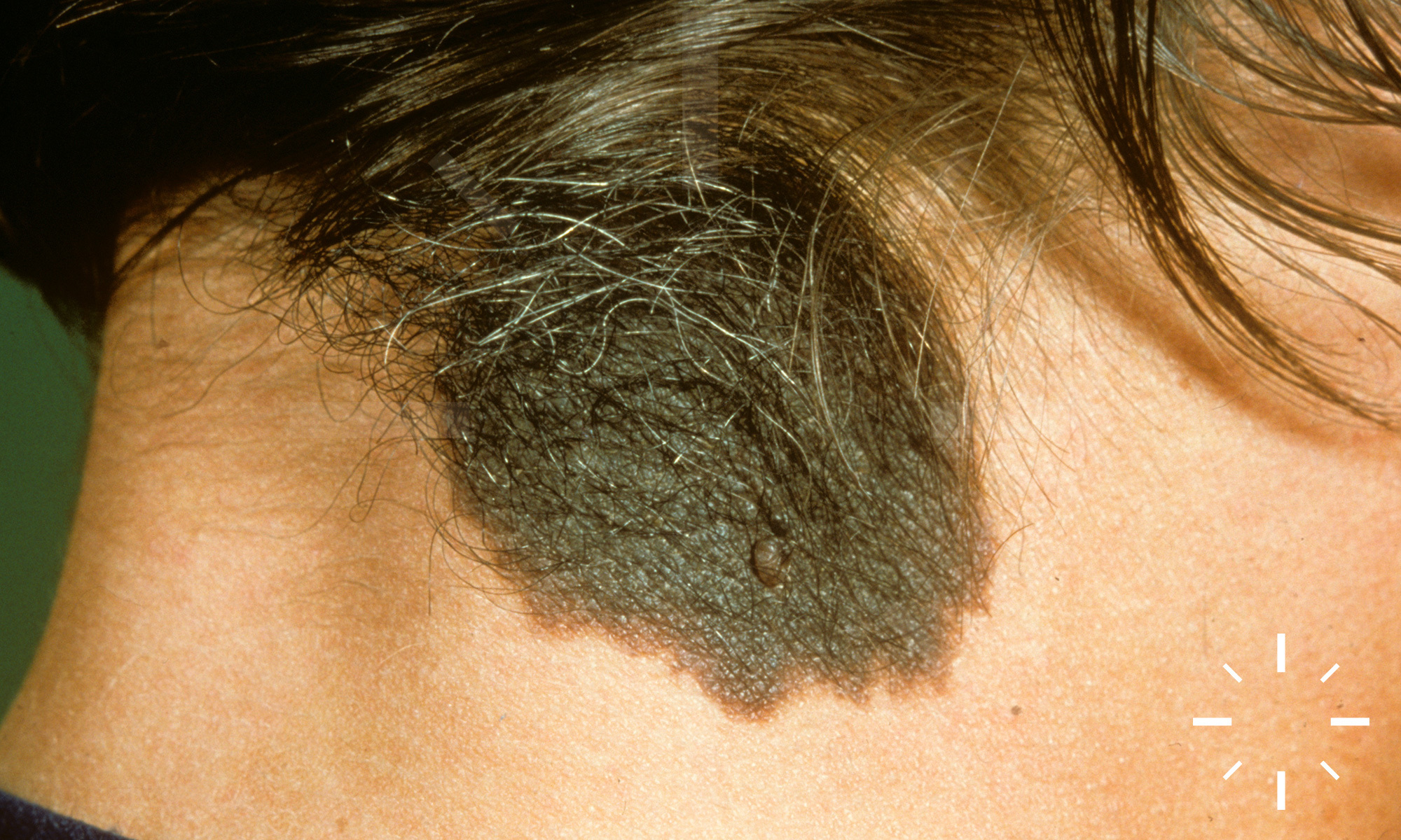
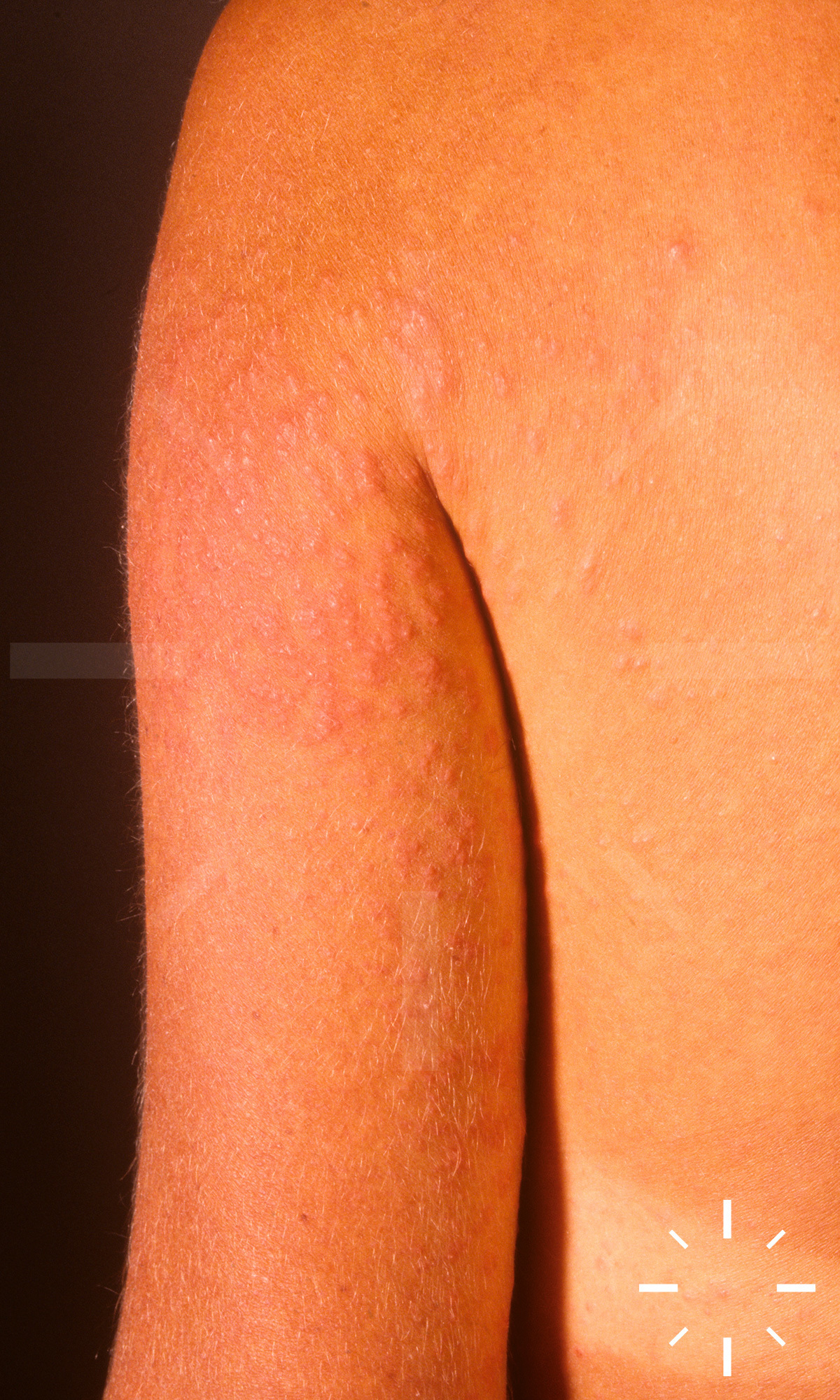
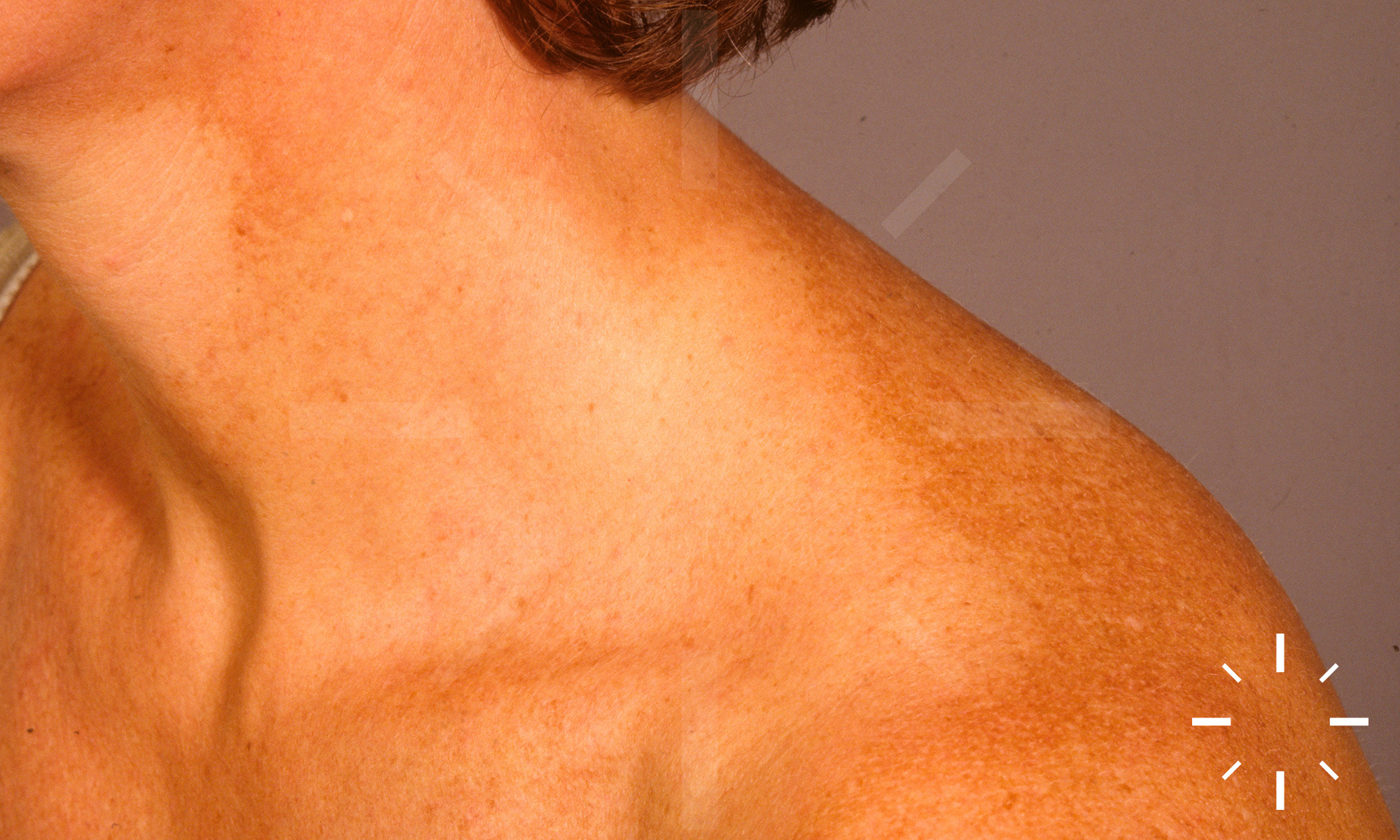
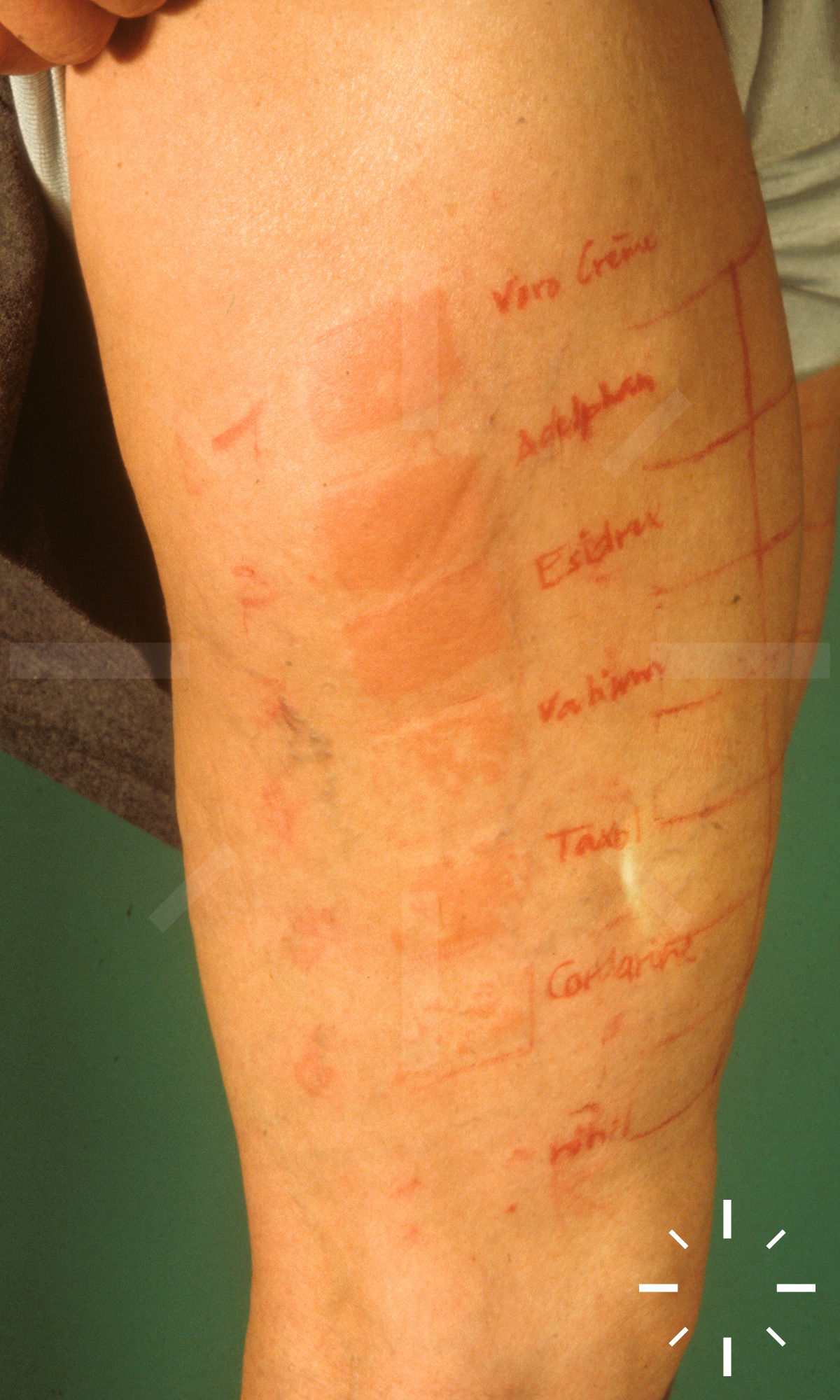
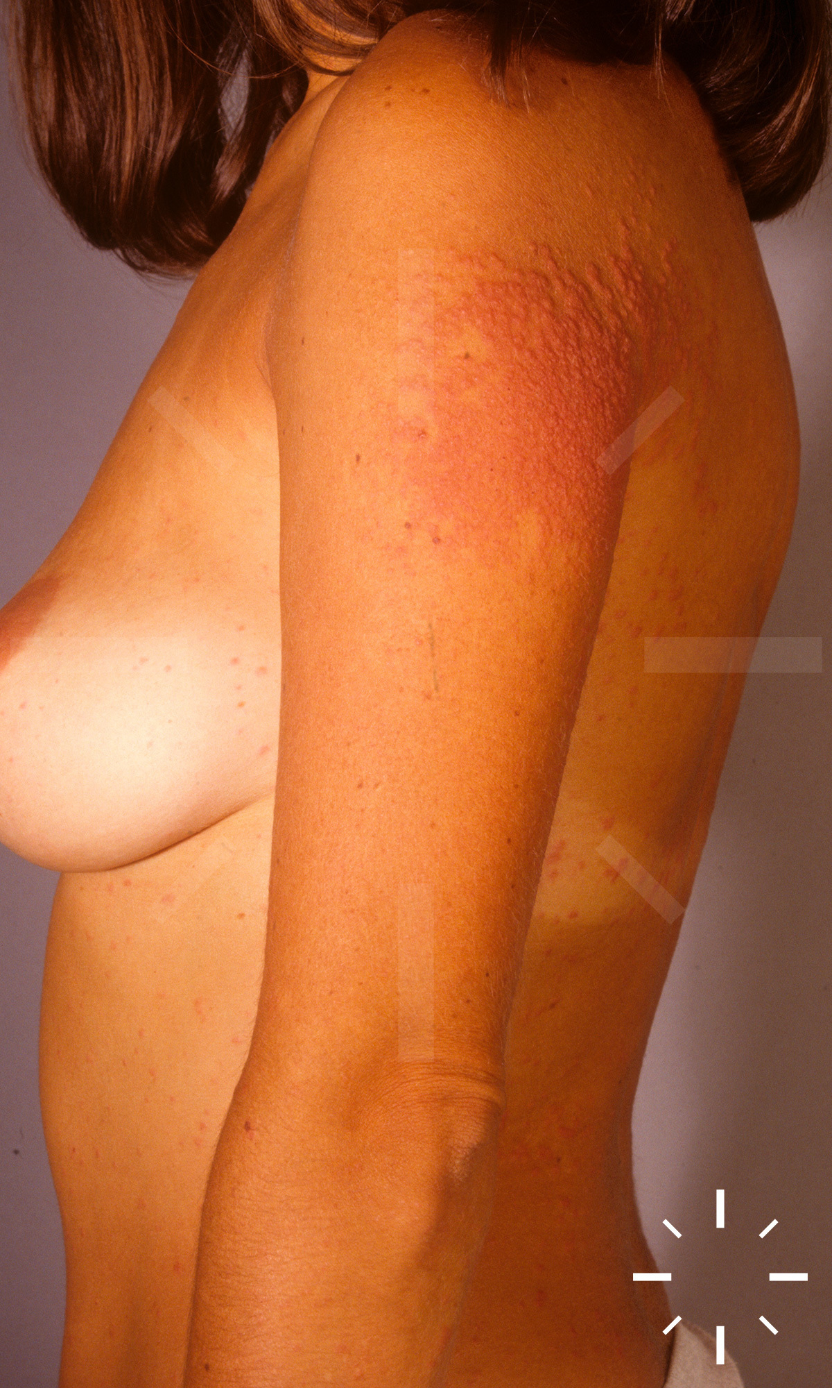
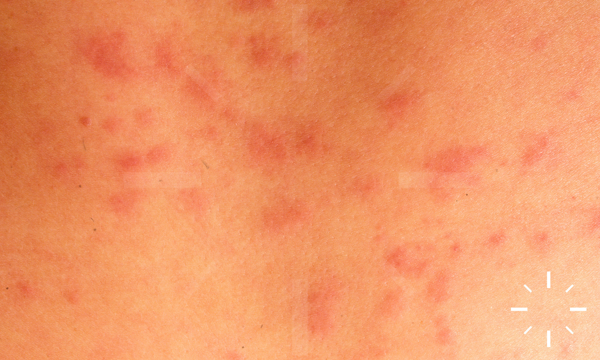
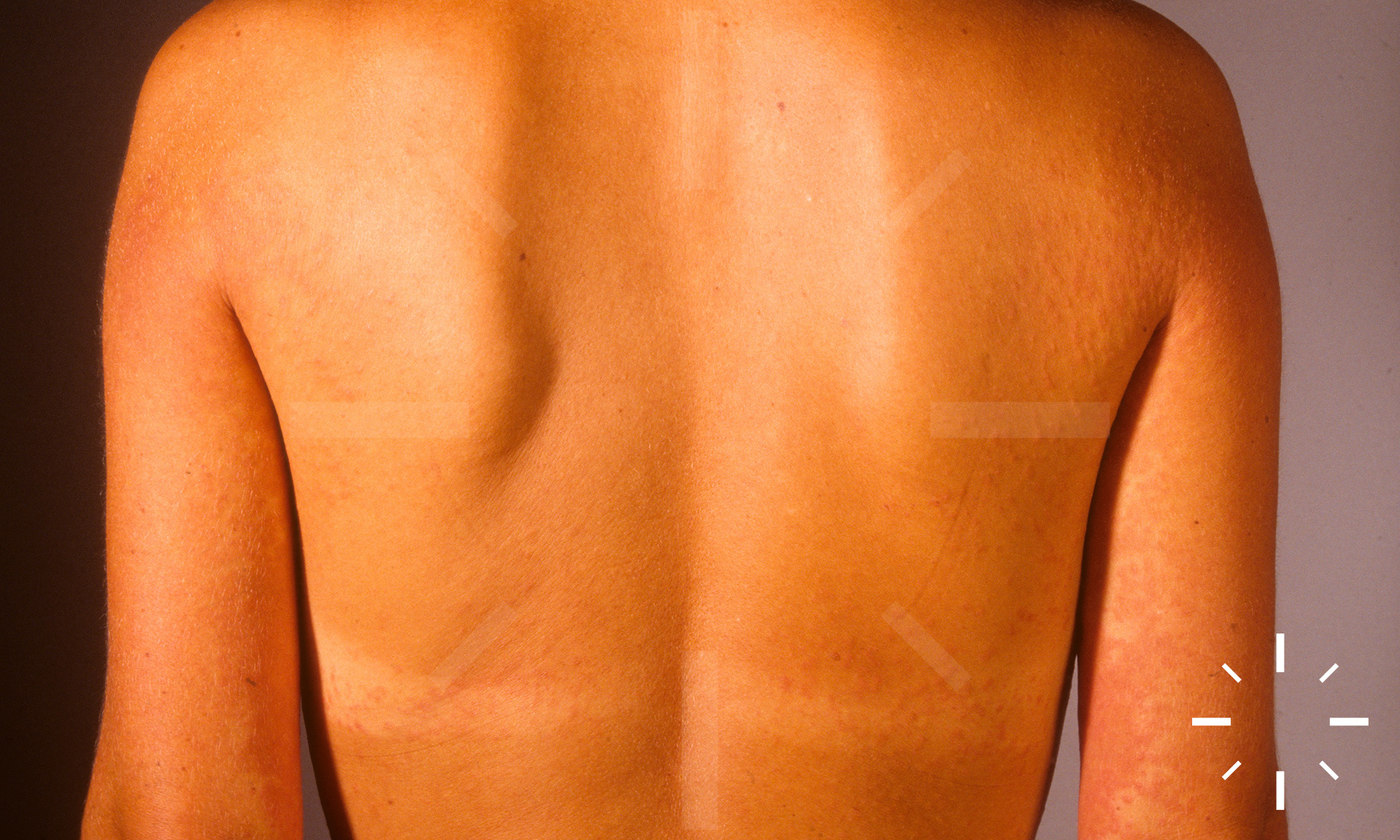
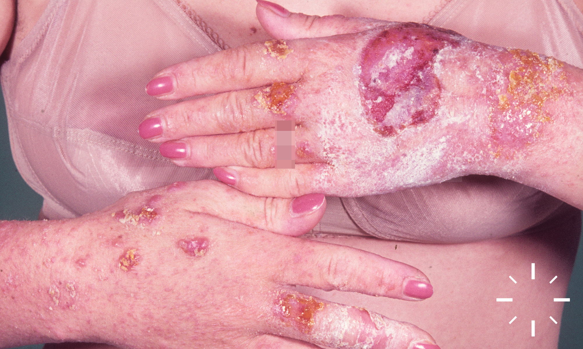
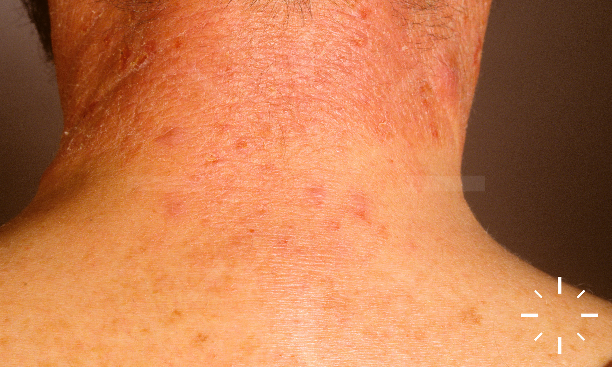
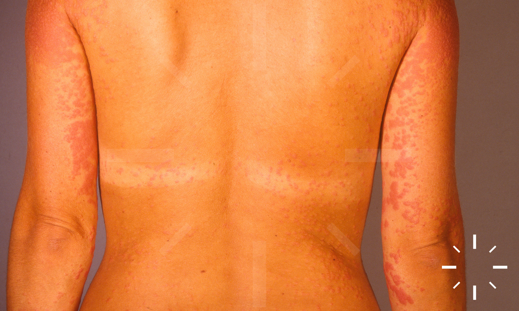
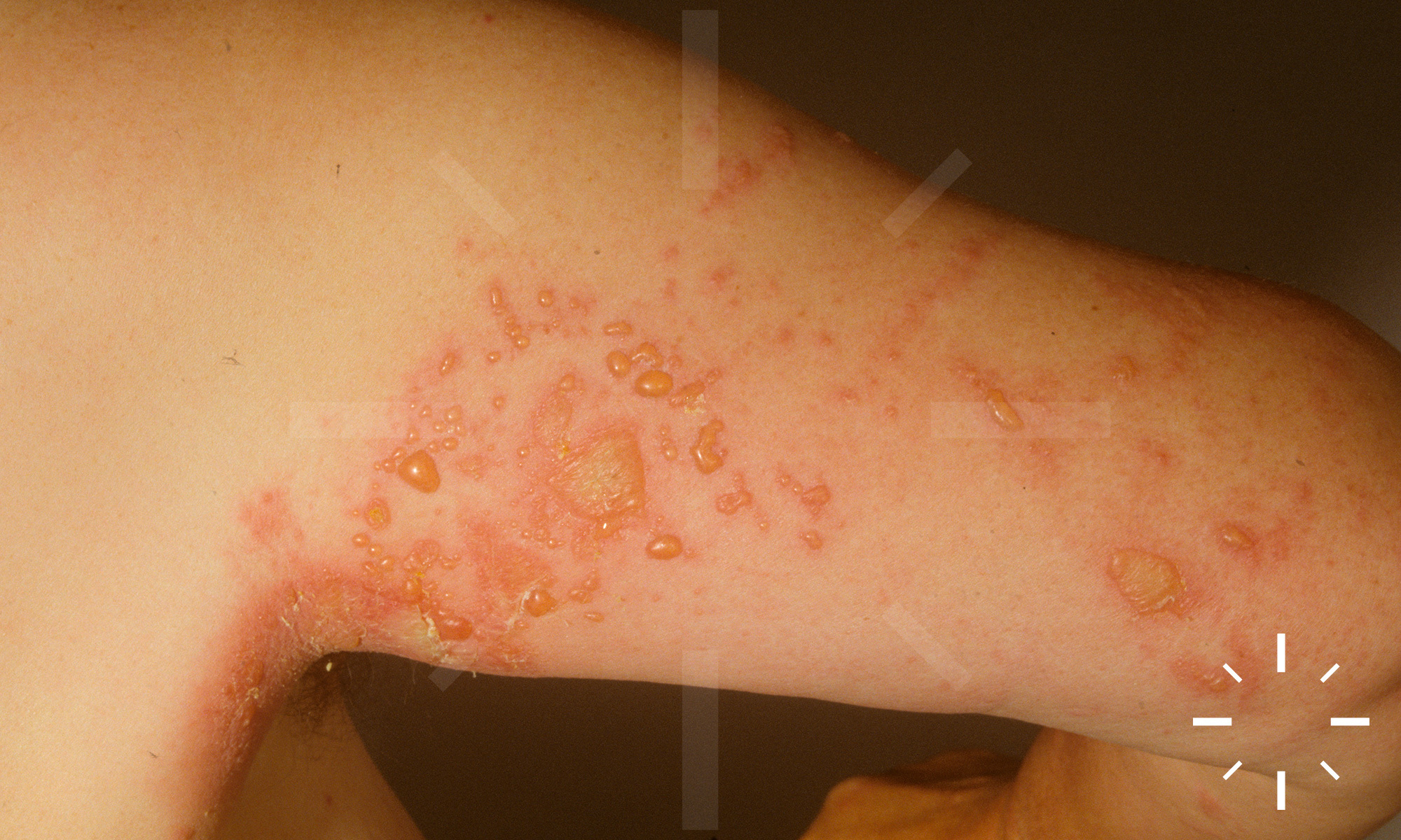
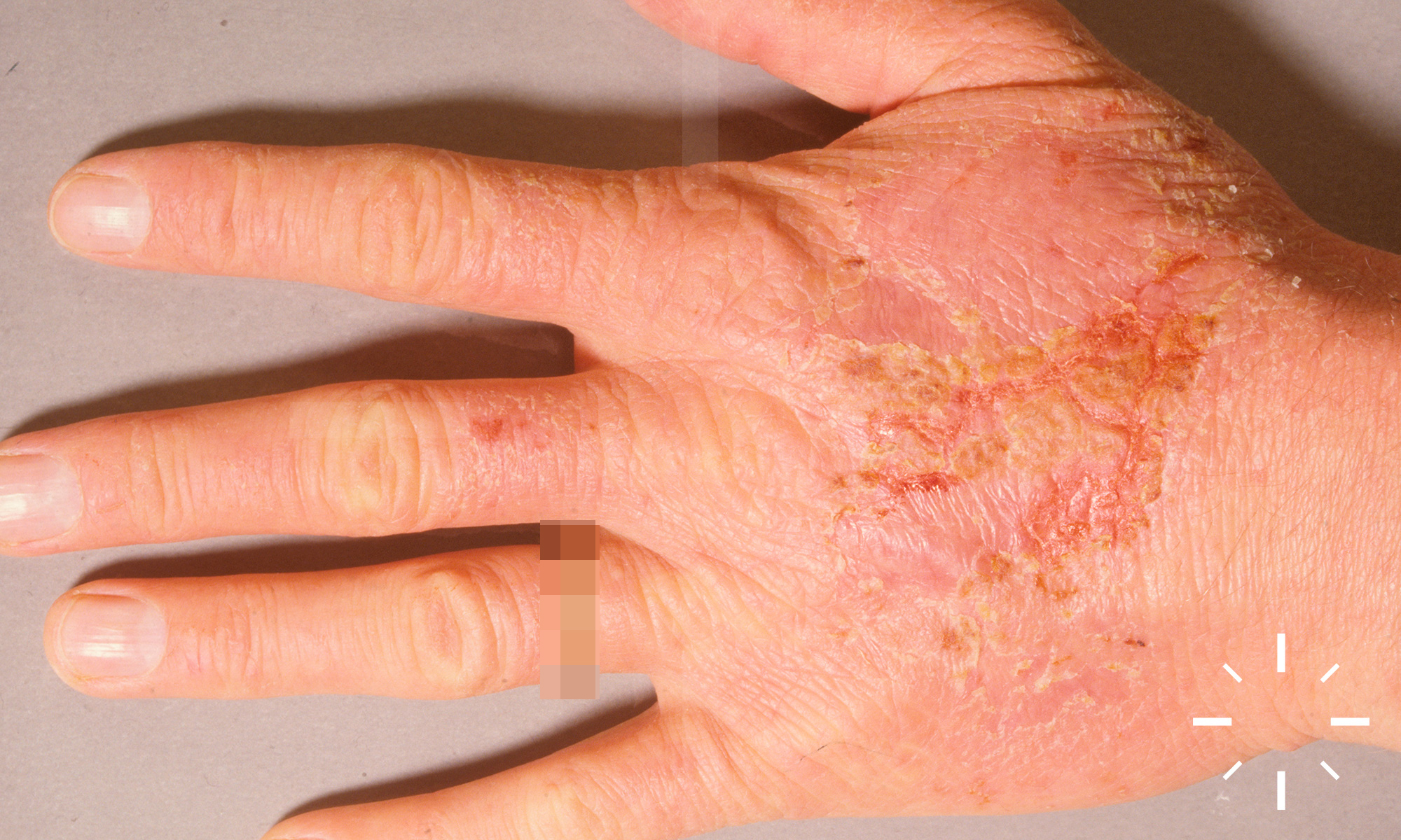
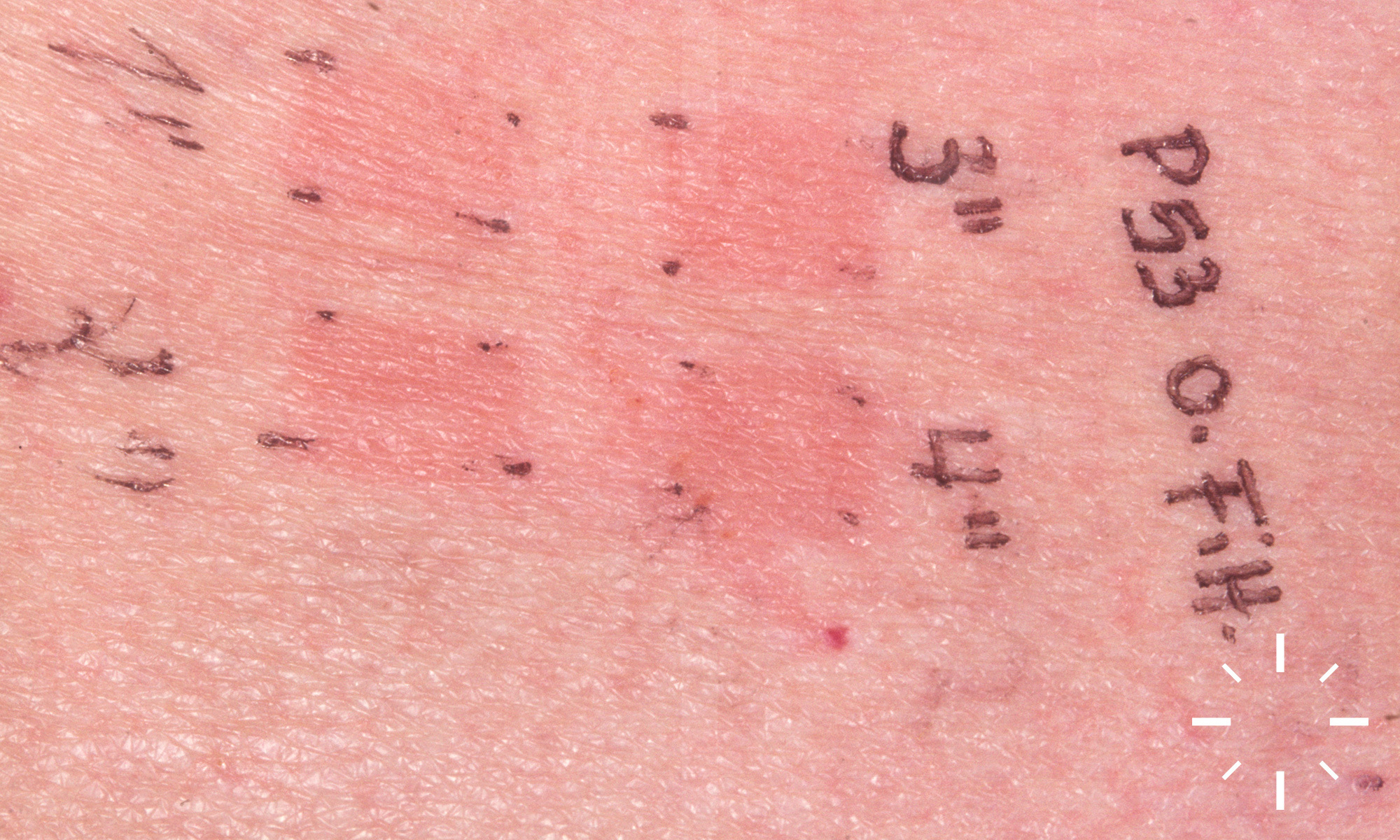
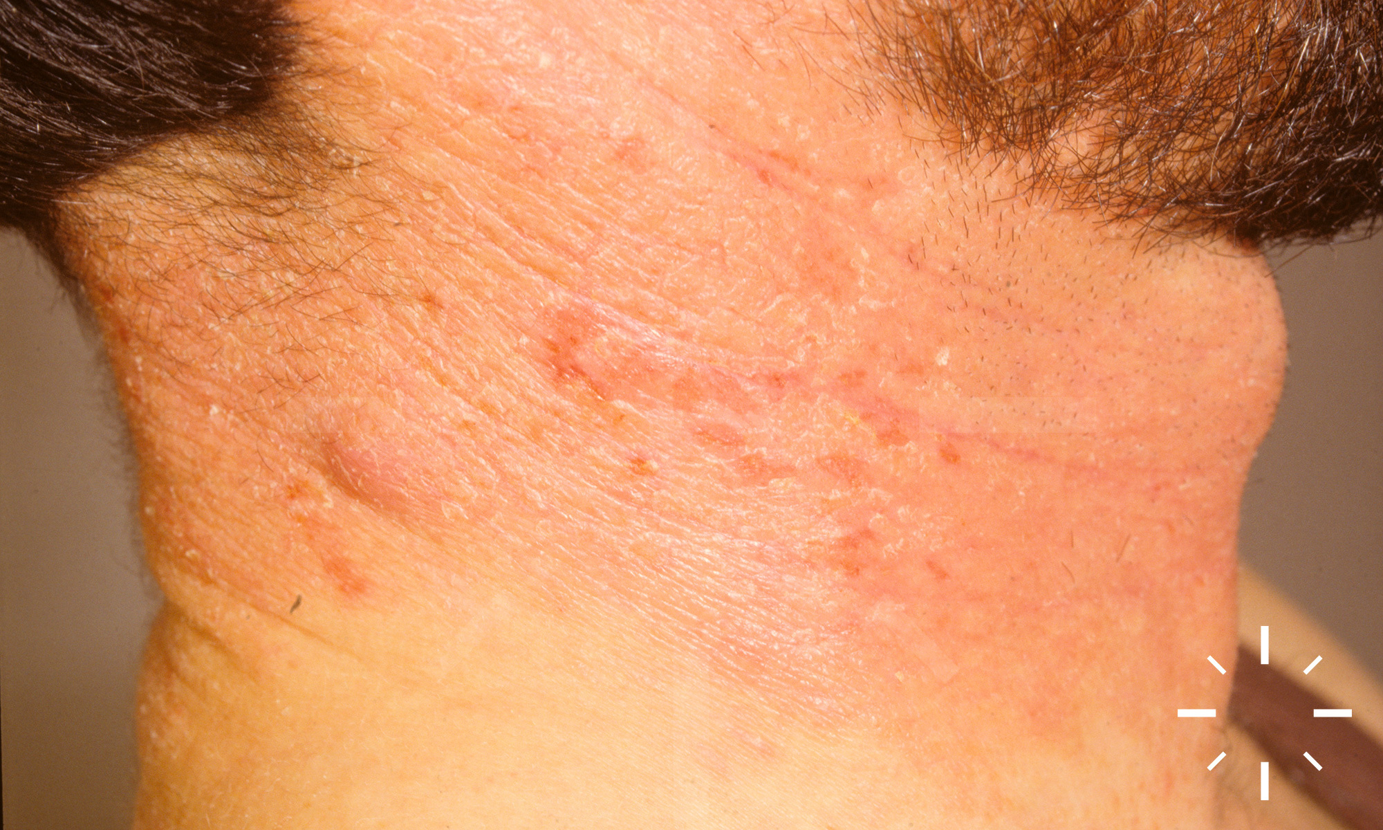
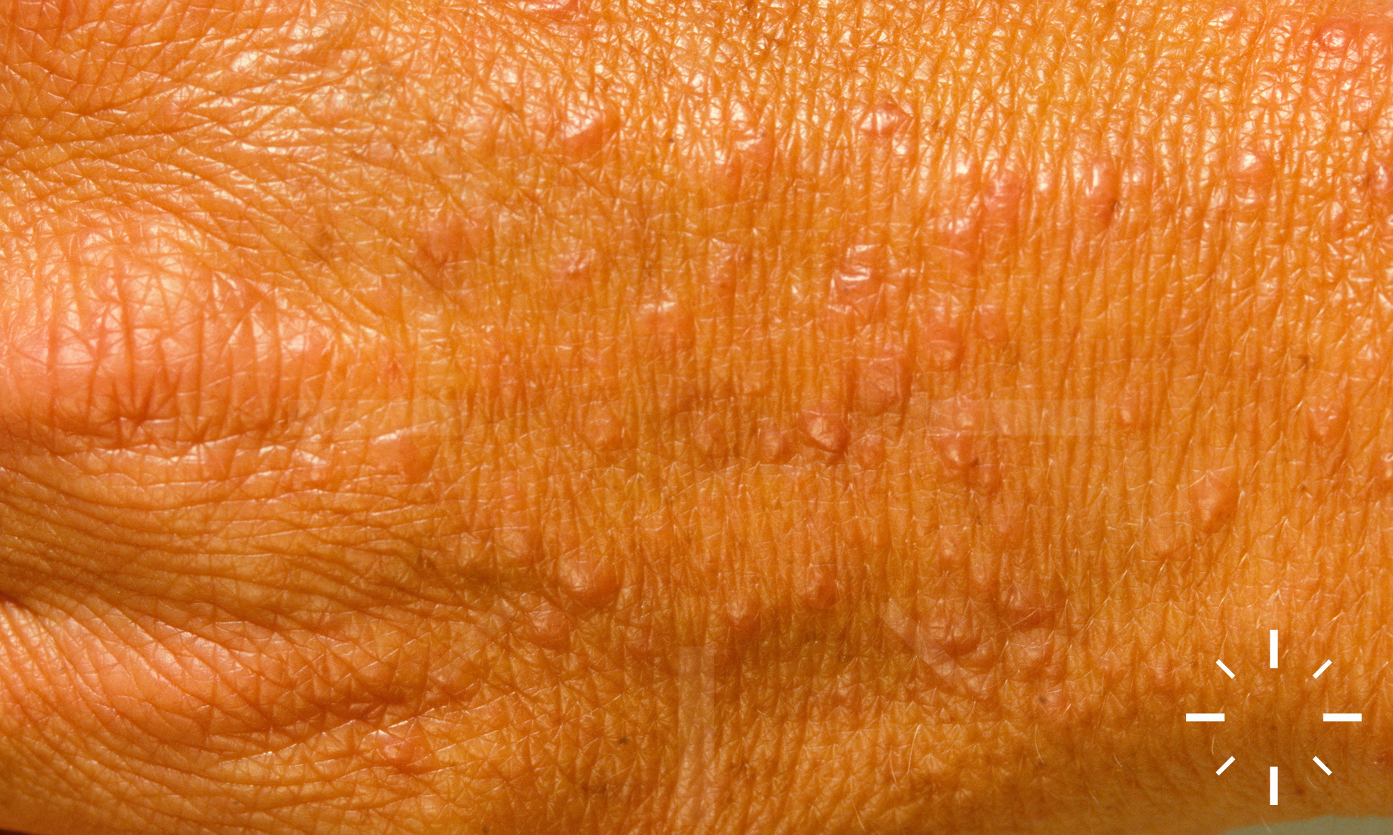
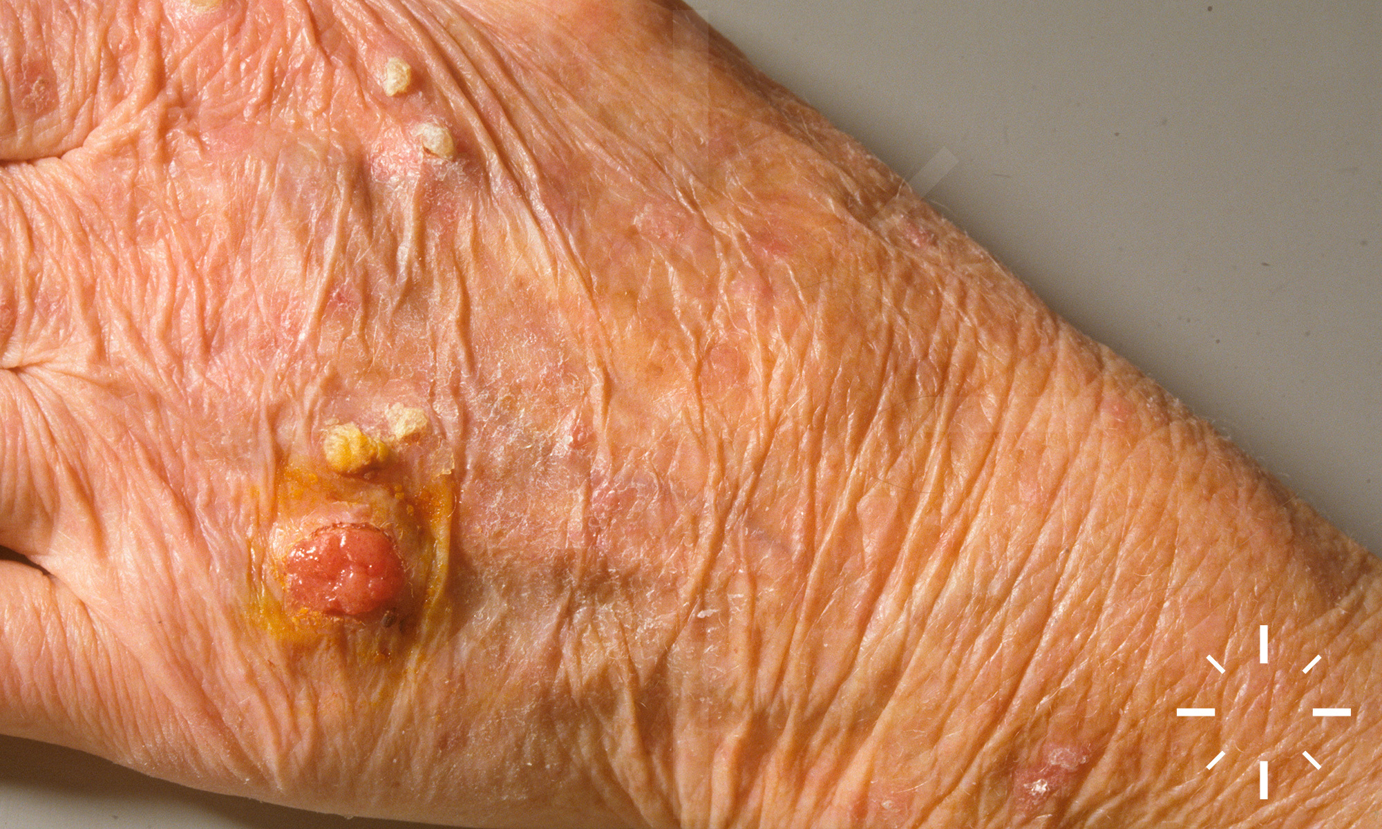
Polymorphous light dermatosis

Last Updated: 2023-07-07

Author(s): Anzengruber F., Navarini A.

ICD11: EJ30.0

  • Bateman 1817
  • Hutchinson 1878
  • Rasch 1900
  • Hausmann and Haxthausen 1929

Sun allergy, photoreactive eczema, polymorphic light eruption (PMLE) prurigo aestivalis, summer prurigo, lupus erythematosus-like light dermatosis, eczema solare

The most common light dermatosis, which is characterised by its intense itching. The appearance is monomorphic within the type of PLD (see below: papules, vesicles, plaques, cocard-like lesions, etc.).

  • Prevalence (depending on data) up to 20%
  • Seasonal accumulation March-June (exception: during holidays in warm countries)
  • Independent of age
  • Women : men = 9:1
  • More common in Caucasians, but also possible in Africans
  • Familial clustering in at least 20%.

  • Triggered by sunlight, more precise aetiology is not known.
  • A genetic disposition probably plays a role.
  • There is an induction of proinflammatory cytokines by UVA (sometimes also UVB) radiation.
  • A delayed-type cell-mediated immunological response occurs due to previously unknown photo-induced antigens.
  • Disruption of UV-induced immunosuppression.

  • Only on sun-exposed areas, individual predilection sites are described.
  • The skin manifestations are polymorphic and can often be classified into different types.
  • Division
    • Papular type
    • Hemorrhagic type
    • Plaque type
    • Erythema exudativum multiforme type
    • Papulo-vesicular type
    • Ictus type
    • Vesiculo-vesicular type
    • Lichenoid type (described in the coloured population)
    • Special forms: Familial variant, occurring in Indians. Juvenile spring eruption (esp. ear helices, hands or nose in young men and children in the early part of the year)
  • First symptoms appear hours to days later. However, manifestation is also possible after several days (cumulative dose). There is often a habituation effect (hardening) in the course. In most cases, there is a pronounced itching.

  • Typical anamnesis
  • Time course (continuous? Relapsing? Depending on the season? Age of first manifestation? - e.g. Hidroa vacciniformia or spring eperniosis), duration of efflorescences? Use of medication? Familial accumulation?
  • Do skin changes also occur behind glass? Indication of triggering by UV-A.
  • Do they occur outdoors but not behind glass? Indication of triggering by UV-B.
  • In room light? Triggering by visible light.
  • After what duration of exposure do the efflorescences develop?
  • Light diagnostics
    • Light staircase
  • Photoprovocation
    • MED is mostly normal
    • UVA provocation more effective than UVB
    • Photopatch test
  • Biopsy
  • if necessary
  • Laboratory
    • ANAs (to look for lupus erythematosus) (plasma fluorescence scan if necessary if there is a reasonable suspicion of porphyria).

Varying patterns according to type. Cuffed, perivascular lymphohistiocytic cell infiltrates are usually seen throughout the dermis. There may also be subepidermal oedema, exocytosis and spongiosis

  • Regular UV hardening in spring (costly)
  • Sun protection, sufficient in the trigger light spectrum

  • Under light abstinence, there is independent healing without residuals.
  • Chronic-recurrent course.

  1. Corbett MF, Hawk JLM, Herxheimer A, Magnus IA. Controlled therapeutic trials in polymorphic light eruption. Br J Dermatol 1982;107:571-81.
  2. Murphy GM, Hawk JLM, Magnus IA. Hydroxychloroquine in polymorphic light eruption: a controlled trial with drug and visual sensitivity monitoring. Br J Dermatol 1987;116:379-86.
  3. Wolf P, Soyer HP, Fink-Puches R, Huff JC, Kerl H. Recurrent post-herpetic erythema multiforme mimicking polymorphic light and juvenile spring eruption: report of two cases in young boys. Br J Dermatol 1994;131:364-7.
  4. Grabczynska, McGregor, Kondeatis, Vaughan, Hawk. Actinic prurigo and polymorphic light eruption: common pathogenesis and the importance of HLA-DR4/DRB1*0407. British Journal of Dermatology 1999;140:232-6.
  5. McGregor JM, Grabczynska S, Hawk JLM, Vaughan R, Lewis CM. Genetic Modeling of Abnormal Photosensitivity in Families with Polymorphic Light Eruption and Actinic Prurigo. Journal of Investigative Dermatology 2000;115:471-6.
  6. Das S, Lloyd JJ, Walshaw D, Farr PM. Provocation testing in polymorphic light eruption using fluorescent ultraviolet (UV) A and UVB lamps. Br J Dermatol 2004;151:1066-70.
  7. Palmer RA, Friedmann PS. Ultraviolet Radiation Causes Less Immunosuppression in Patients with Polymorphic Light Eruption Than in Controls. Journal of Investigative Dermatology 2004;122:291-4.
  8. Richards HL, Ling TC, Evangelou G, Brooke RCC, Fortune DG, Rhodes LE. Evidence of high levels of anxiety and depression in polymorphic light eruption and their association with clinical and demographic variables. British Journal of Dermatology 2008;159:439-44.
  9. Lebwohl, Mark. Treatment of Skin Disease: Comprehensive Therapeutic Strategies. Elsevier, 2014. Print.