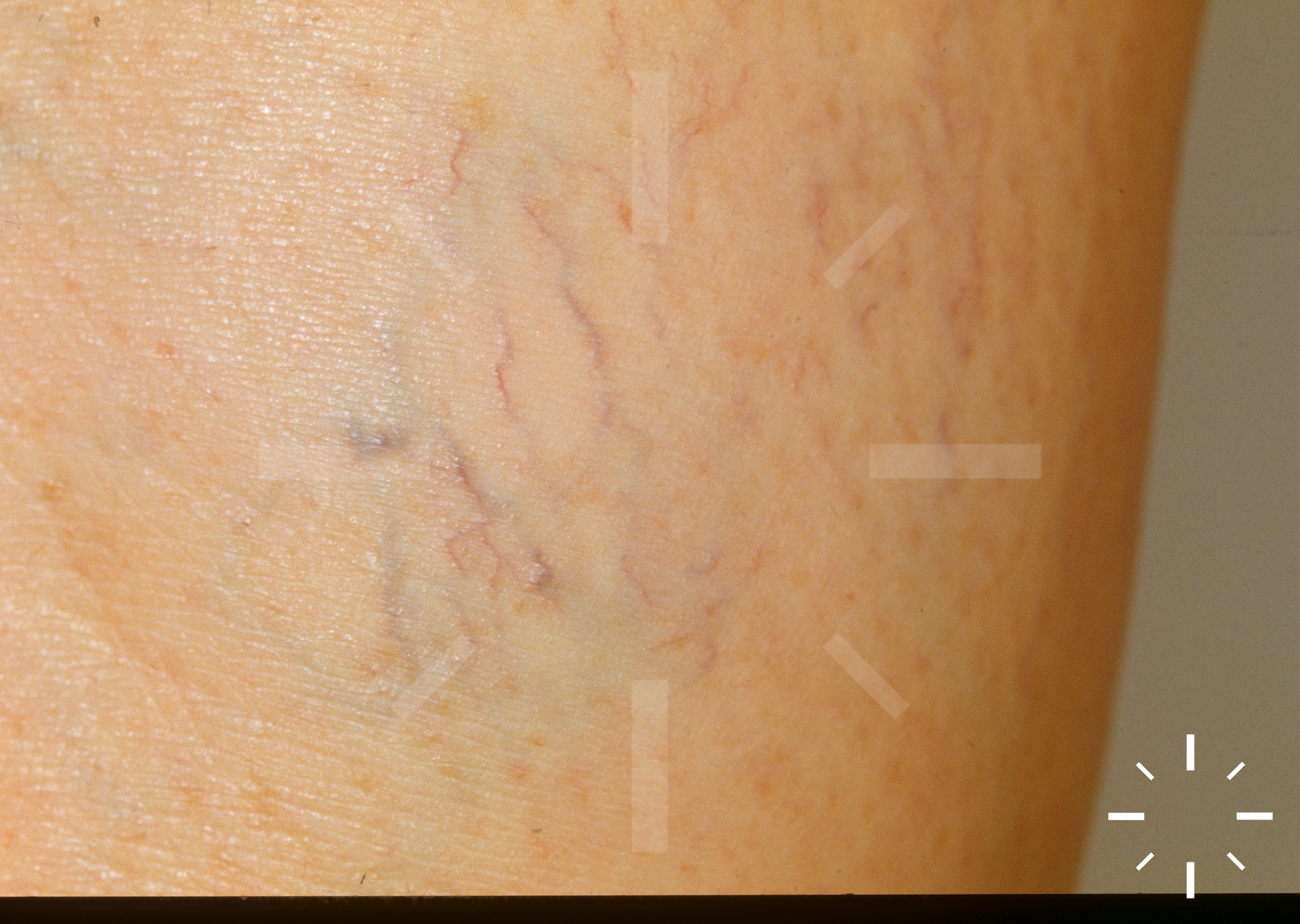
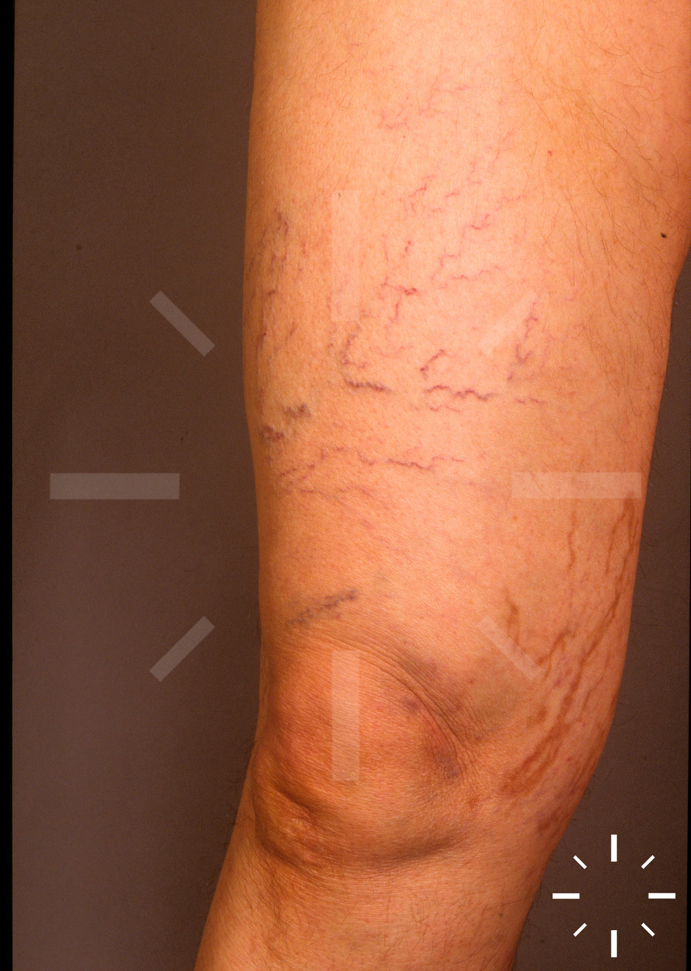
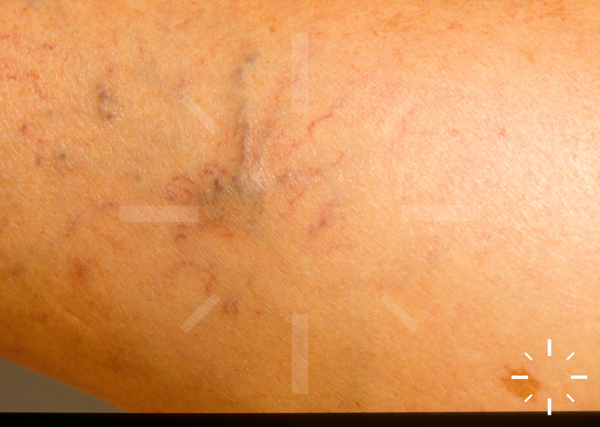
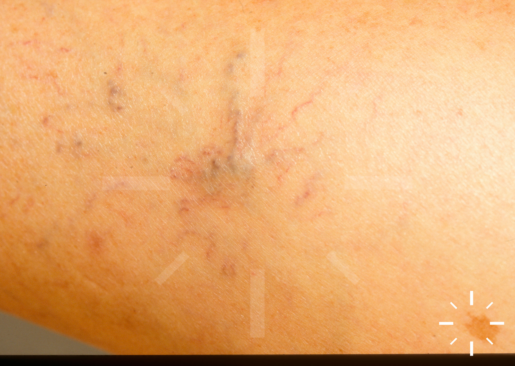
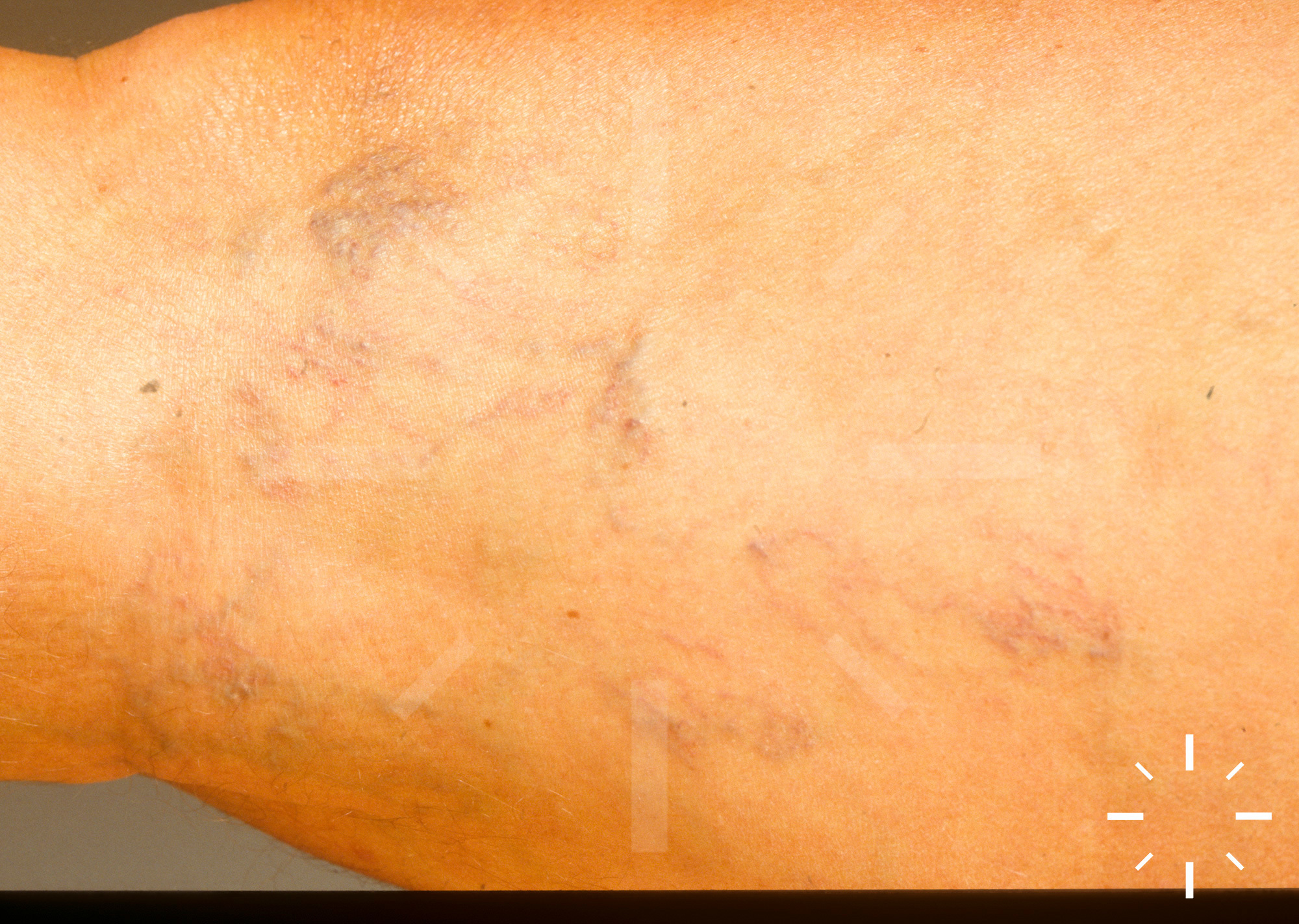
Varicose veins

Last Updated: 2021-10-15

Author(s): Anzengruber F., Navarini A.

ICD11: -

Varicose veins, spider veins, varicosis, varices, reticular varices, varicose veins. 

Degenerative disease of the vein walls leads to pathologically dilated, epifascial veins, usually in the course of the saphenous veins magna and parva.

  • Women > Men
  • There are different data regarding the incidence. It can be assumed that about 1/3 of the population suffer from relevant varicose veins. There is a significant increase in old age. The first symptoms can appear as early as 20 years of age.

  • Primary varicosis: Degeneration of the vascular wall.

  • Secondary varicosis: Vascular wall caused by another disease. 
    • Valvular agenesis 
    • Arteriovenous fistula

    • Klippel-Trénaunay syndrome 

  • Spider vein varices (telangiectasias): Dilatations of the subpapillary intradermal vein plexus caused by localized venous hypertension or primary vascular wall weakness.

  • Reticular varices: Net-like areas with broom tears on the lateral thigh or lower leg and in the hollow of the knee.

  • Stem varices: Varicose alteration of V. saphena magna (80-85%) or V. saphena parva (15-20%).

  • Lateral branch varices: From the branches of Vv. saphenae.

  • Perforating veins:  Insufficiency of the Boyd or Dodd perforating vein can lead to varicosis.

  • Pregnancy varicosis: Mostly reversible varicoses occurring during pregnancy (in about 1/3 of first pregnant women, in 2/3 of multiparous women.

Severity according to Widmer/Partsch 

Varicosities 

Subjective complaints (pruritus, heaviness, dysesthesia, swelling)

Hypostatic eczema, hyperpigmentation, atrophy 

Ulcers 
Degree 1 + - - -
Degree 2 + + - -
Degree 3 + + + -
Degree 4 + + + +


  • Due to remodeling processes, contractile abilities, vasodilatation and finally valve insufficiency decrease.

Predisposing factors 

  • Genetics 
  • Increasing age 
  • Standing
  • Gestation
  • Adiposity 
  • Family disposition (genetic factors)

  • Hormones

  • Depending on the clinic, telangiectasias to dilated vessels may be visible.
  • Classification by Hach 
    • V. saphena magna
      • Grade I: Insufficiency of the krosse (reflux up to approx. hand width below the groin)

      • Grade II: Insufficiency of the krosse (reflux to hand width above the knee)

      • Grade III: Insufficiency of the krosse (reflux to below the knee)

      • Grade IV: Insufficiency of the krosse (reflux to the ankle)

    • V. saphena parva 
      • Grade I: Insufficiency of the krosse at the sluice flap/junction.

      • Grade II: Insufficiency of krosse up to the middle of the US.

      • Grade III: Insufficiency of krosse to the lat. ankle.

  • Medical history 
  • Phlebological examination 
    • Doppler sonography 
    • Duplex sonography 
    • Light reflection rheography 

  • Chronic venous insufficiency 
  • Variceal hemorrhage 
  • Thromboses 

  1. Agu, O., et al., Endothelin Receptors in the Aetiology and Pathophysiology of Varicose Veins. European Journal of Vascular and Endovascular Surgery, 2002. 23(2): p. 165-171.
  2. Baccaglini, U., et al., Consensus Conference on Sclerotherapy of Varicose Veins of the Lower Limbs, in Phlebology ’95. 1995, Springer Science + Business Media. p. 604-605.
  3. Barrett, J.M., et al., Microfoam Ultrasound-Guided Sclerotherapy of Varicose Veins in 100 Legs. Dermatologic Surgery, 2004. 30(1): p. 6-12.
  4. Beebe-Dimmer, J.L., et al., The Epidemiology of Chronic Venous Insufficiency and Varicose Veins. Annals of Epidemiology, 2005. 15(3): p. 175-184.
  5. Bergan, J.J., Ambulatory phlebectomy: A practical guide for treating varicose veins. Journal of Vascular Surgery, 1995. 22(4): p. 510-511.
  6. Bradbury, A., et al., What are the symptoms of varicose veins? Edinburgh vein study cross sectional population survey. BMJ, 1999. 318(7180): p. 353-356.
  7. Butie, A., Clinical Examination of Varicose Veins. Dermatologic Surgery, 1995. 21(1): p. 52-56.
  8. Darke, S.G. and S.J.A. Baker, Ultrasound-guided foam sclerotherapy for the treatment of varicose veins. British Journal of Surgery, 2006. 93(8): p. 969-974.
  9. Darvall, K.A.L., et al., Higher prevalence of thrombophilia in patients with varicose veins and venous ulcers than controls. Journal of Vascular Surgery, 2009. 49(5): p. 1235-1241.
  10. Garde, C., Cryosurgery of Varicose Veins. The Journal of Dermatologic Surgery and Oncology, 1994. 20(1): p. 56-58.
  11. Goldman, M.P., Intravascular lasers in the treatment of varicose veins. J Cosmet Dermat, 2004. 3(3): p. 162-166.
  12. Goldman, M.P., R.A. Weiss, and J.J. Bergan, Diagnosis and treatment of varicose veins: A review. Journal of the American Academy of Dermatology, 1994. 31(3): p. 393-413.
  13. Guex, J.-J., Sclerofoam for Treatment of Varicose Veins, in The Vein Book. 2007, Elsevier BV. p. 201-207.
  14. Jukkola, T.M., et al., The effects of parity, oral contraceptive use and hormone replacement therapy on the incidence of varicose veins. Journal of Obstetrics and Gynaecology, 2006. 26(5): p. 448-451.
  15. London, N.J.M., ABC of arterial and venous disease: Varicose veins. BMJ, 2000. 320(7246): p. 1391-1394.
  16. Marley, W., Fading Rate of Pigmentation Induced by Sotradecol for Sclerotherapy of Varicose Veins, in Phlebology ’95. 1995, Springer Science + Business Media. p. 618-618.
  17. Nishibe, T., Stripping operation with preservation of the calf saphenous veins for primary varicose veins: hemodynamic evaluation. Cardiovascular Surgery, 2003. 11(5): p. 341-345.
  18. Orbach, E.J., Sclerotherapy of varicose veins. The American Journal of Surgery, 1944. 66(3): p. 362-366.
  19. Pfisterer, L., et al., Pathogenesis of varicose veins - lessons from biomechanics. Vasa, 2014. 43(2): p. 88-99.
  20. Raju, S. and P. Neglén, Chronic Venous Insufficiency and Varicose Veins. New England Journal of Medicine, 2009. 360(22): p. 2319-2327.
  21. Rautio, T., et al., Endovenous obliteration versus conventional stripping operation in the treatment of primary varicose veins: A randomized controlled trial with comparison of the costs. Journal of Vascular Surgery, 2002. 35(5): p. 958-965.
  22. Raymond-Martimbeau, P., Technical Feasibility and Early Results of Radiologic-Guided Foam Sclerotherapy for Treatment of Varicose Veins. Dermatologic Surgery, 2011. 37(8): p. 1196-1197.
  23. Ruckley, C.V., Varicose Veins and Telangiectasias — Diagnosis and Treatment. J. J. Bergan and M. P. Goldman (eds). 261 × 185 mm. Pp. 448. Illustrated. 1993. St Louis, Missouri: Quality Medical Publishing. £104. British Journal of Surgery, 1994. 81(3): p. 477-477.
  24. Sadick, N.S., Treatment of Varicose and Telangiectatic Leg Veins with Hypertonic Saline: A Comparative Study of Heparin and Saline. The Journal of Dermatologic Surgery and Oncology, 1990. 16(1): p. 24-28.
  25. Sadick, N.S., Sclerotherapy of Varicose and Telangiectatic Leg Veins: Minimal Sclerosant Concentration of Hypertonic Saline and Its Relationship to Vessel Diameter. The Journal of Dermatologic Surgery and Oncology, 1991. 17(1): p. 65-70.
  26. Sadick, N.S. and H. Schanzer, Combined High Ligation and Stab Avulsion for Varicose Veins in an Outpatient Setting. Dermatologic Surgery, 1998. 24(4): p. 475-479.
  27. Stansby, G., Women, pregnancy, and varicose veins. The Lancet, 2000. 355(9210): p. 1117-1118.
  28. van der Velden, S.K., et al., Management Strategies for Patients with Varicose Veins (C2–C6): Results of a Worldwide Survey. European Journal of Vascular and Endovascular Surgery, 2015. 49(2): p. 213-220.
  29. Weiss, M.A., Sclerotherapy: Treatment of Varicose and Telangiectatic Leg Veins. Arch Dermatol, 1997. 133(1): p. 117.
  30. Weiss, R.A. and M.P. Goldman, Endovenous Ablation Techniques with Ambulatory Phlebectomy for Varicose Veins, in Surgery of the Skin. 2010, Elsevier BV. p. 589-600.
  31. Weiss, R.A. and M.A. Weiss, Continuous Wave Venous Doppler Examination for Pretreatment Diagnosis of Varicose and Telangiectatic Veins. Dermatologic Surgery, 1995. 21(1): p. 58-62.
  32. Weiss, R.A. and M.A. Weiss, Controlled Radiofrequency Endovenous Occlusion Using a Unique Radiofrequency Catheter Under Duplex Guidance to Eliminate Saphenous Varicose Vein Reflux. Dermatologic Surgery, 2002. 28(1): p. 38-42.
  33. Wolf, M.A.X., LICHEN AMYLOIDOSUS ASSOCIATED WITH VARICOSE VEINS. Arch Dermatol, 1953. 67(4): p. 407.
  34. Kahle, B. and M. Stücker, Diagnostik und Therapie der Varikosis. Aktuelle Dermatologie, 2009. 35(06): p. 243-255.
  35. Lorenz, M.B., P. Gkogkolou, and T. Goerge, Sklerotherapie der Varikosis in der Dermatologie. JDDG: Journal der Deutschen Dermatologischen Gesellschaft, 2014. 12(5): p. 391-394.
  36. Nüllen, H., T. Noppeney, and W. Lang, Chirurgie der Perforans-Varikosis, in Diagnostik und Therapie der Varikose. 2010, Springer Science + Business Media. p. 329-342.
  37. Schultz-Ehrenburg, U., G. Gallenkemper, and A. Miller, Diagnostik der chronischen venösen Insuffizienz und Therapie der Varikosis, in Dermatologie an der Schwelle zum neuen Jahrtausend. 2000, Springer Science + Business Media. p. 613-615.