Actinomycosis

Last Updated: 2023-07-07

Author(s): Anzengruber F., Navarini A.

ICD11: 1C10.Z

  • Bollinger 1877
  • Israel 1878

Lumpy Jaw, Streptotrichosis, Radiation Fungal Disease, Leptotrichosis.

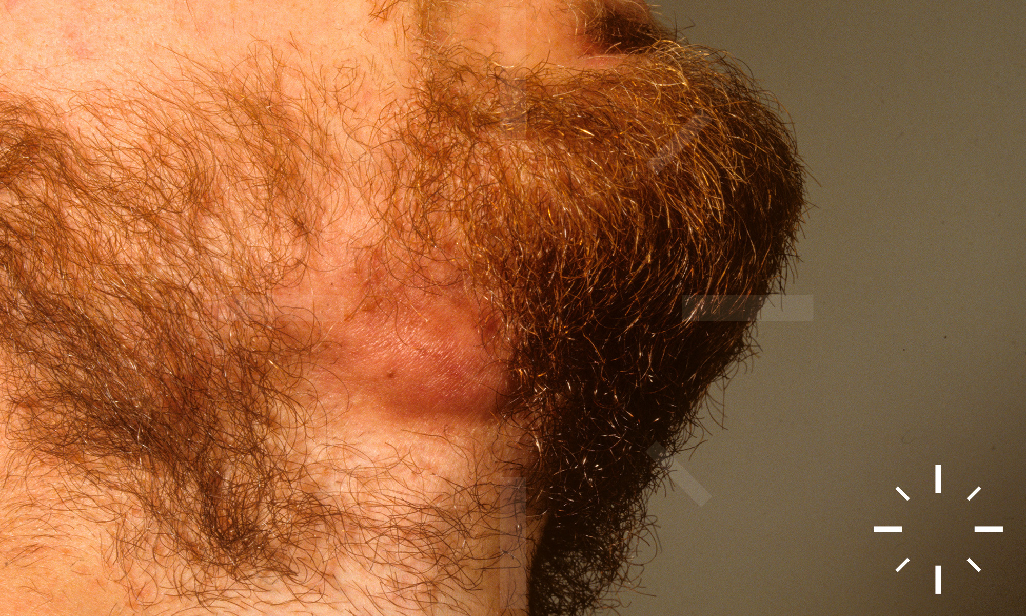
Infection by the facultative pathogenic, anaerobic, pleomorphic, non-acid-fast, gram-positive pathogens Actinomyces israelii, rarely also Actinomyces naeslundi, Actinomyces radingae and in individual cases also Actinomyces bovi. Frequently, there are mixed infections (Actinobacillus actinomycetemcomitans, staphylococci, streptococci, etc.).

  • Klineli
    • Clinical classification
      • Cervicofacial actinomycosis (80-95% of all cases): mandible and oral mucosa.
      • Abdominal actinomycosis (approx. 3% of all cases): mostly after operations (e.g.: appendicitis).
      • Thoracic actinomycosis (up to 15% of all cases).
    • Occurring worldwide.
    • More common in rural populations.
    • Men > women.
    • Most prevalent in developing countries.
    • Peak in incidence 20th-40th yrs.

    • Predisposing factors
    • Bone fractures
    • Tooth extractions
    • Actinomyces israelii is physiologically present in the mouth or pharynx (in 50% of excised tonsils). Occasionally, it can also be detected in the colon. Infection is thus endogenous. The bacteria penetrate into the deeper tissue. It can be assumed that the assistance of other bacteria is necessary for this.

    • Tough, blue-red, overheated, hard as a board, bulging, painful nodules and indurations, sometimes ulcerations. Fistulous openings may release a serous to purulent content on pressure, in which yellow granules, the actinomyces drusen, can be seen. The affected region is oedematously altered. Spontaneous healing does not occur. Periostitits, ostemyelitis.
    • Reduced general condition, fever, locoregional lymphadenopathy, weight loss and night sweats.
    • Cervicofacial actinomycosis: Carious teeth or mucosal defects act as the main portal of entry.
    • Abdominal actinomycosis: The cause is usually a previous operation (e.g. appendicitis). Clinically, there is an intra-abdominal mass, and often a psoas abscess can be diagnosed as a secondary finding. Secondary skin involvement is possible.
    • Thoracic actinomycosis: Characteristic are fistulas and abscesses, pleural pain as well as productive cough with sputum

    • Clinic.
    • Dermatopathological backup (deep biopsy, punch biopsy is not sufficient).
    • Bact. smear.

    Nonspecific granulation tissue with plasma cells or eosinophilic granulocytes, leucocyte-rich melting necrosis. The acidophilic granules (drusen) are detectable in HE staining and in Grocott's method. Filamentous filaments are seen in the periphery.

    • Mouth phlegmon
    • Miliar seeding
    • Jaw clamp

    Frequently chronic form of progression, but with timely diagnosis and therapy there is a favourable prognosis.

    1. Barabás J, Suba Z, Szabó G, Németh Z, Bogdán S, Huszár T. False Diagnosis Caused by Warthin Tumor of the Parotid Gland Combined with Actinomycosis. Journal of Craniofacial Surgery 2003;14:46-50.
    2. Gahrn-Hansen B, Frederiksen W. Human infections with Actinomyces pyogenes (Corynebacterium pyogenes). Diagn Microbiol Infect Dis 1992;15:349-54.
    3. Lerner PI. Susceptibility of pathogenic actinomycetes to antimicrobial compounds. Antimicrob Agents Chemother 1974;5:302-9.
    4. Sudhakar SS, Ross JJ. Short‐Term Treatment of Actinomycosis: Two Cases and a Review. Clinical Infectious Diseases 2004;38:444-7.
    5. Warren NG. ACTINOMYCOSIS, NOCARDIOSIS, AND ACTINOMYCETOMA. Dermatologic Clinics 1996;14:85-95.
    6. Yeruham I, Elad D, Perl S, Avidar Y, Israeli B, Shlosberg A. Isolation of Corynebacterium pilosum and Actinomyces pyogenes from cystitis and vulvovaginitis infection in a 2-month-old female calf. Zentralbl Veterinarmed B 1999;46:127-30.