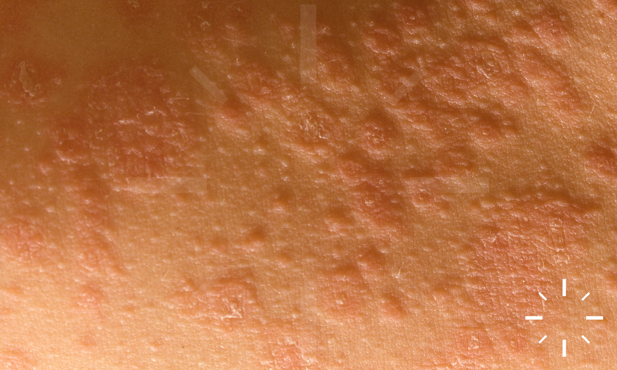
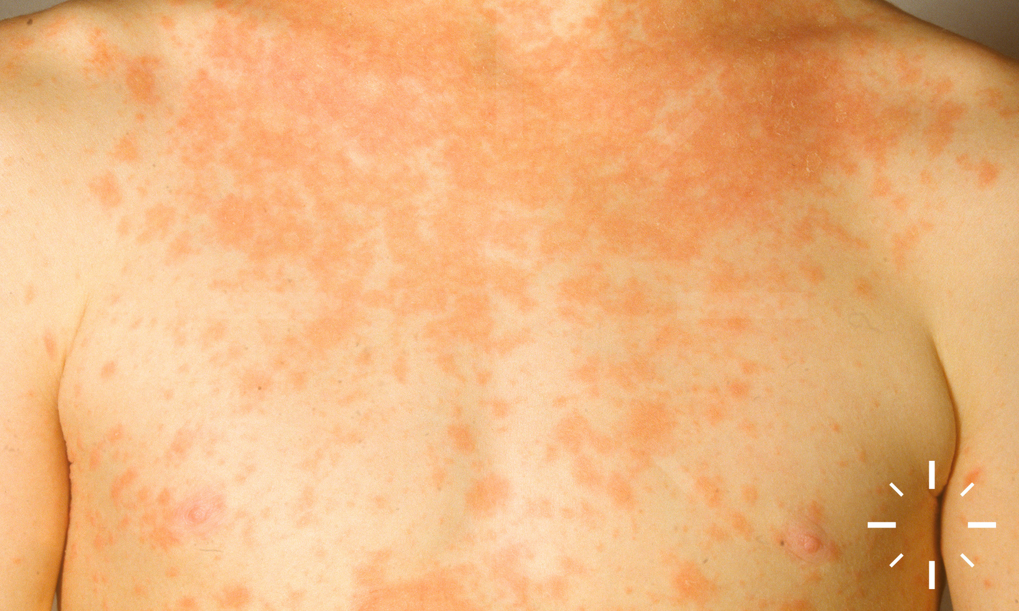
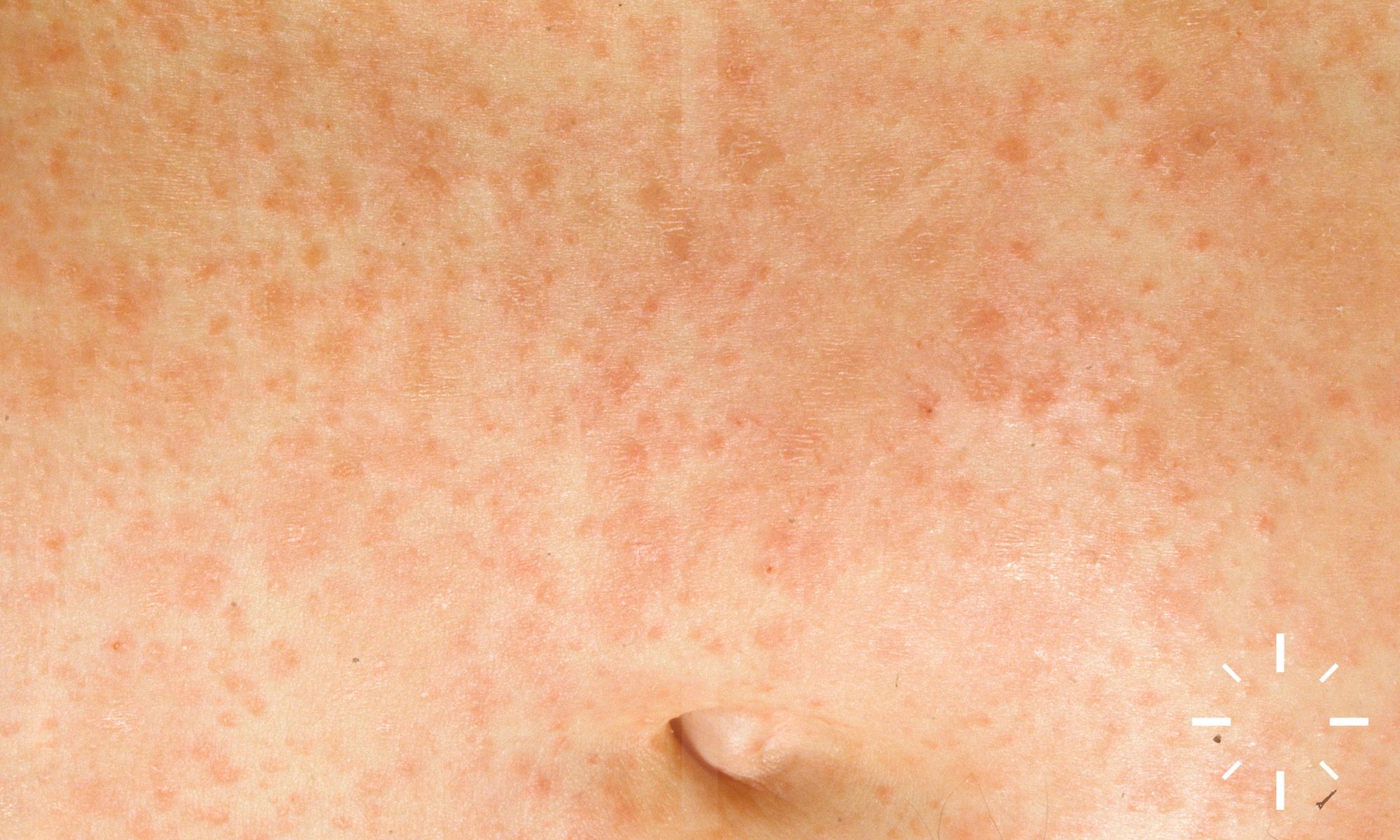
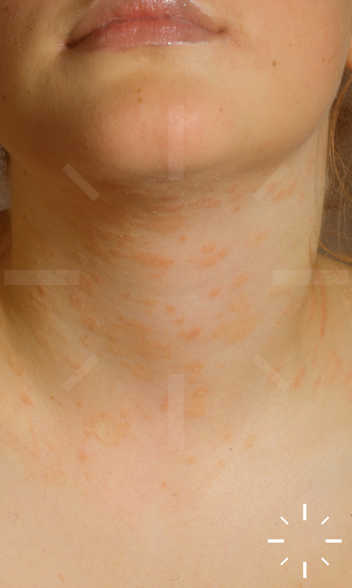
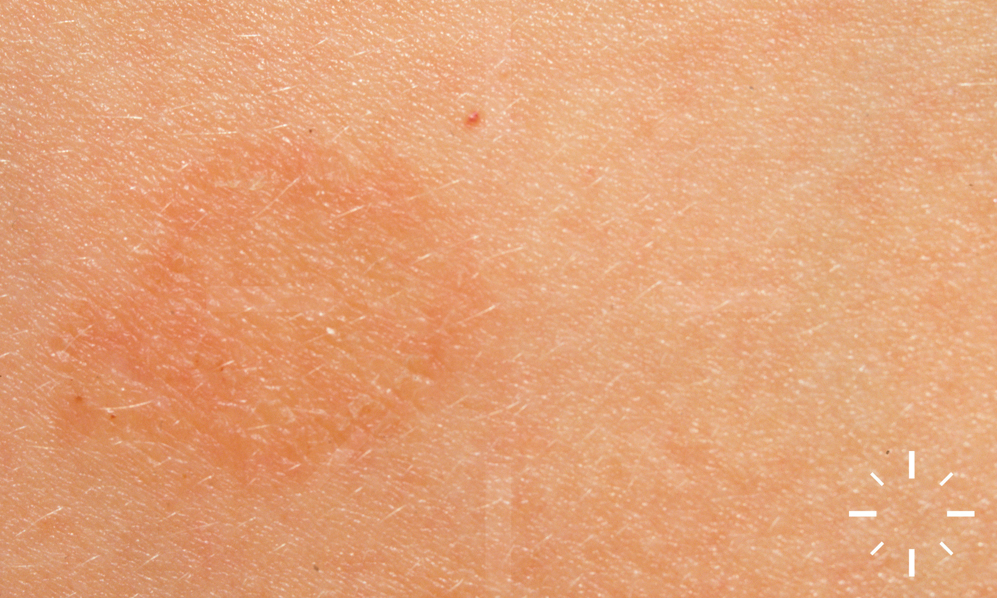
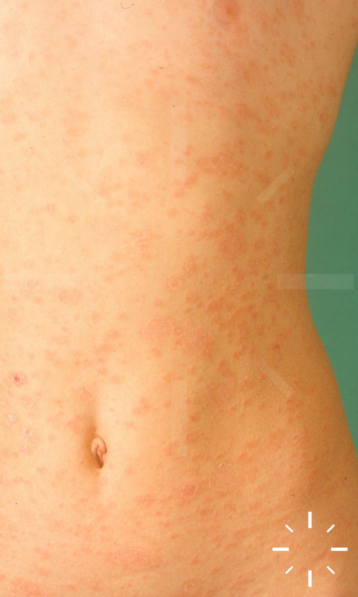
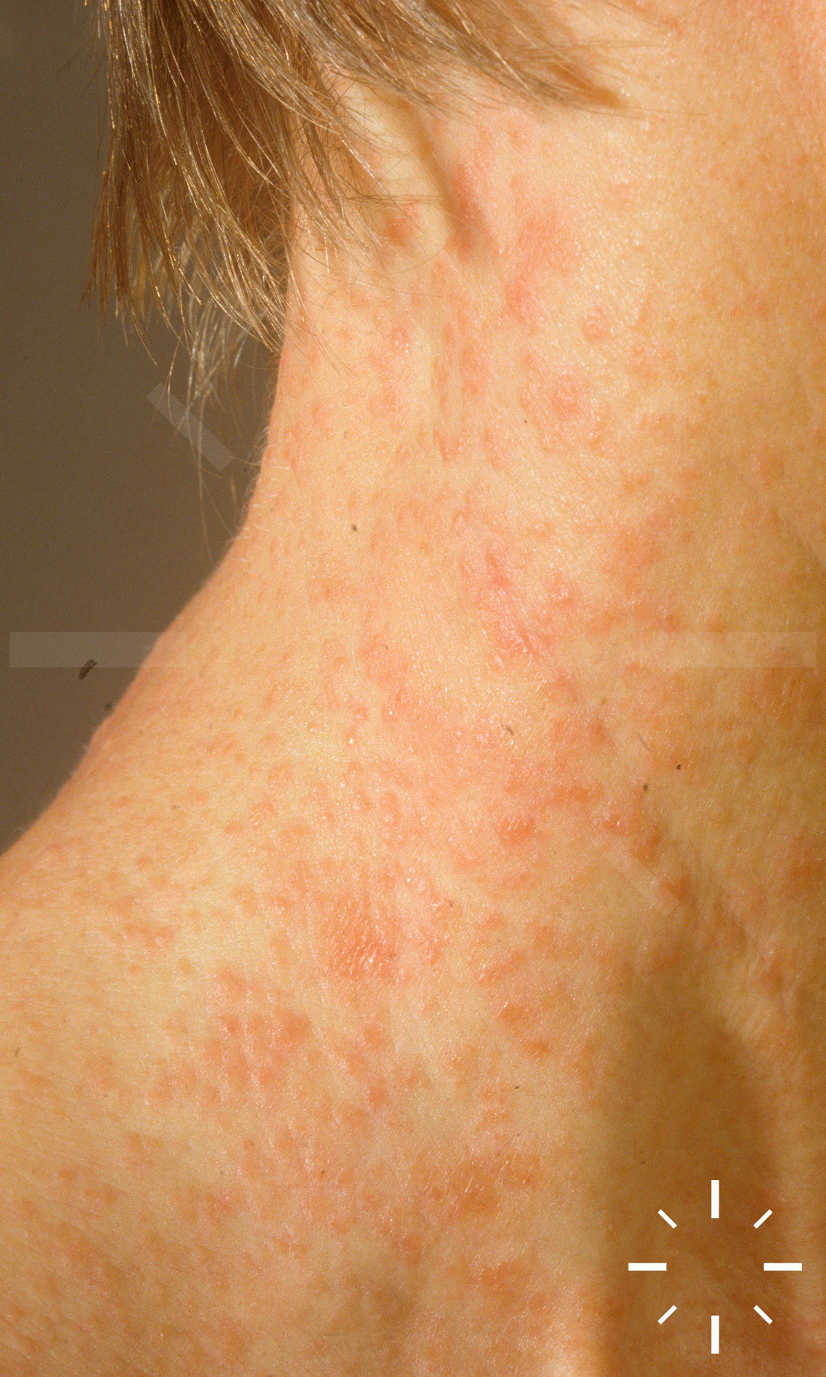
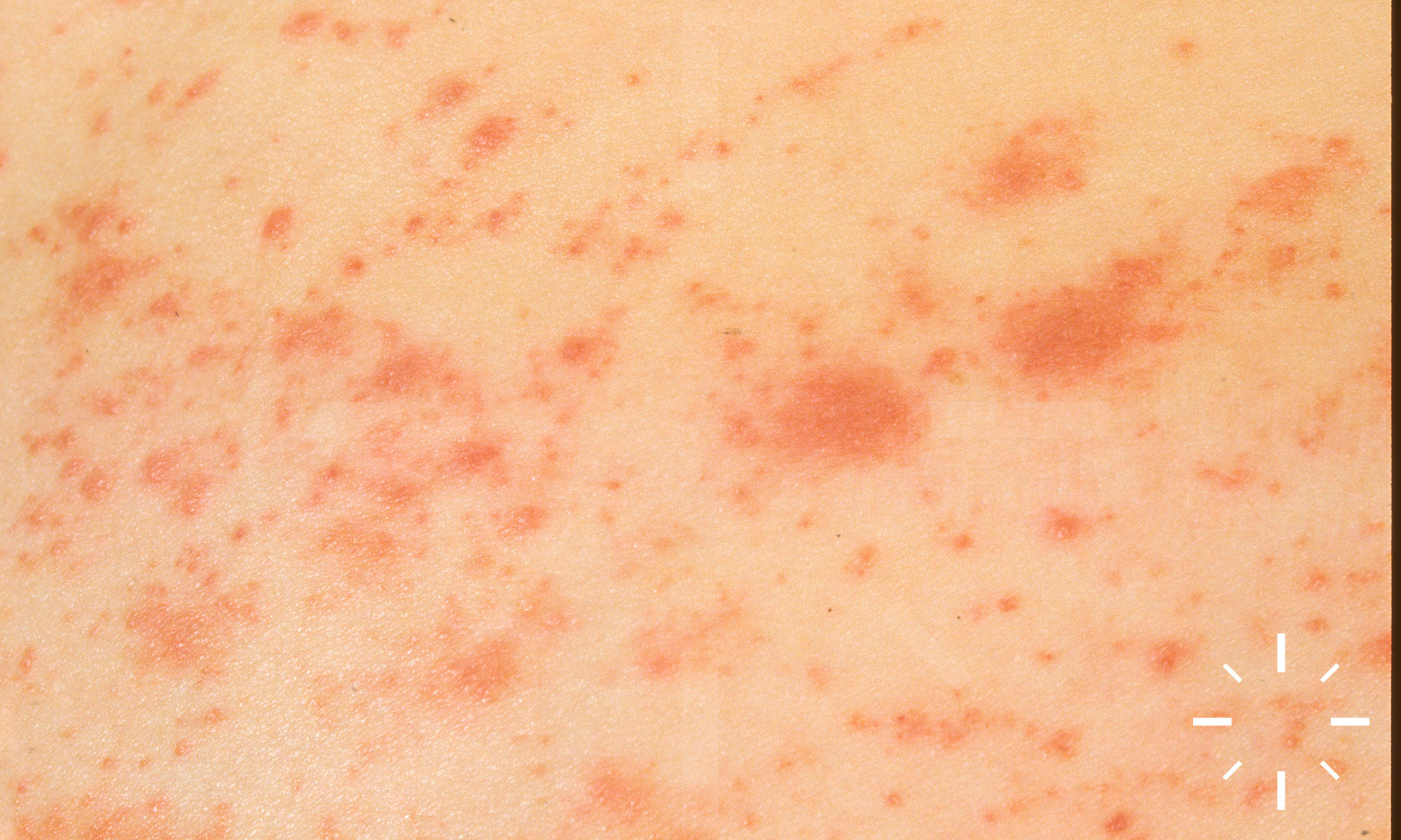
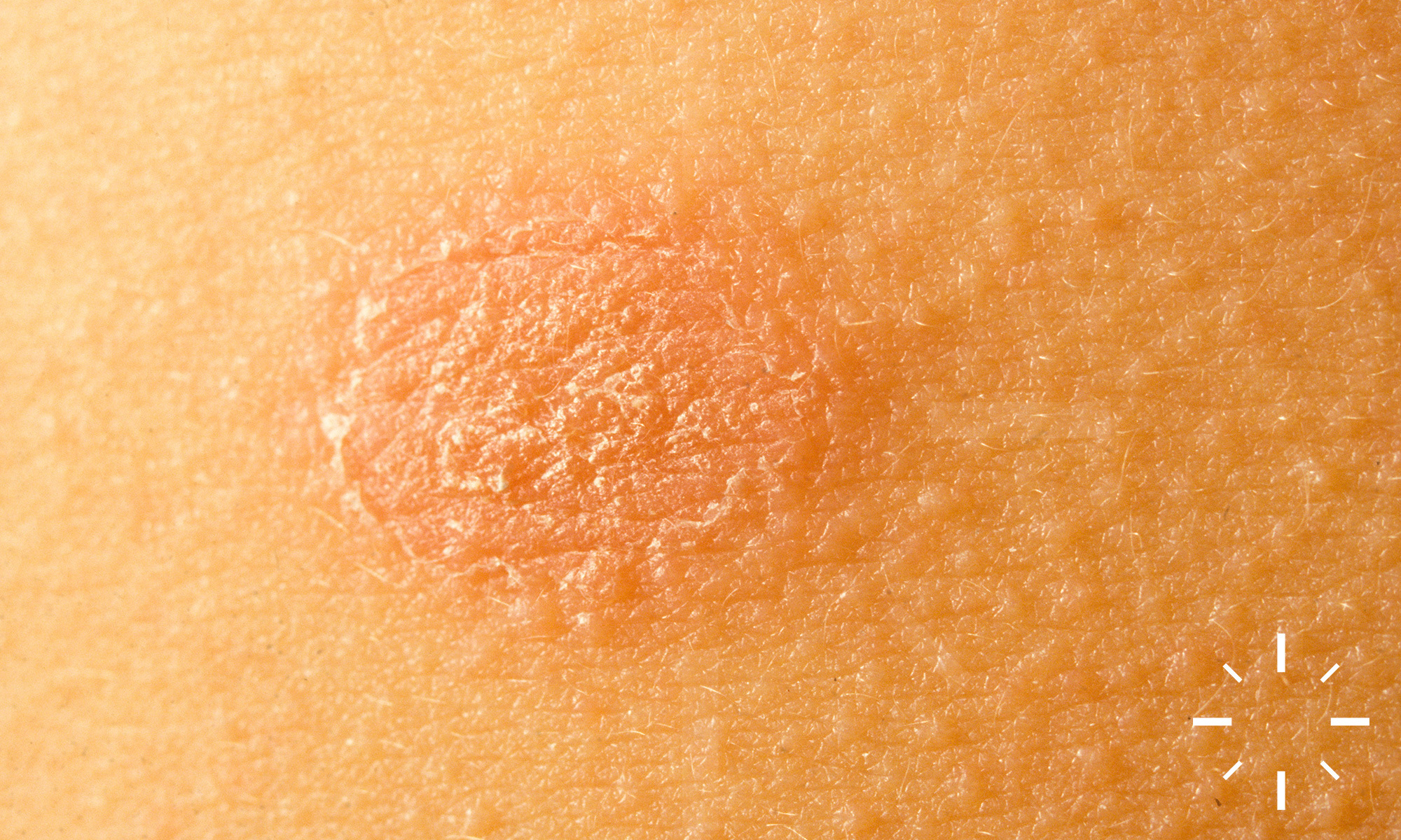
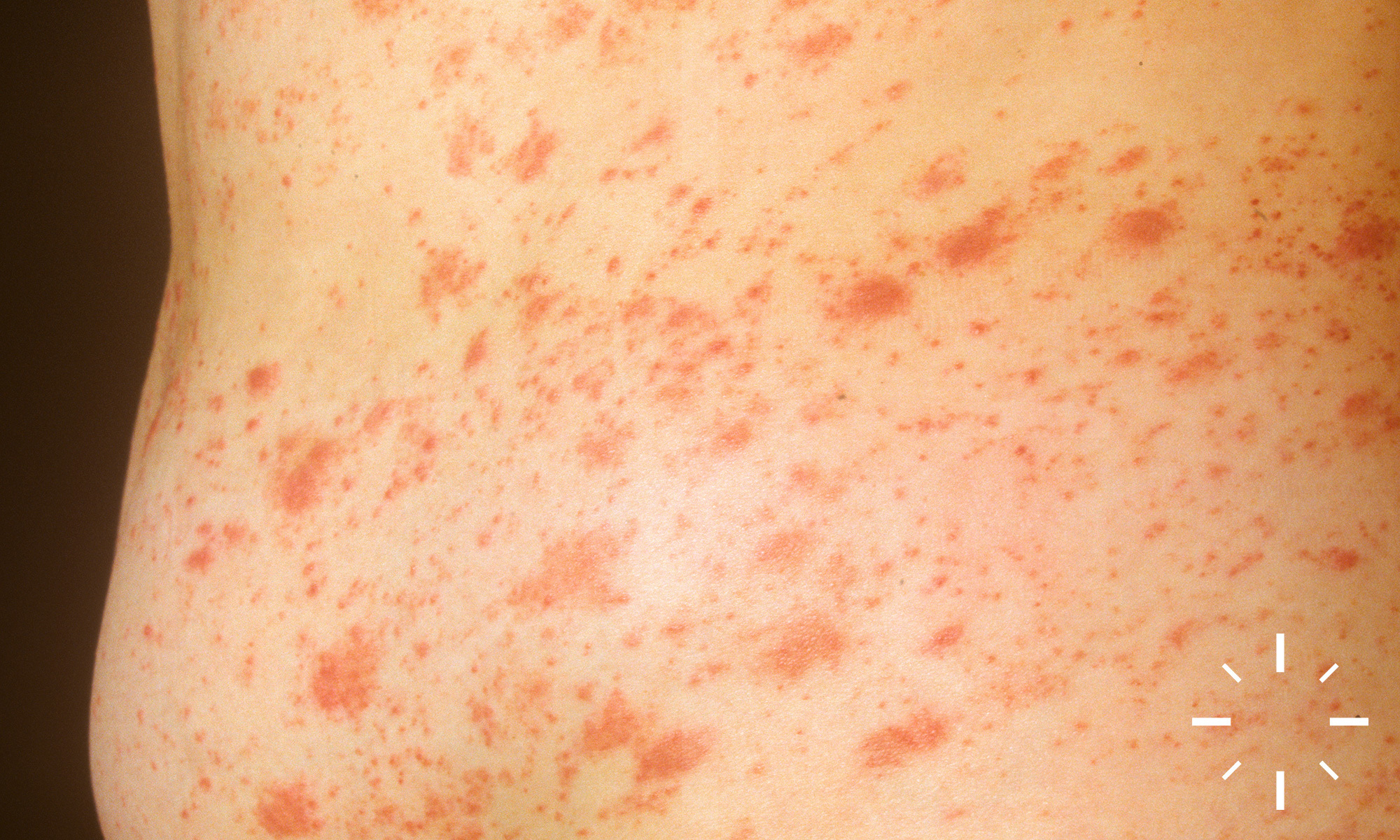
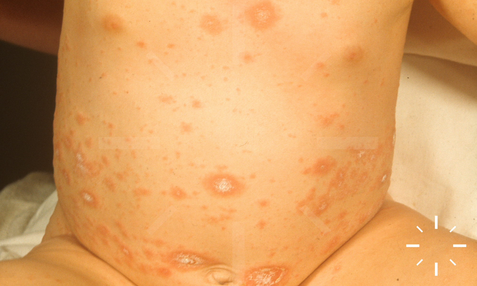
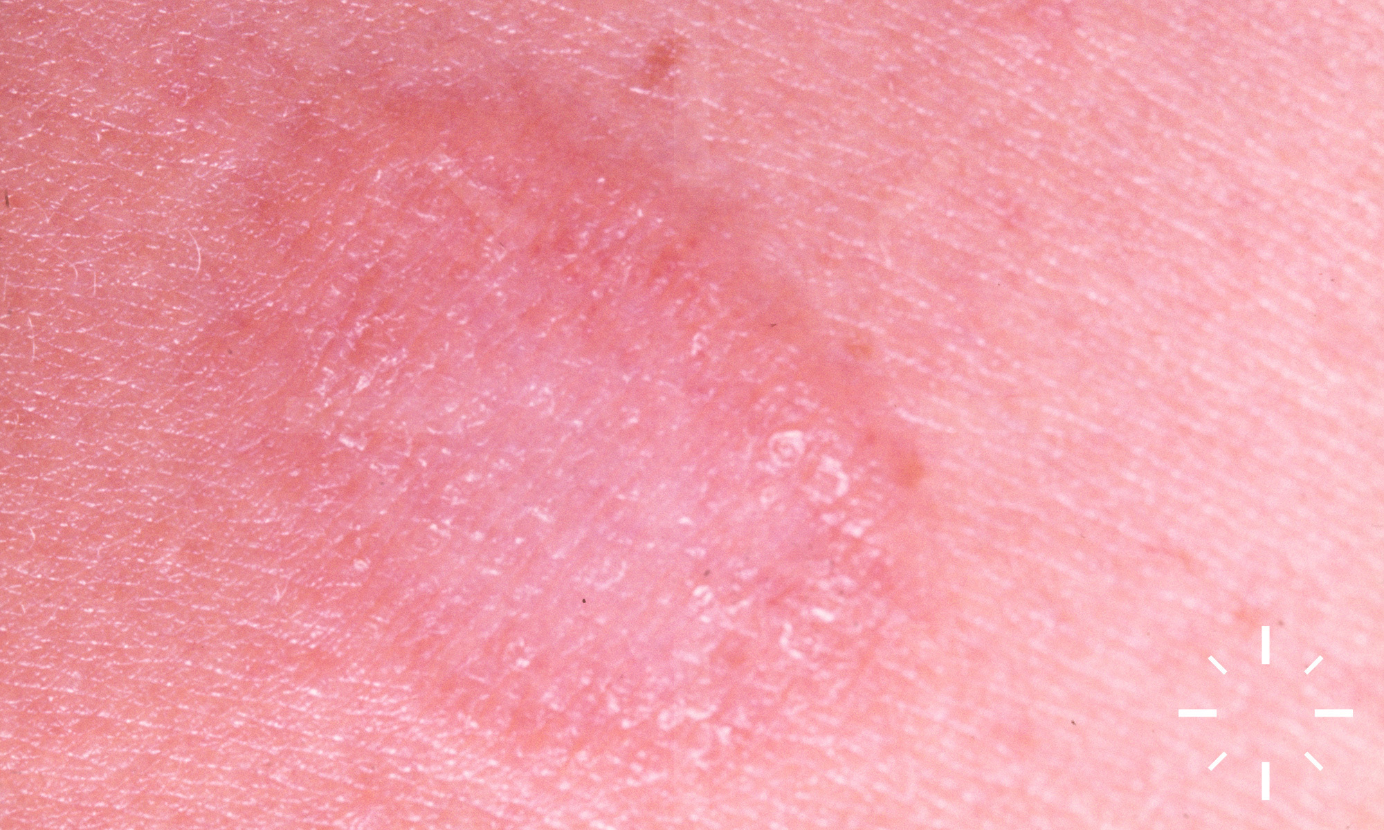
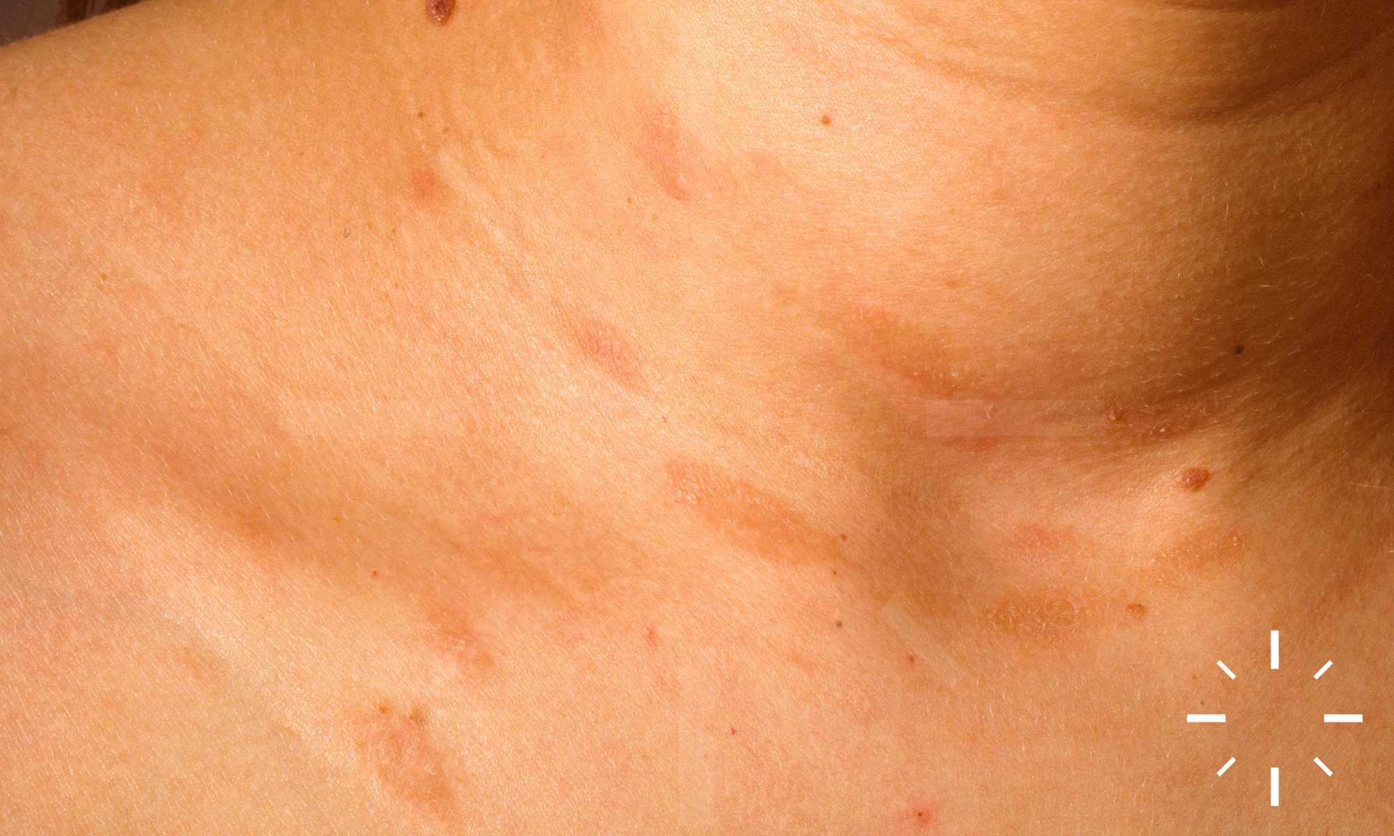
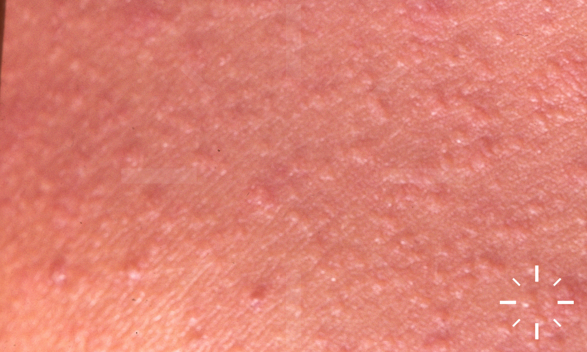
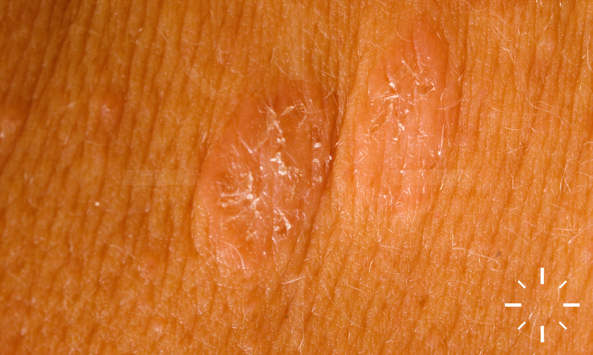
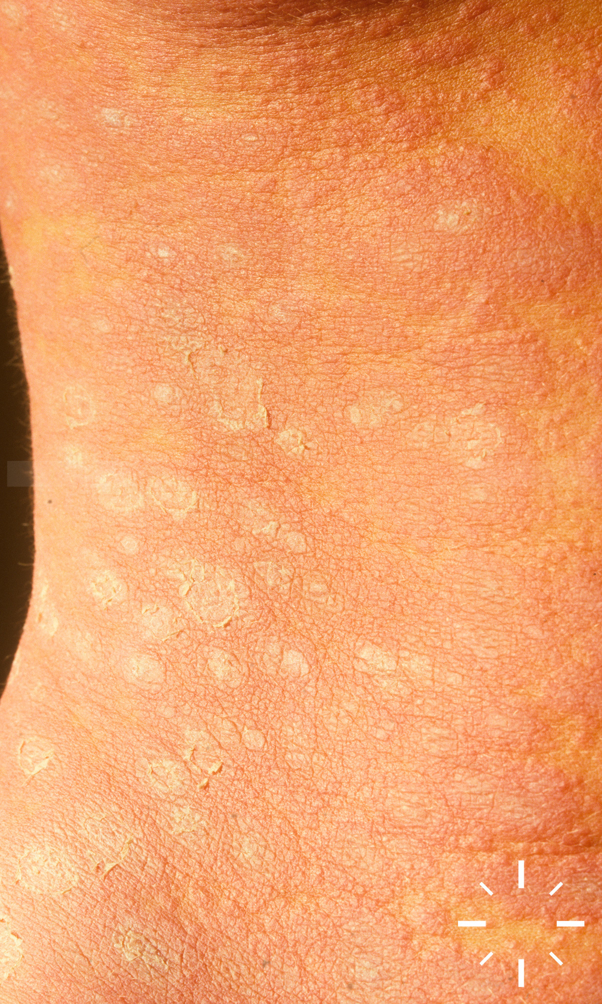
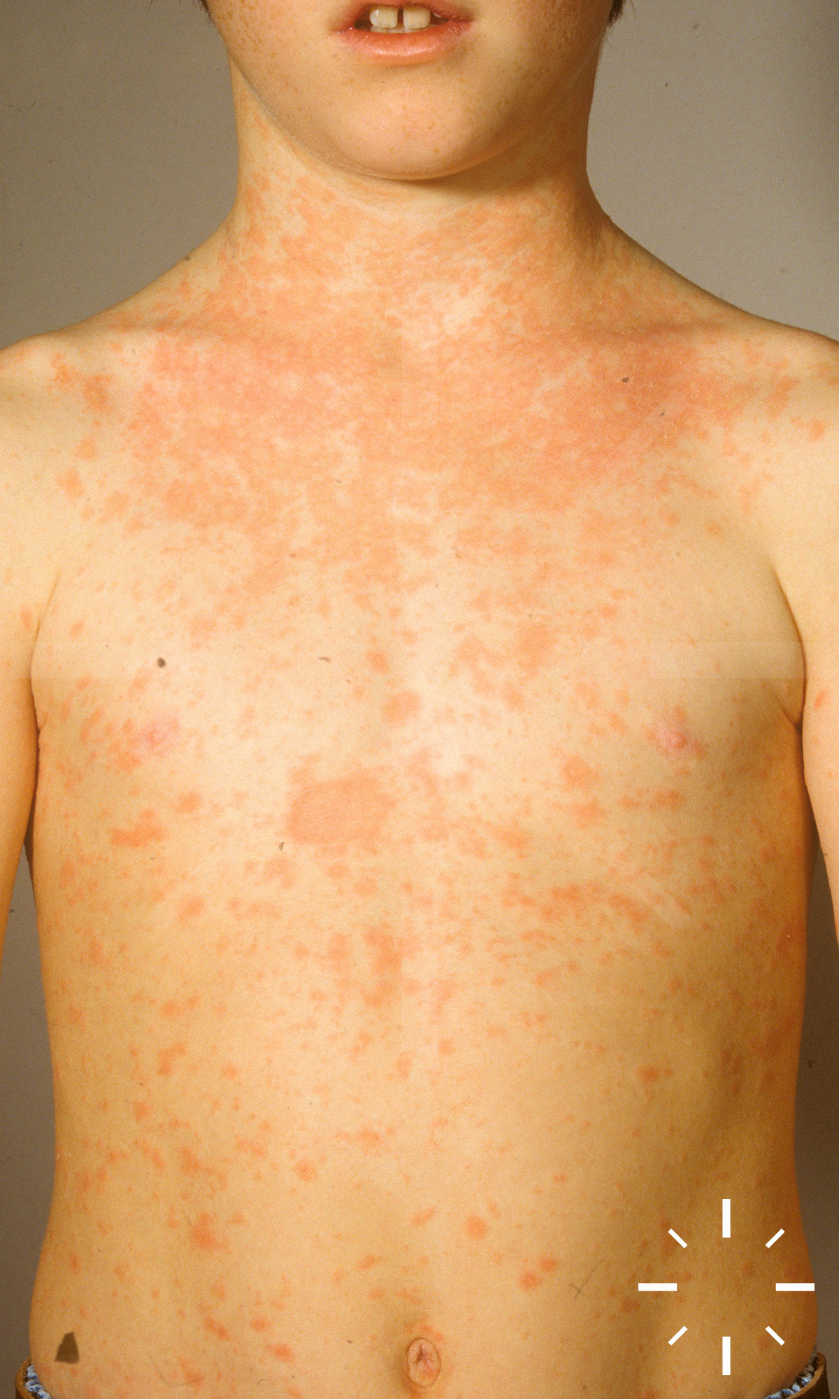
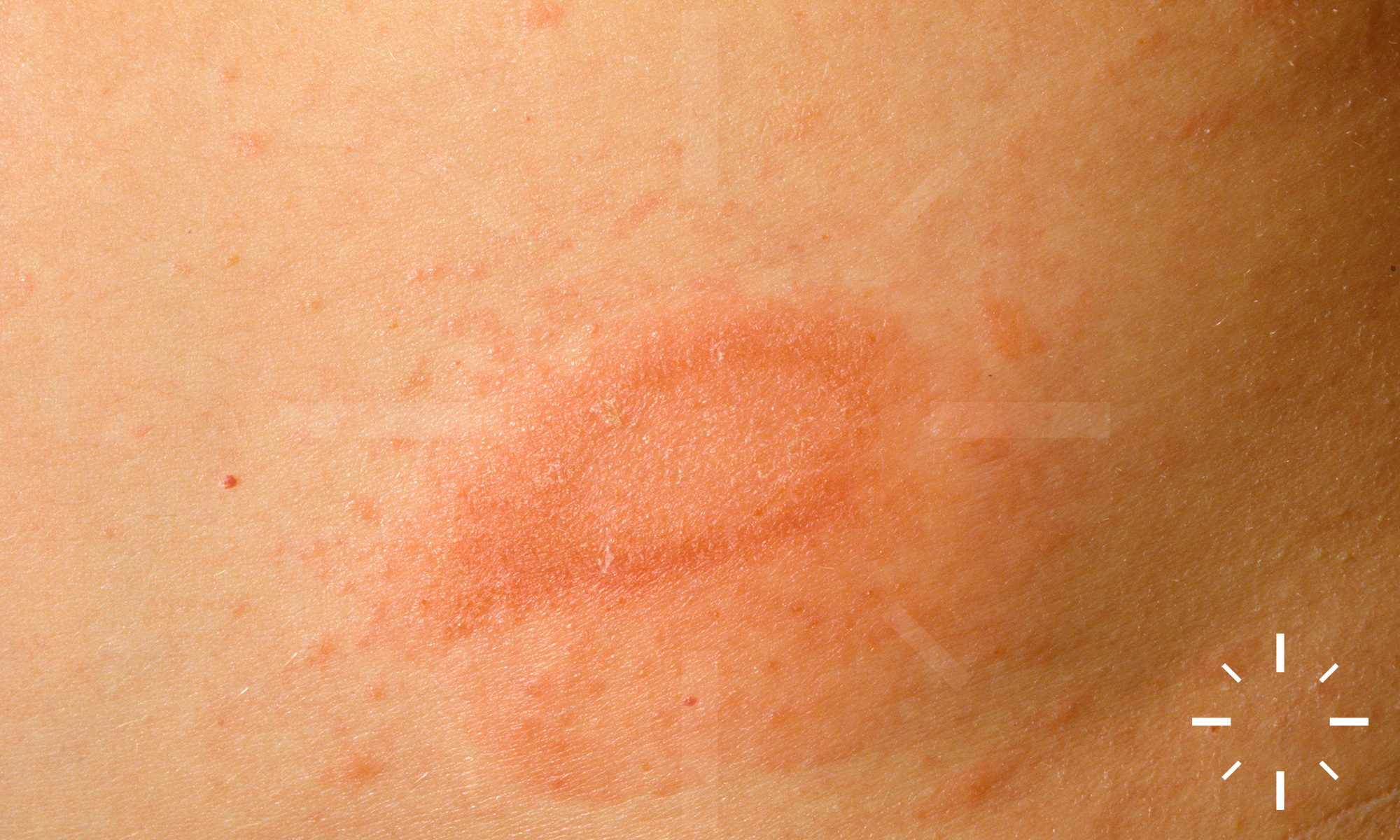
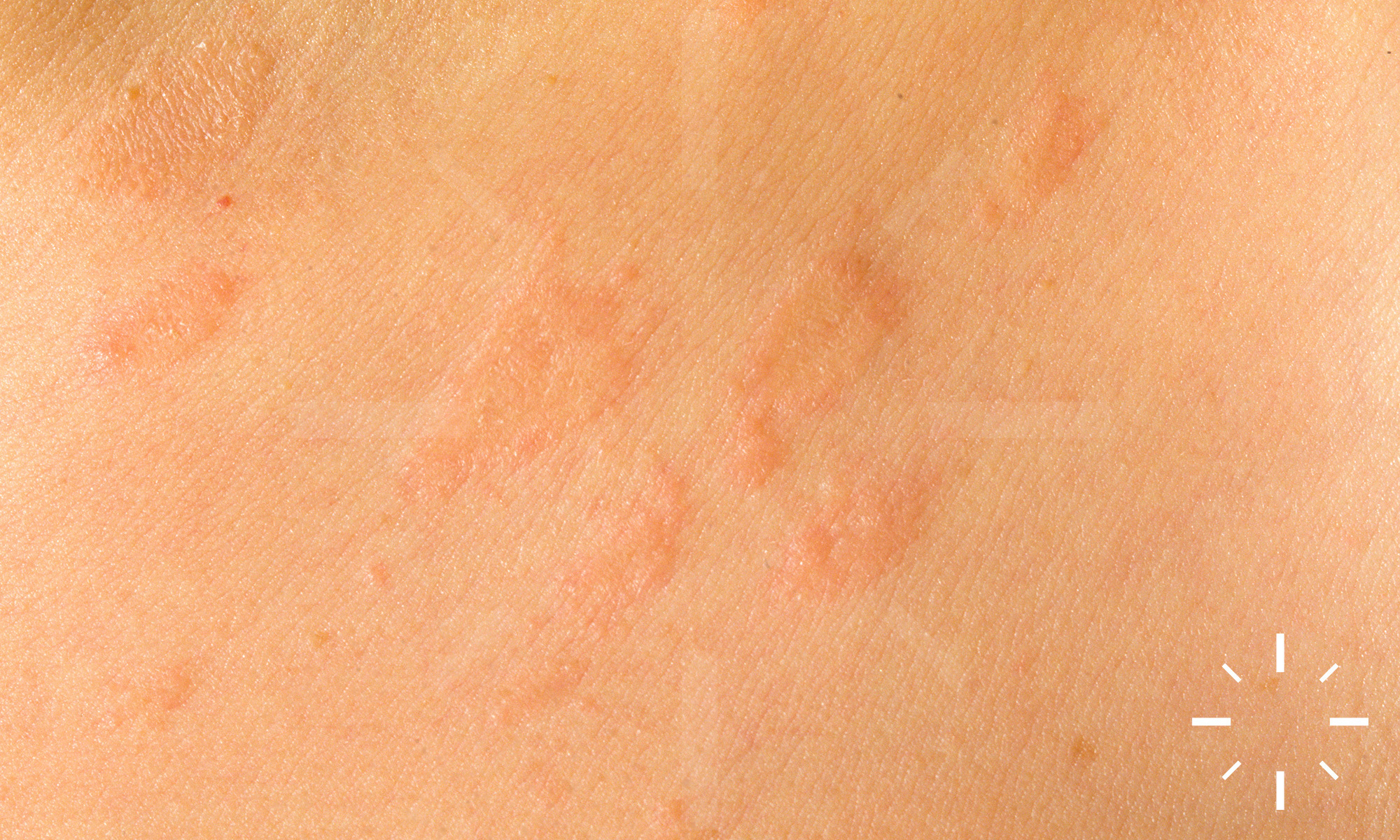
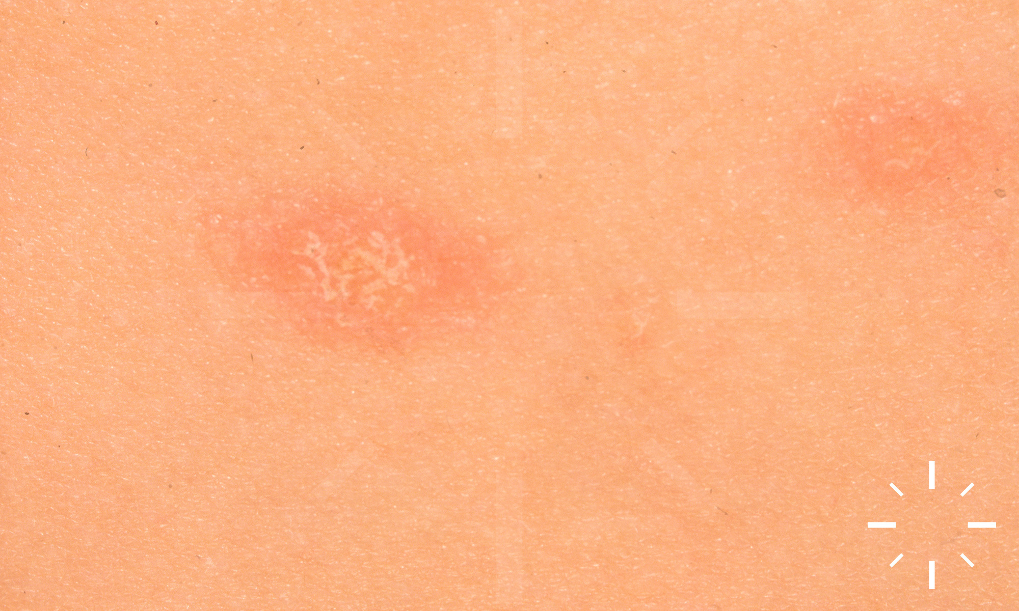
Pityriasis rosea

Last Updated: 2023-07-07

Author(s): Anzengruber F., Navarini A.

ICD11: EA10

Gibert, 1860.

Röschenflechte, Gibert's disease.

Self-limiting, exanthematous, inflammatory skin disease.

  • Age peak: 10-35 years
  • Women > men
  • Occurring in all ethnicities
  • Prevalent mainly in spring and autumn

  • The cause is not yet known
  • Possible triggers:
    • Viral infections (HHV-6, HHV-7, H1N1, EBV, CMV)
    • Drugs
    • Stress
  • Increases in tissue-derived interleukin-17 (IL-17) and -22, interferon-γ, vascular endothelium growth factor (VEGF) and CXCL10 (IP-10) have been shown

  • Prodromi
    • ↓ general condition, nausea, (sub-)febrile temperatures, cephalgia
  • Sharply demarcated, erythematous, oval plaques with collerett-like scaling. A tache mère (sometimes called plaque mère, herald patch or primary medallion) manifests first. This is usually located on the trunk and is larger than all the others
  • As it progresses, the skin lesions spread along the cleavage lines (Christmas tree pattern)
  • The face and distal halves of the extremities are usually left out. The oral mucosa (enanthema) is involved only in exceptional cases
  • Special forms:
    • Pityriasis rosea inversa: It mainly affects the axillae and the groin region. An inverse form may be seen in children or African-Americans
    • The skin lesions may also be atypical: urticarial, haemorrhagic, follicular bound, vesicular, psoriasiform and circine

  • Anamnesis (previous infections?)
  • Clinical image (search for plaque mère)

Discreet focal spongiosis and parakeratosis are visible in the epidermis. Erythrocyte extravasations are typical but rare. A perivascular lymphocytic infiltrate is seen in the dermis.

  • Healing after 3-8 weeks
  • Disease can lead to abortion or premature birth in pregnant women (up to 15 weeks gestation)

  1. Pityriasis rosea, in SpringerReference. Springer Science + Business Media.
  2. Chuang, T.-Y.I., et al., Pityriasis rosea in Rochester, Minnesota, 1969 to 1978. Journal of the American Academy of Dermatology, 1982. 7(1): p. 80-89.
  3. Chuang, T.-Y., et al., Recent upper respiratory tract infection and pityriasis rosea: a case-control study of 249 matched pairs. Br J Dermatol, 1983. 108(5): p. 587-591.
  4. Drago, F., et al., Human Herpesvirus 7 in Patients with Pityriasis rosea. Dermatology, 1997. 195(4): p. 374-378.
  5. Drago, F., et al., Human herpesvirus 7 in pityriasis rosea. The Lancet, 1997. 349(9062): p. 1367-1368.
  6. Kempf, W., et al., Pityriasis Rosea Is Not Associated With Human Herpesvirus 7. Arch Dermatol, 1999. 135(9).
  7. Kempf, W. and G. Burg, Pityriasis rosea - a virus-induced skin disease? An update. Archives of Virology, 2000. 145(8): p. 1509-1520.
  8. Watanabe, T., et al., Pityriasis Rosea is Associated with Systemic Active Infection with Both Human Herpesvirus-7 and Human Herpesvirus-6. Journal of Investigative Dermatology, 2002. 119(4): p. 793-797.
  9. Chuh, A.A.T. and H.H.L. Chan, Effect on quality of life in patients with pityriasis rosea: Is it associated with rash severity? International Journal of Dermatology, 2004. 0(0): p. 060720080827124.
  10. Broccolo, F., et al., Additional Evidence that Pityriasis Rosea Is Associated with Reactivation of Human Herpesvirus-6 and -7. Journal of Investigative Dermatology, 2005. 124(6): p. 1234-1240.
  11. Drago, F., F. Vecchio, and A. Rebora, Use of high-dose acyclovir in pityriasis rosea. Journal of the American Academy of Dermatology, 2006. 54(1): p. 82-85.
  12. Drago, F., et al., Pregnancy outcome in patients with pityriasis rosea. Journal of the American Academy of Dermatology, 2008. 58(5): p. S78-S83.
  13. Drago, F., F. Broccolo, and A. Rebora, Pityriasis rosea: An update with a critical appraisal of its possible herpesviral etiology. Journal of the American Academy of Dermatology, 2009. 61(2): p. 303-318.
  14. Rassai, S., et al., Low dose of acyclovir may be an effective treatment against pityriasis rosea: a random investigator-blind clinical trial on 64 patients. Journal of the European Academy of Dermatology and Venereology, 2010. 25(1): p. 24-26