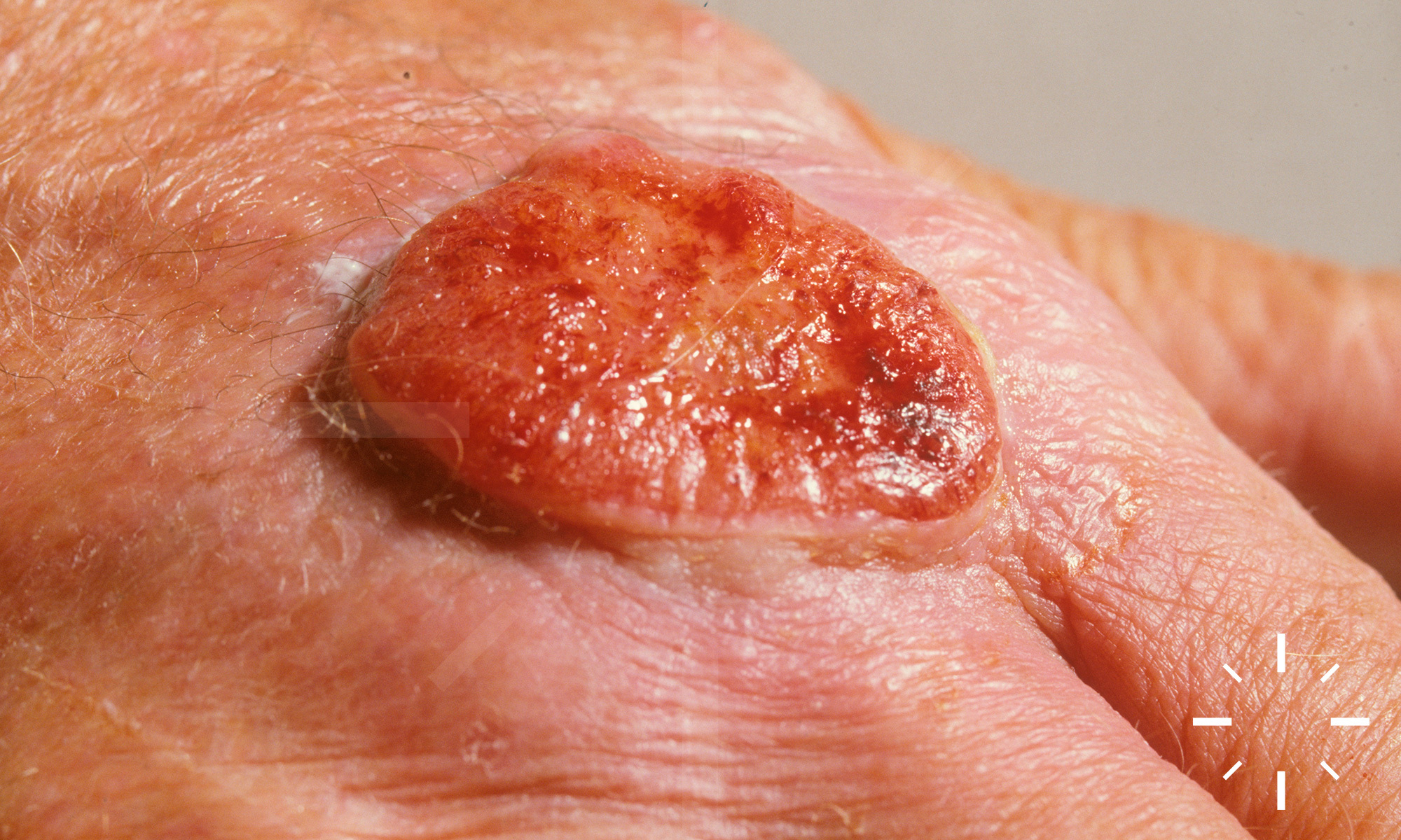
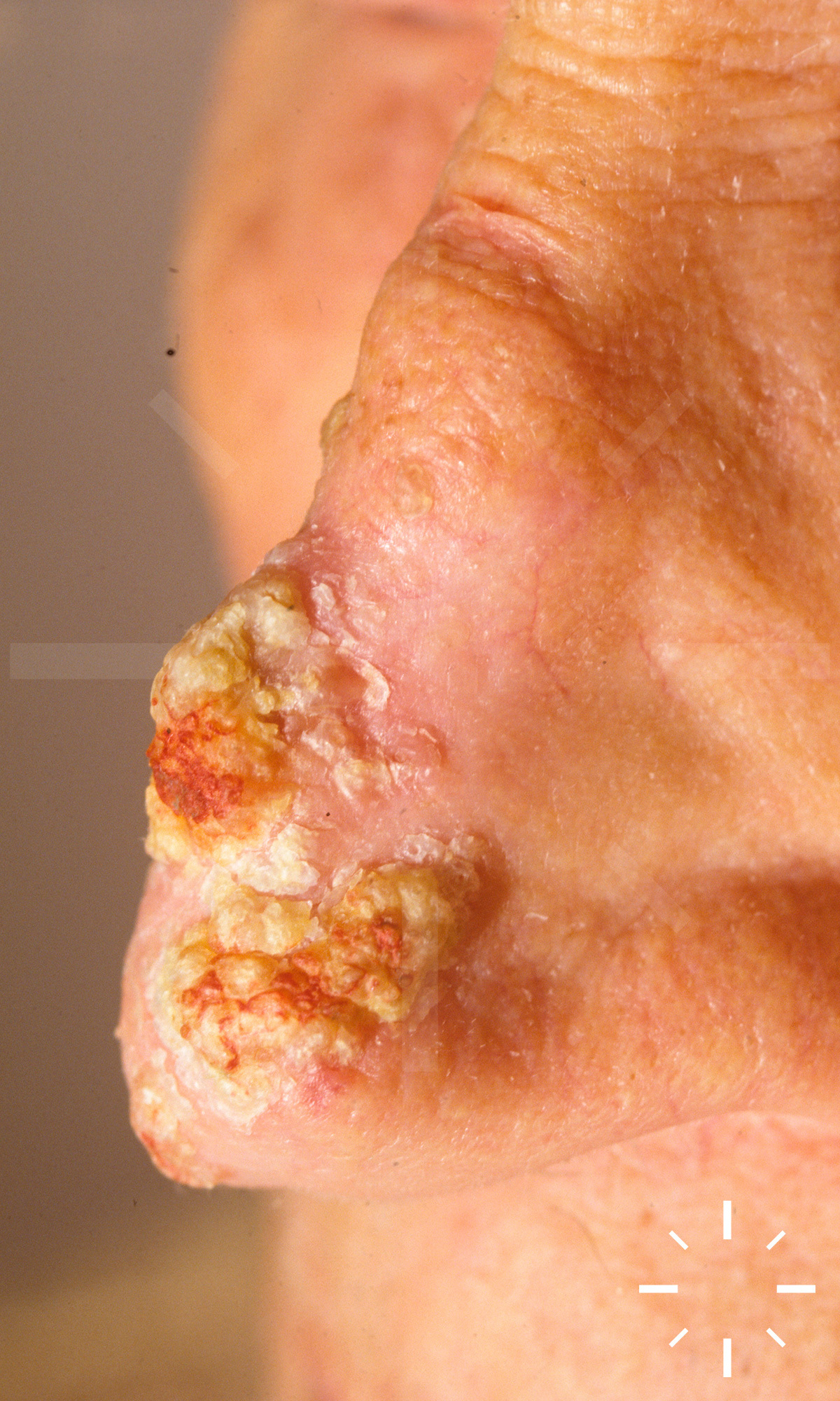
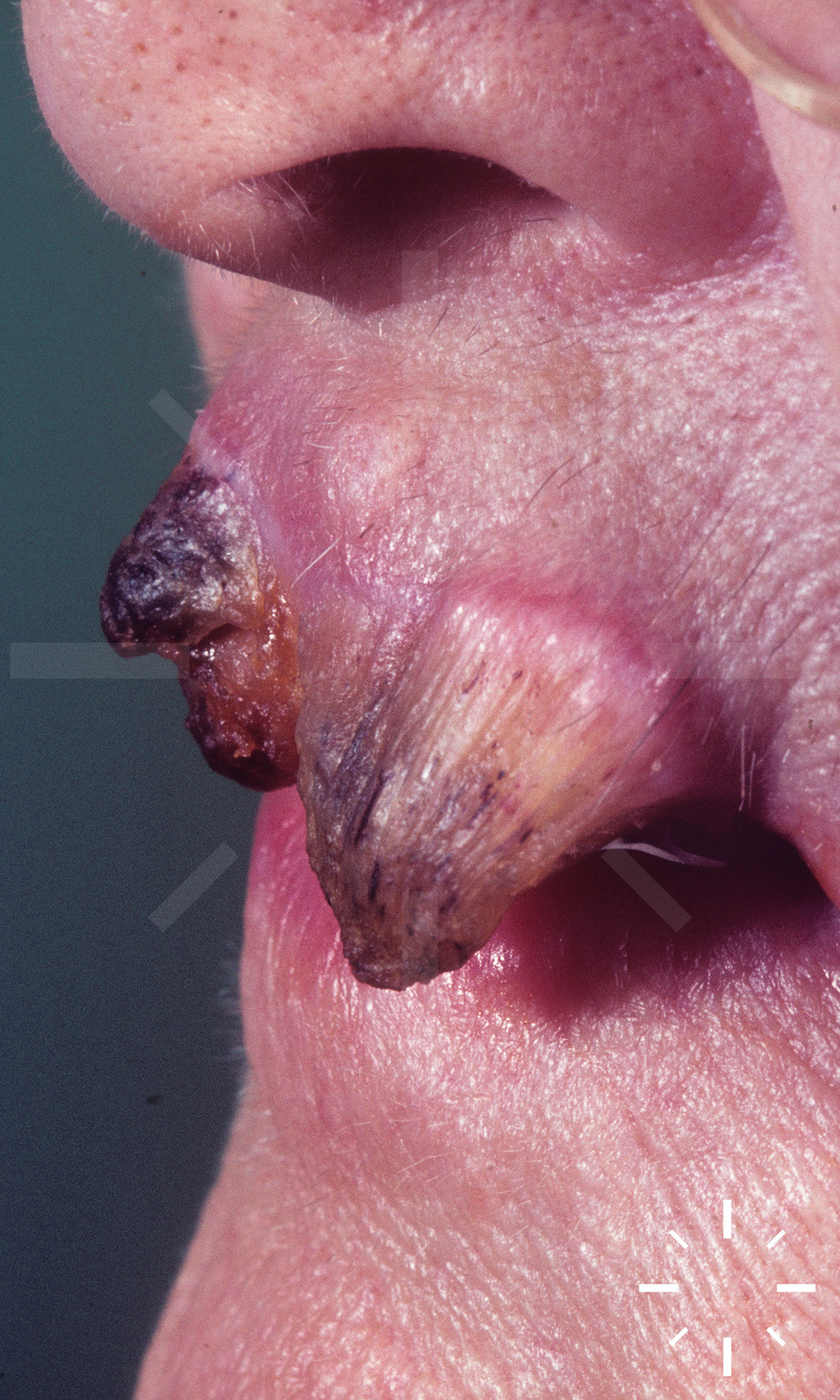
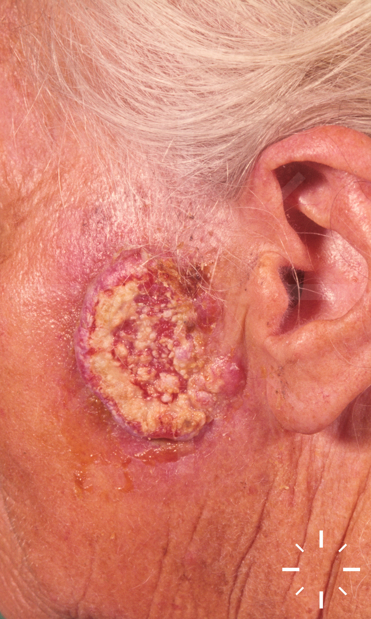
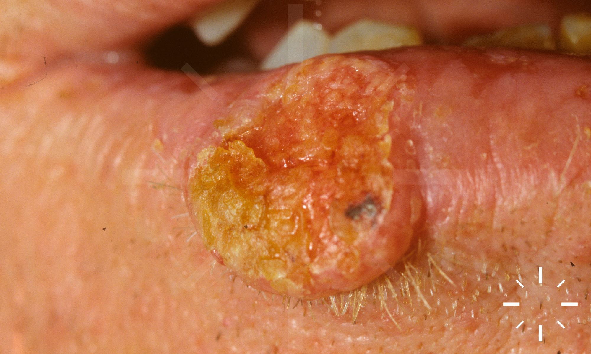
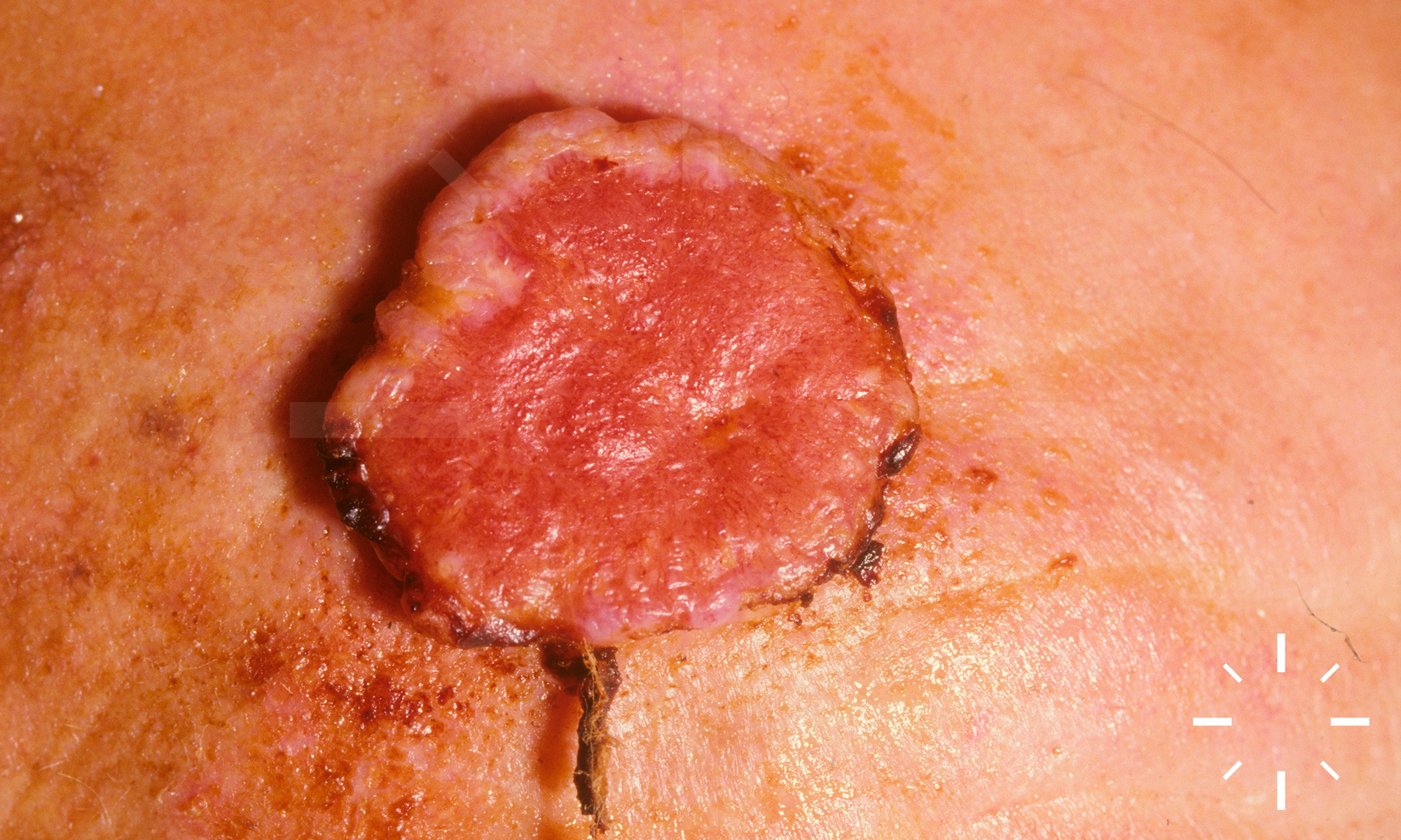
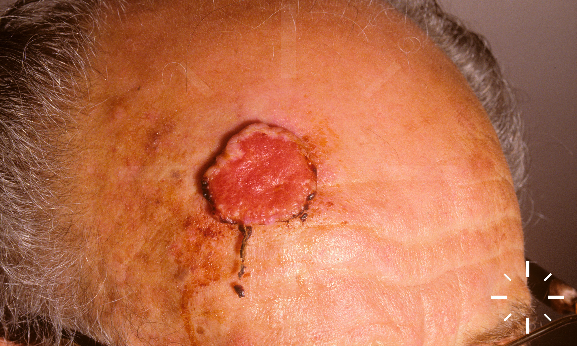
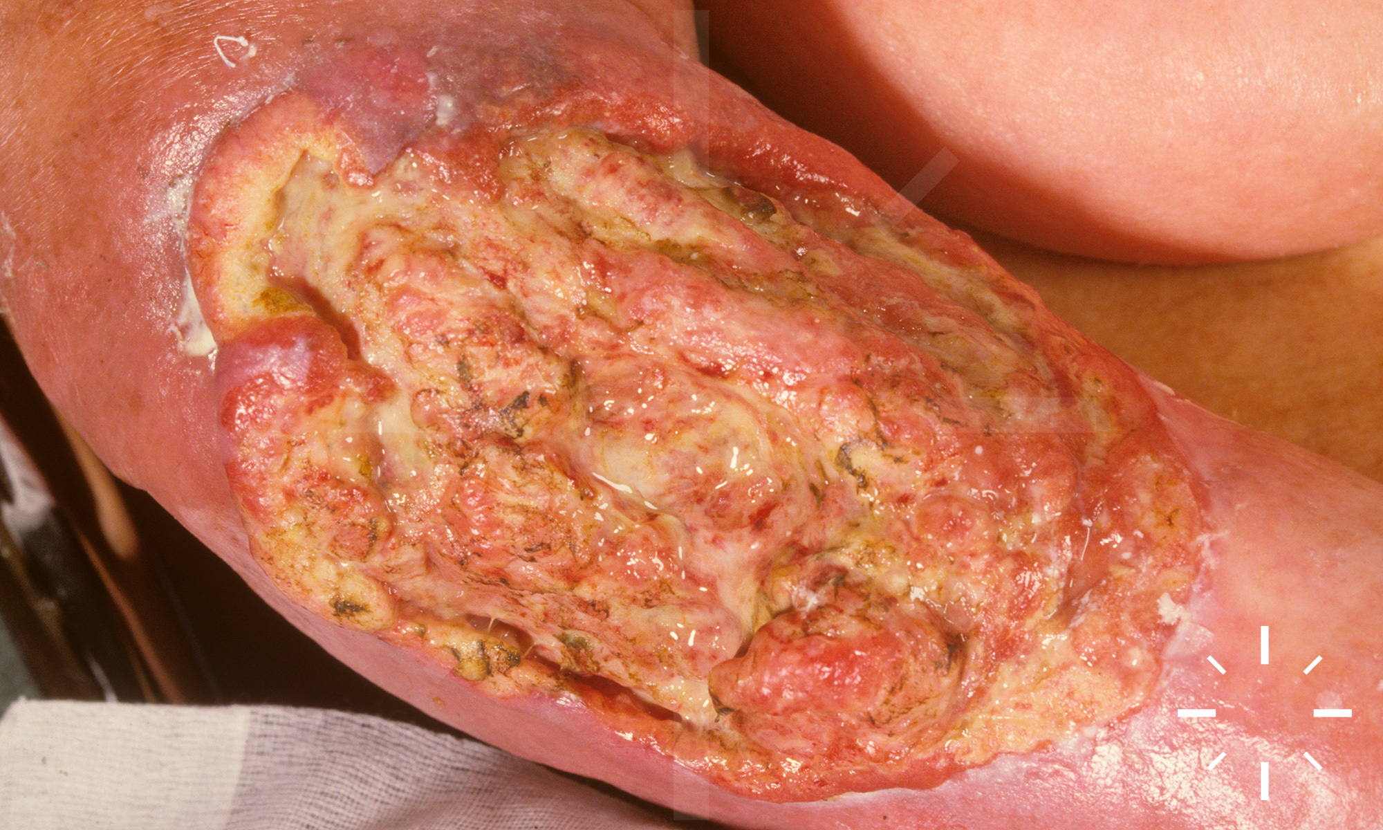
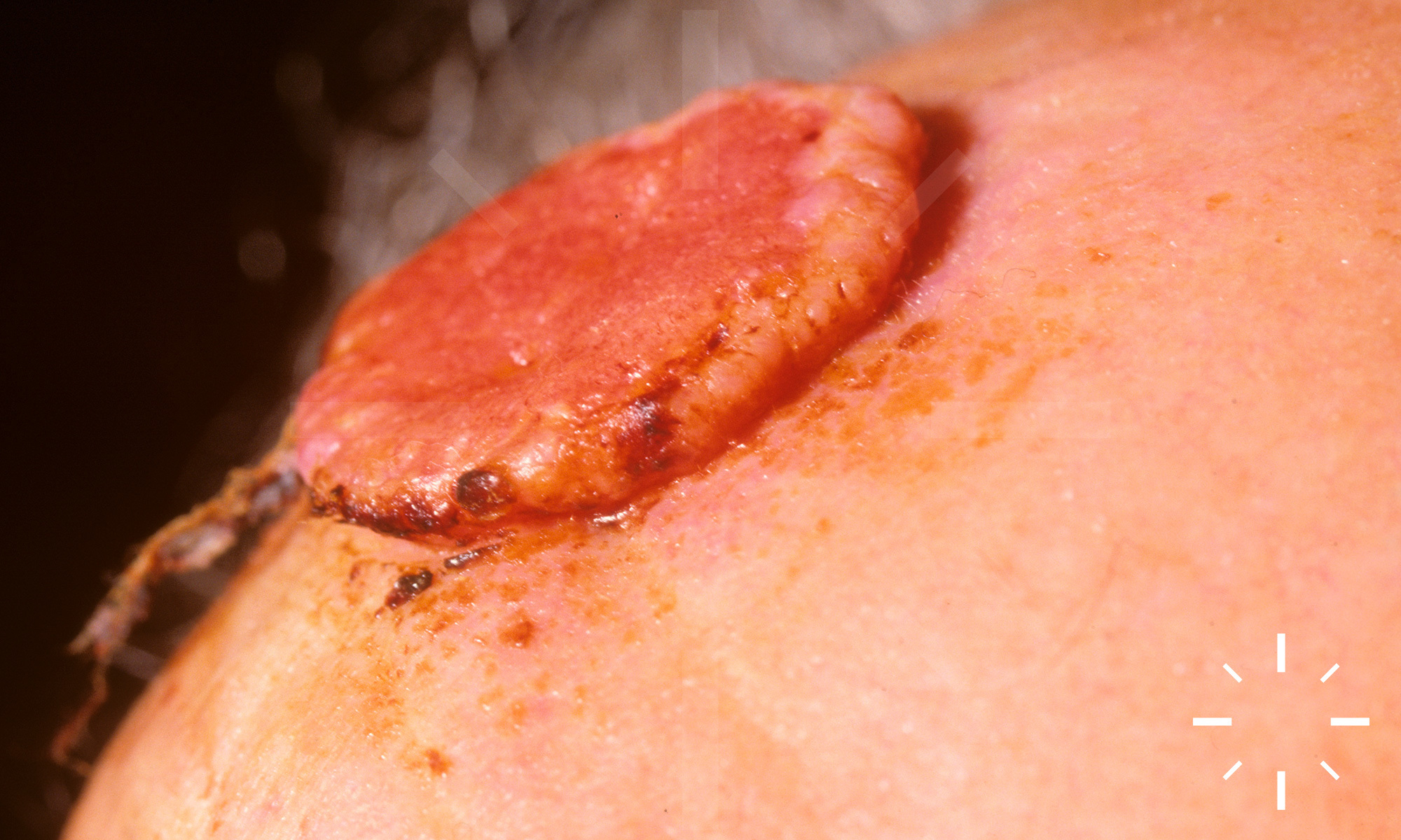
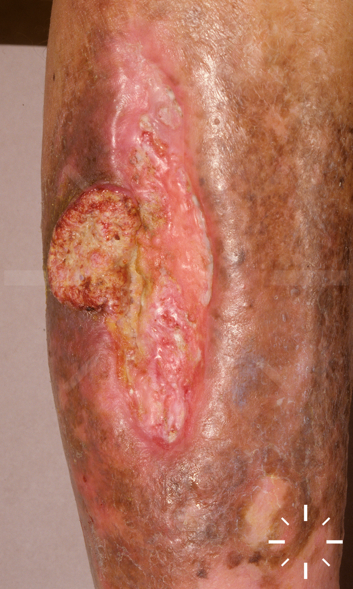
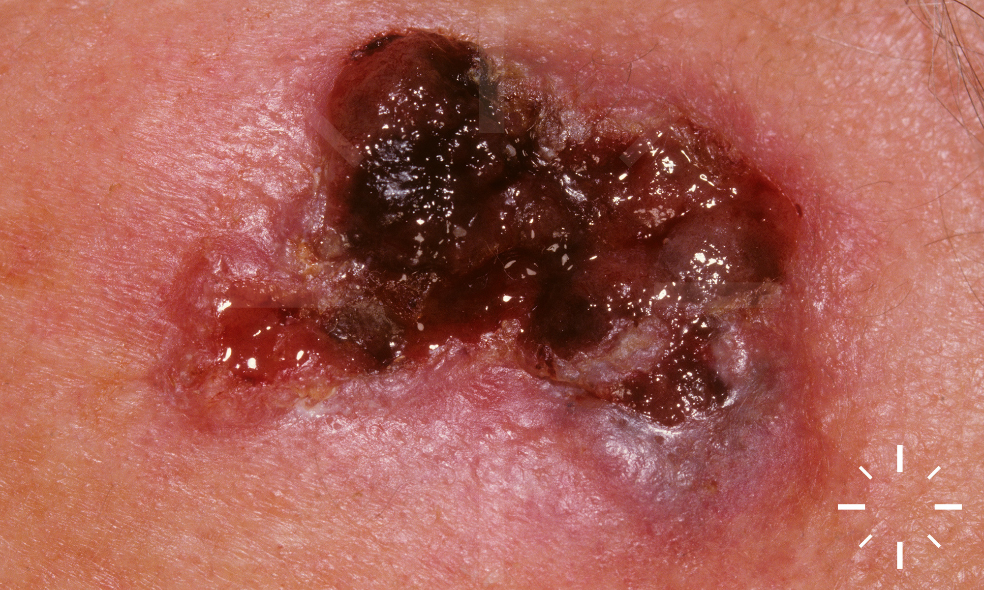
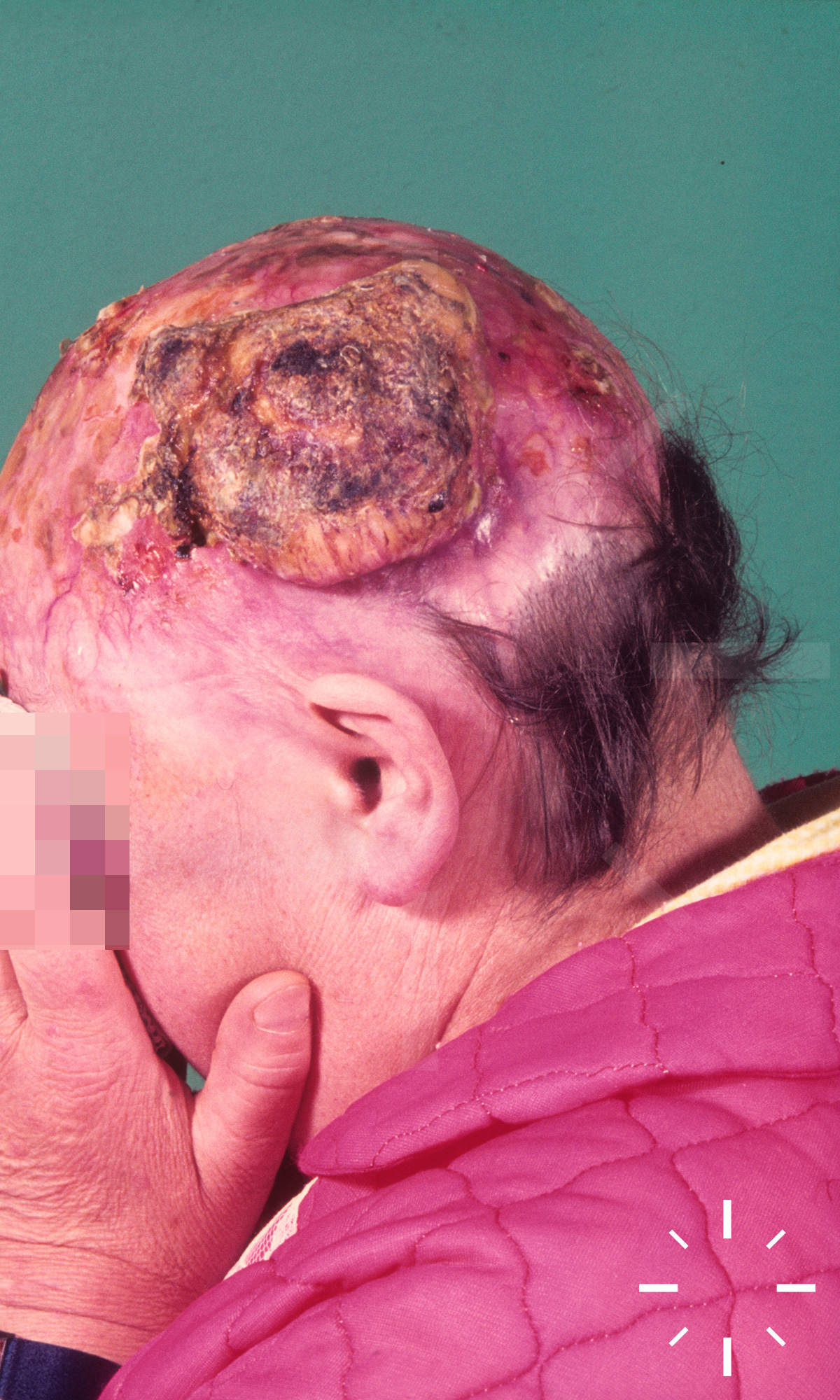
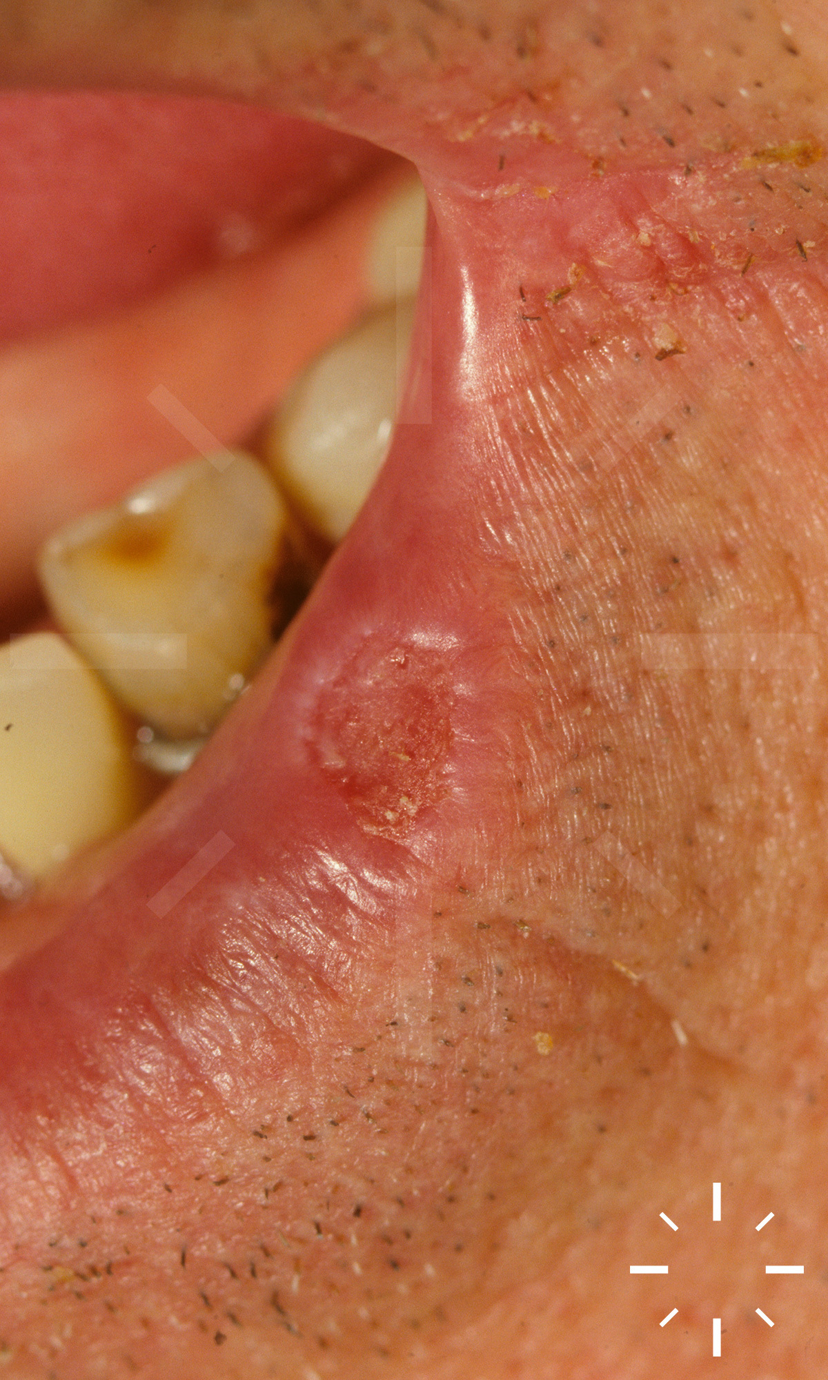
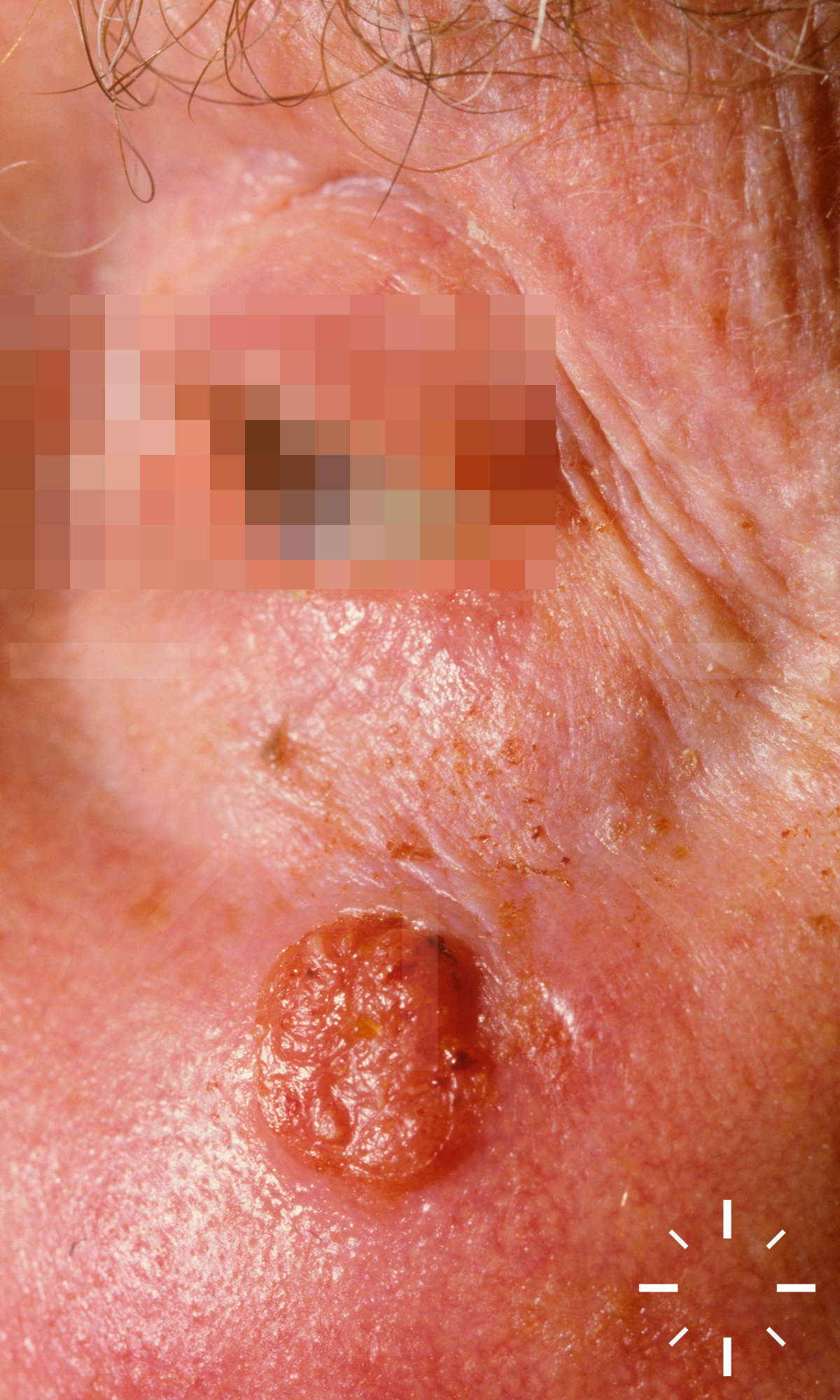
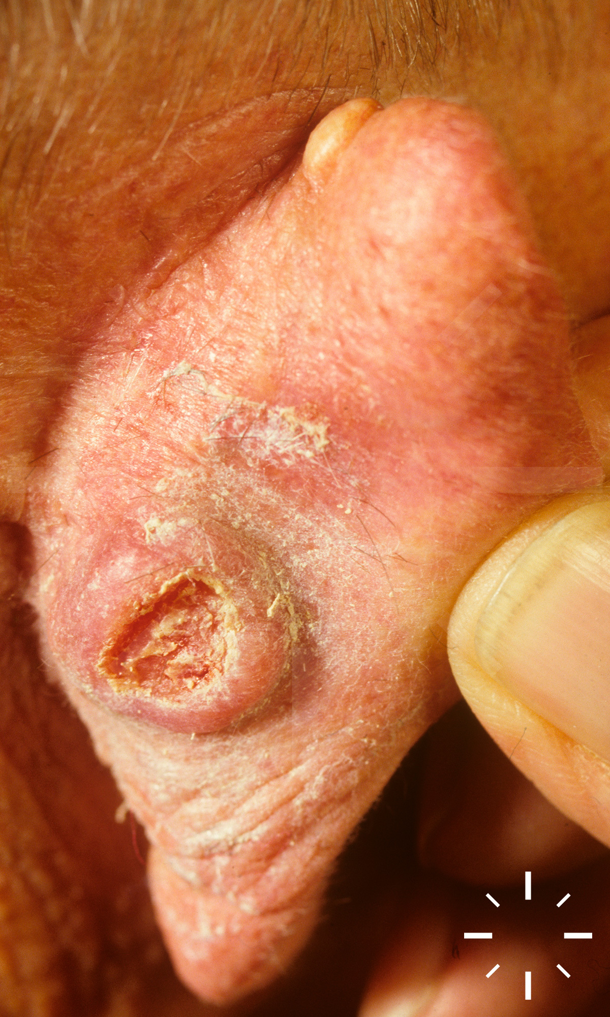
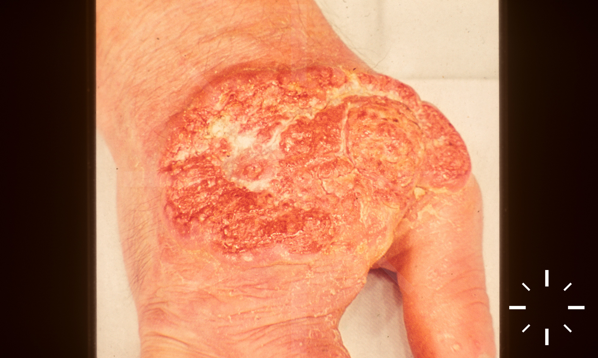
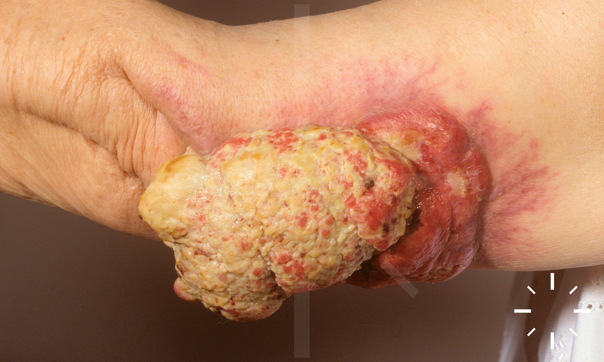
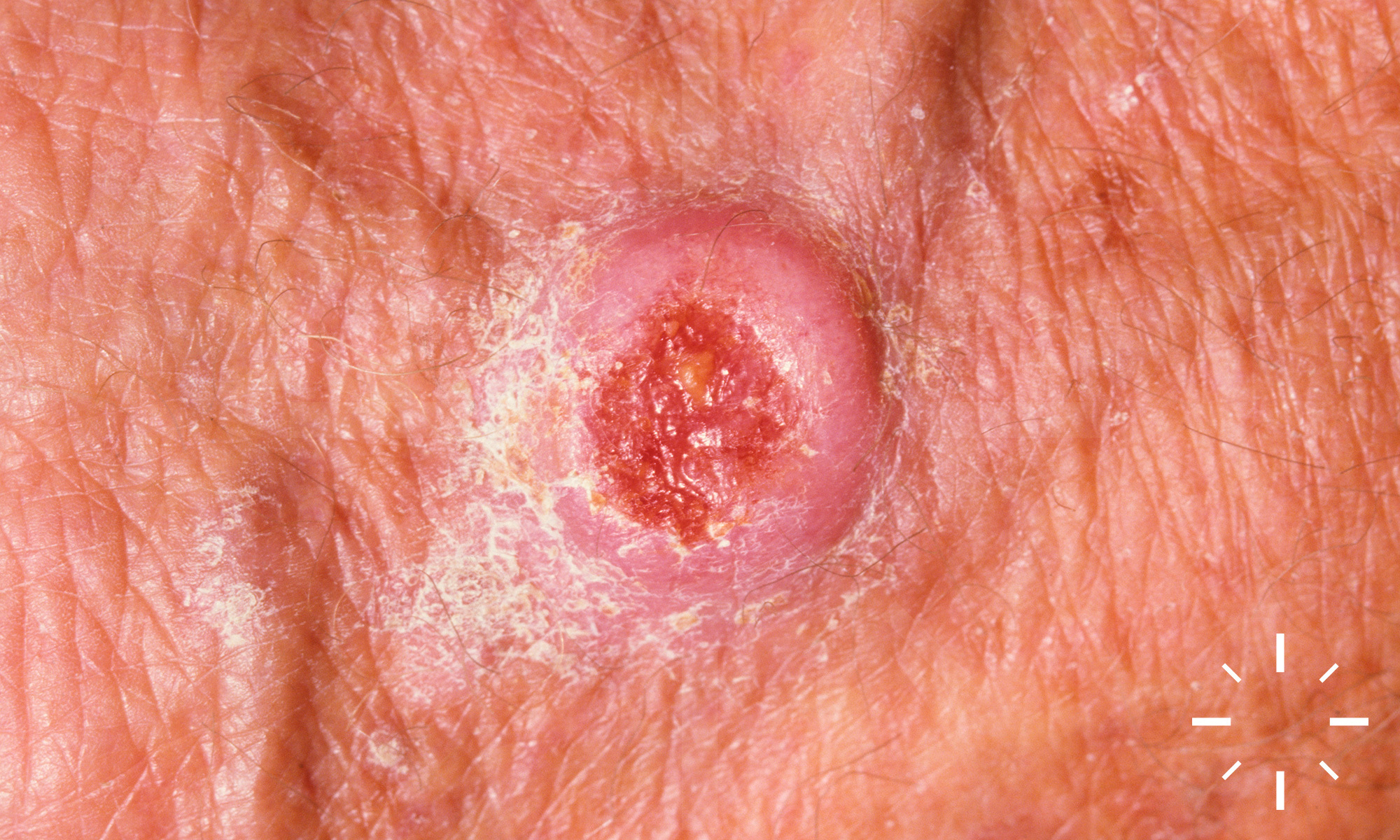
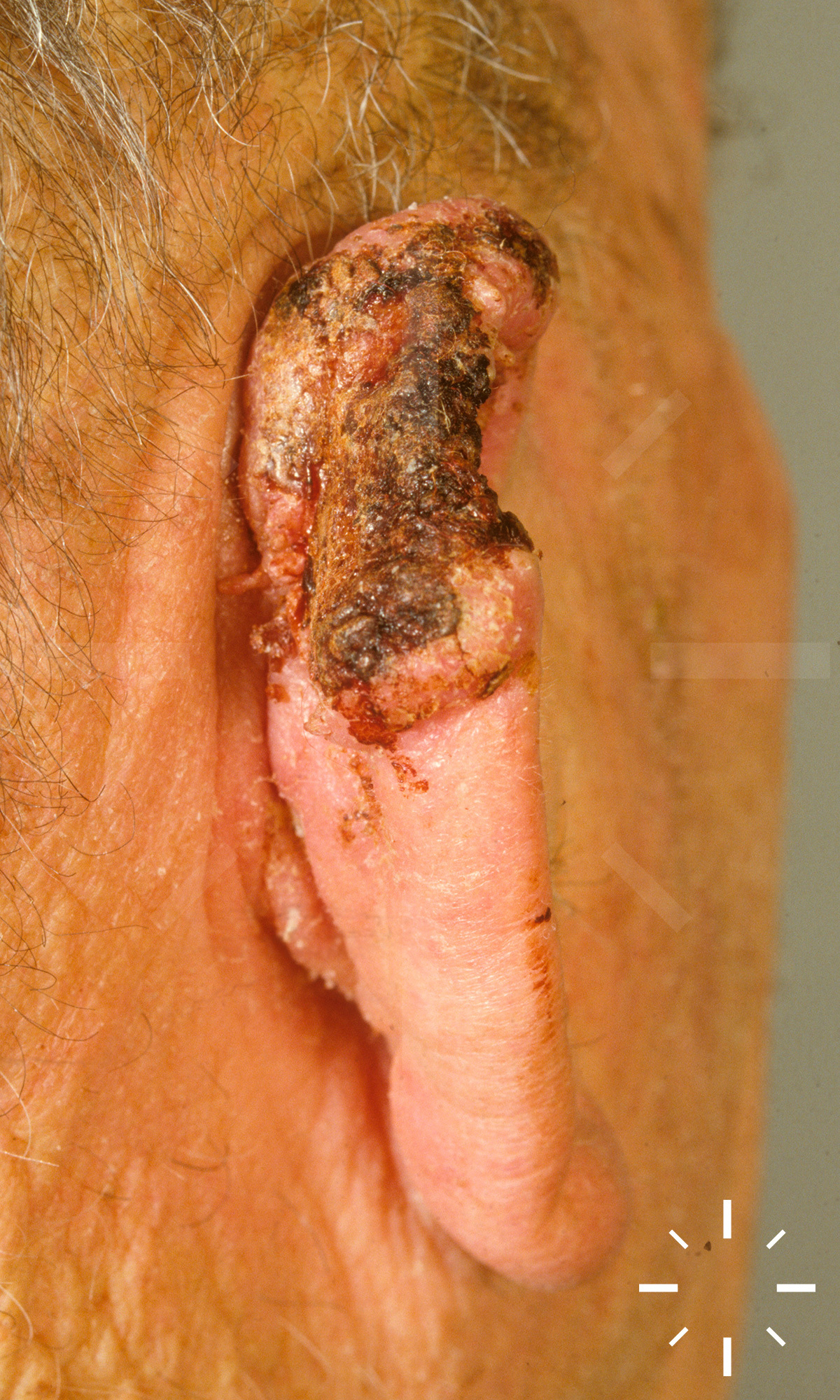
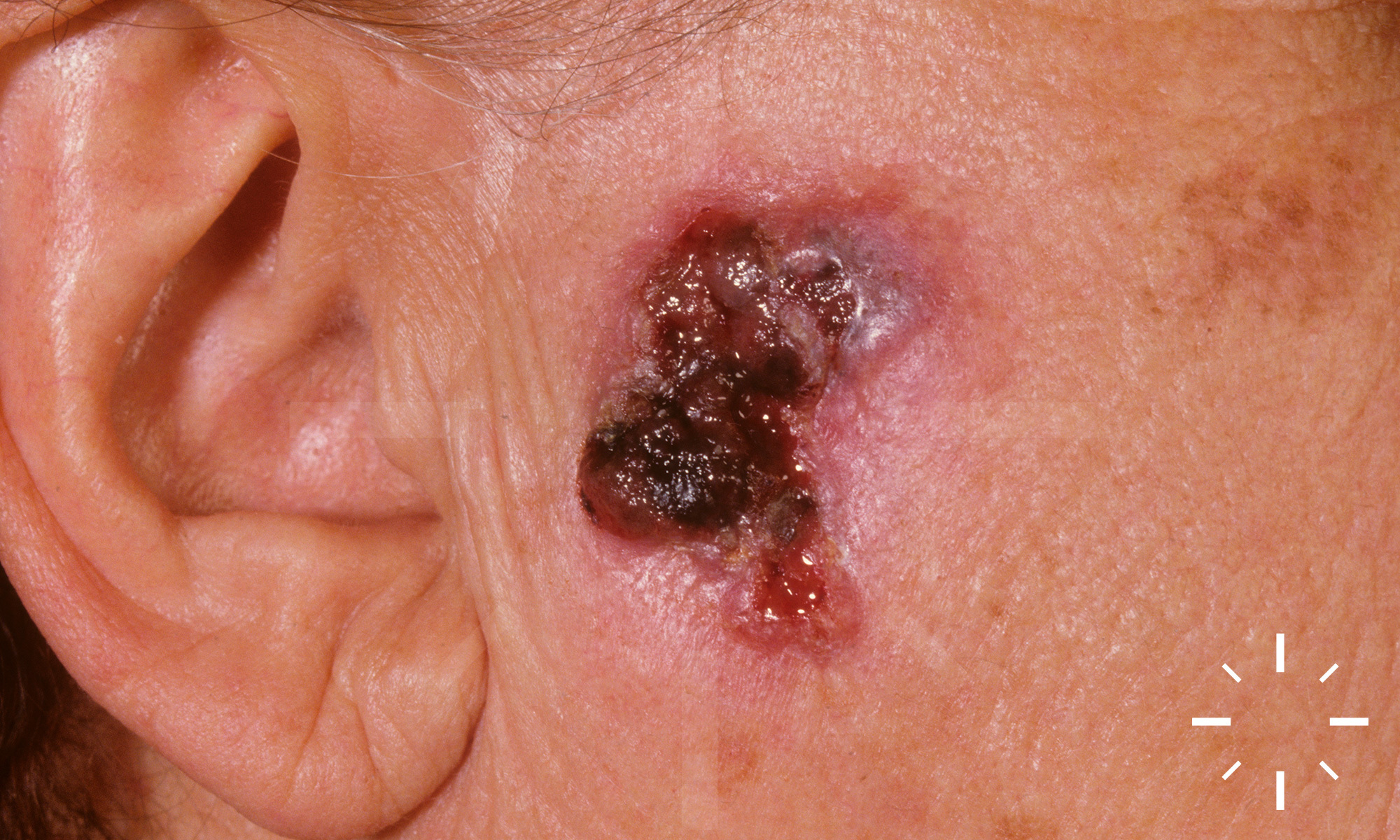
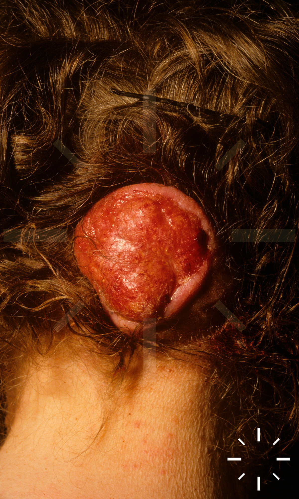
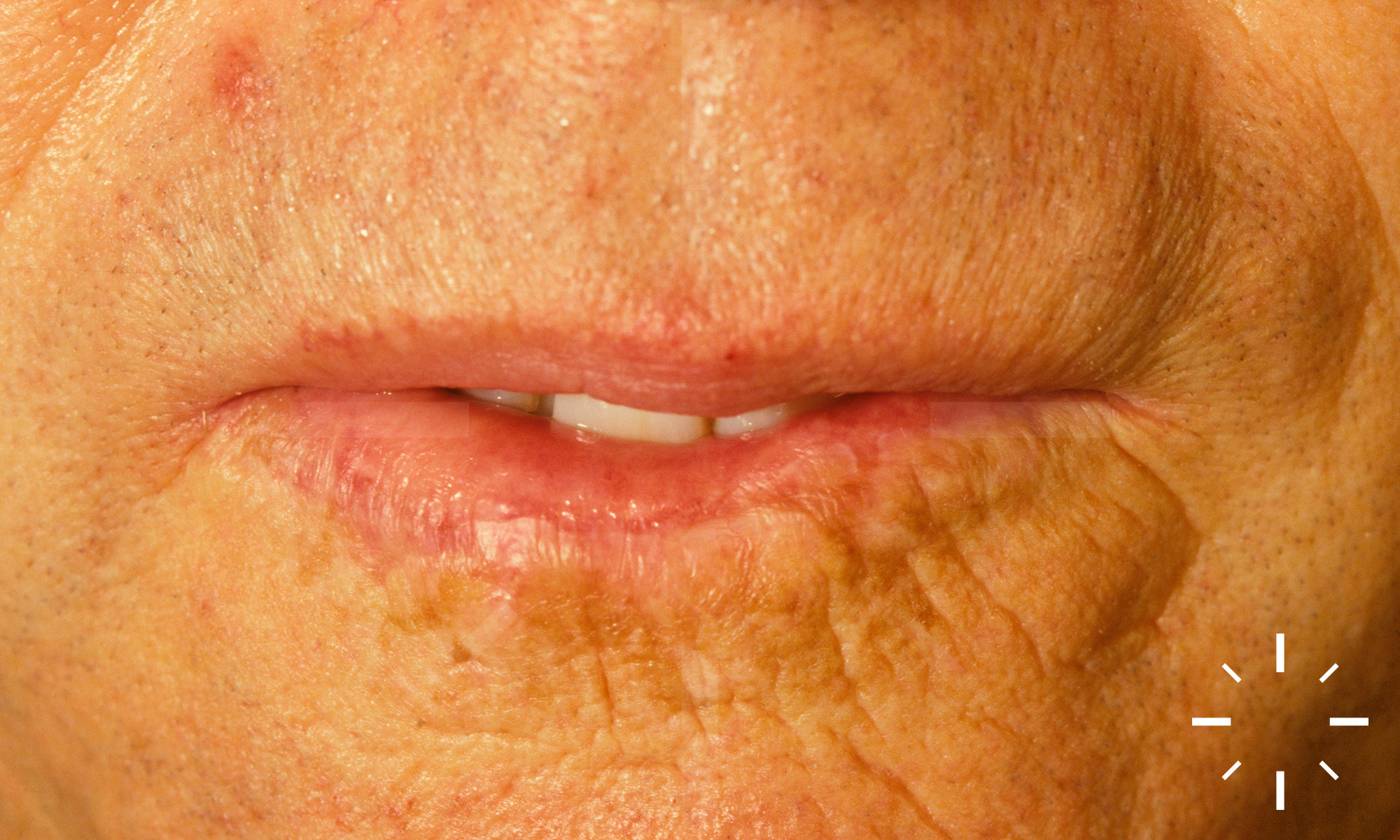
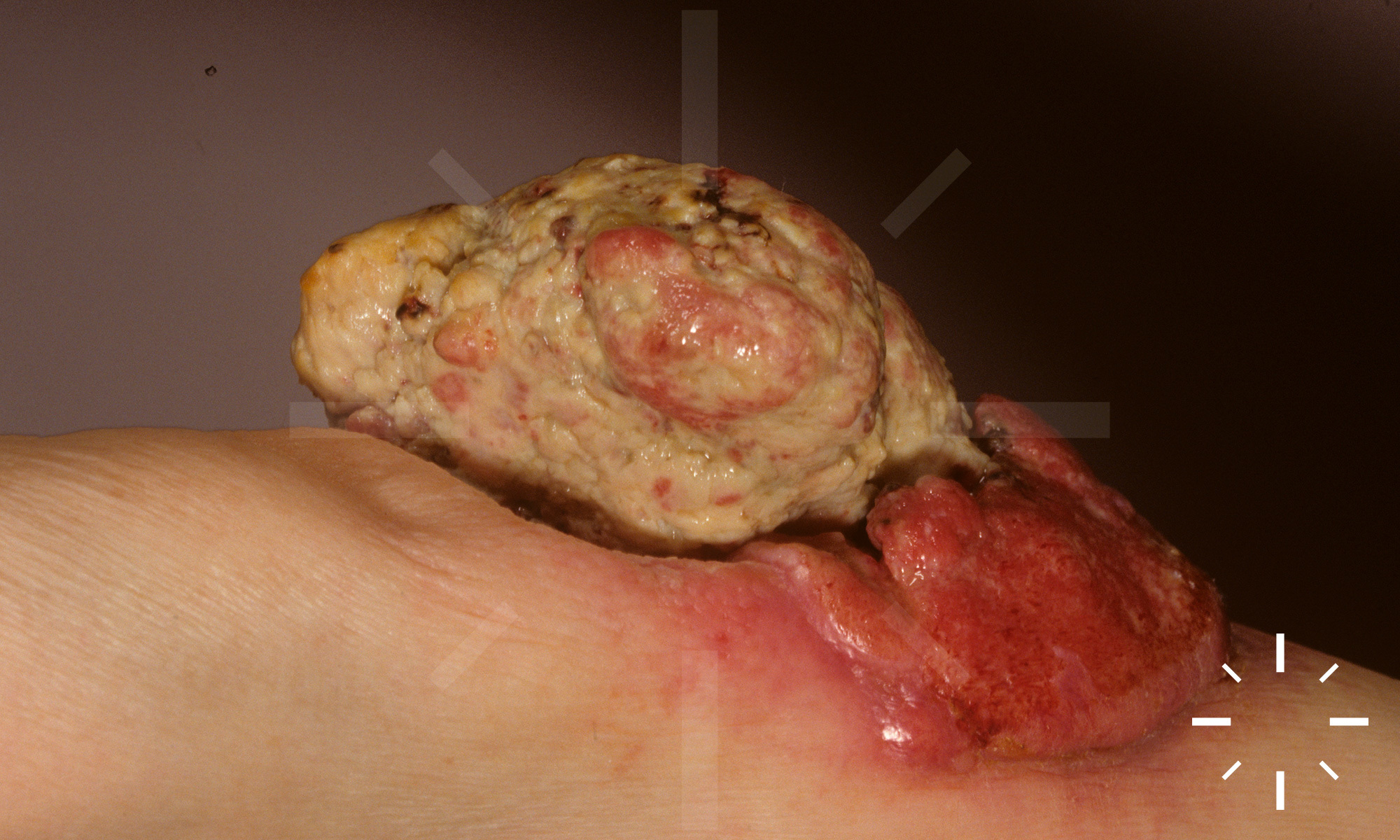
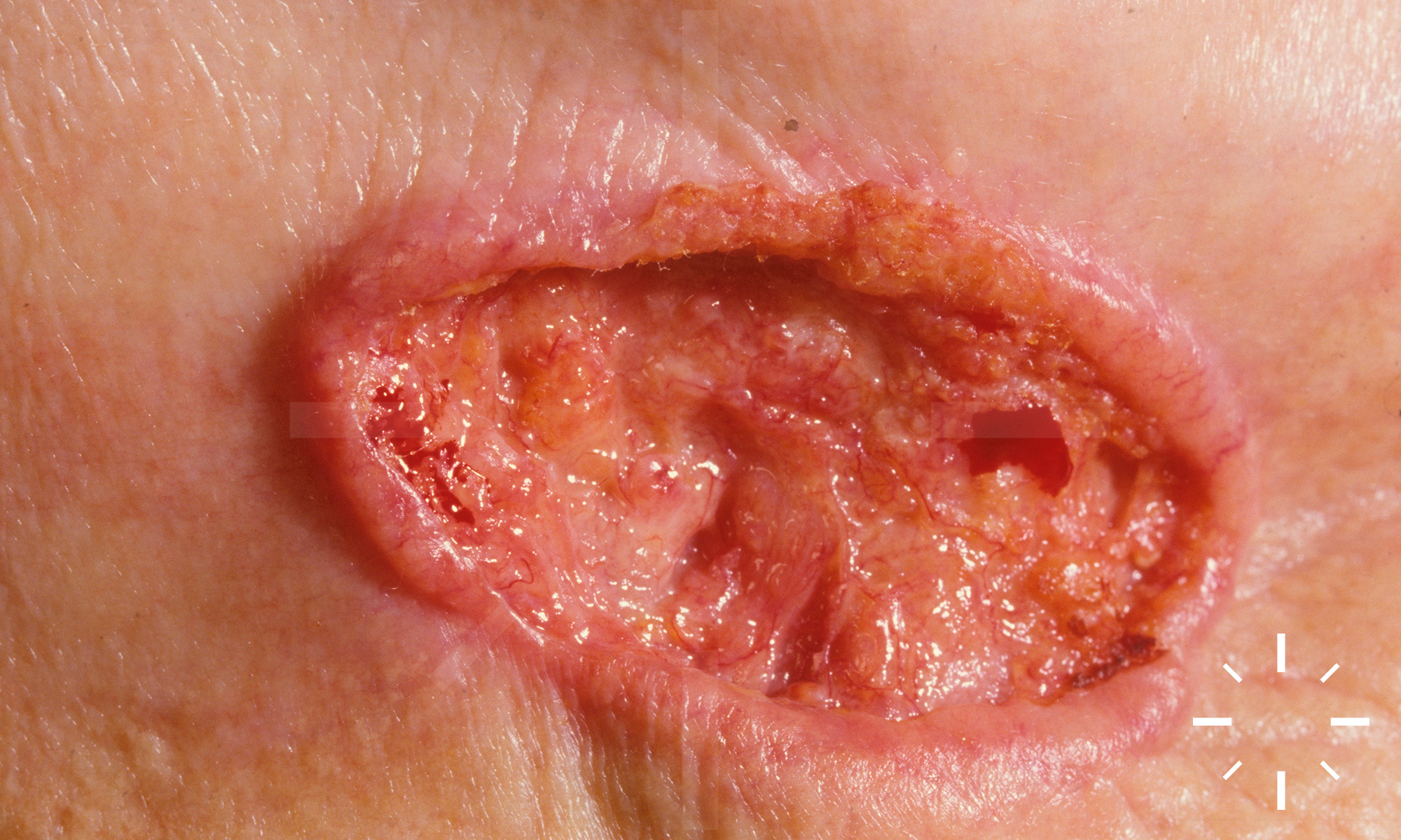
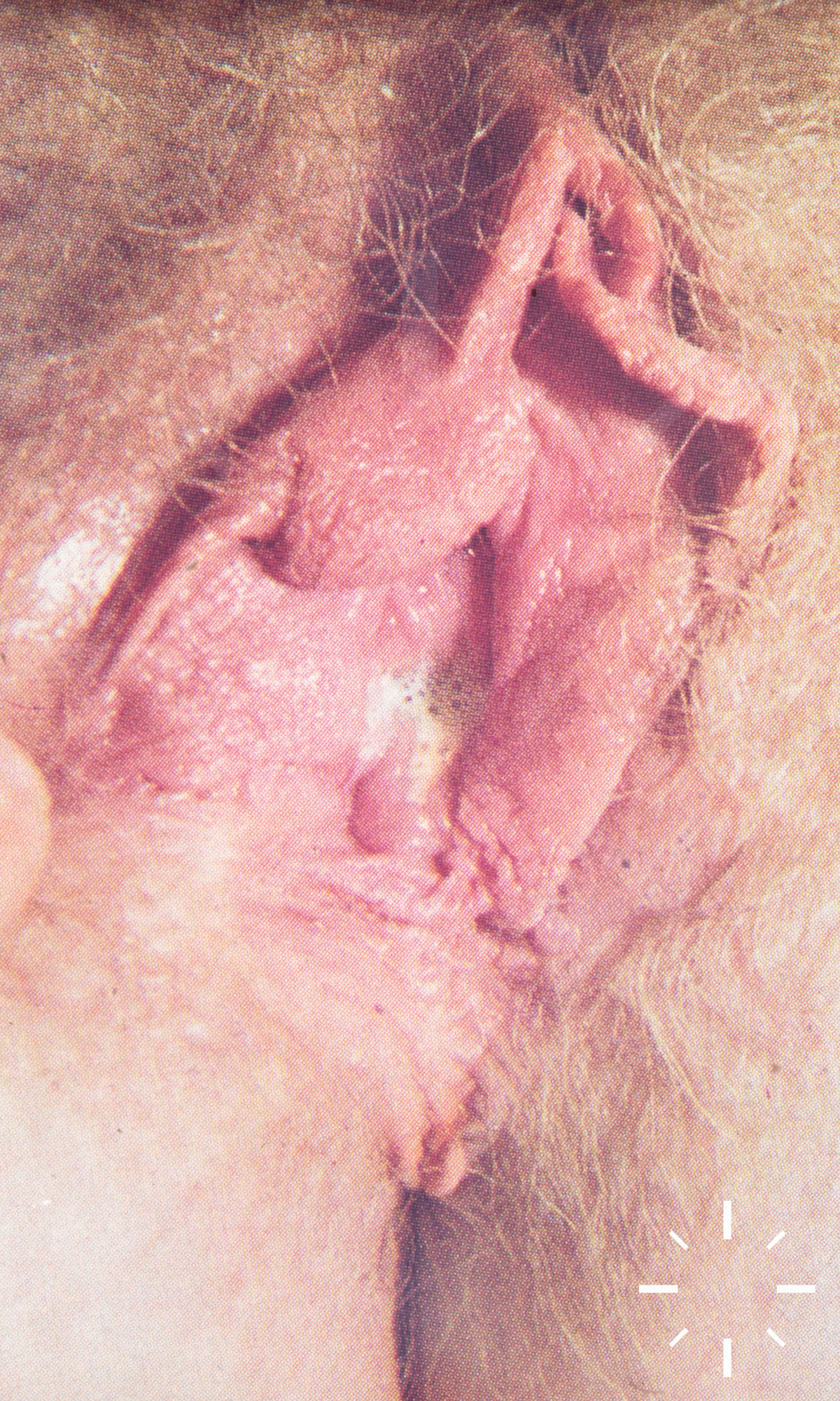
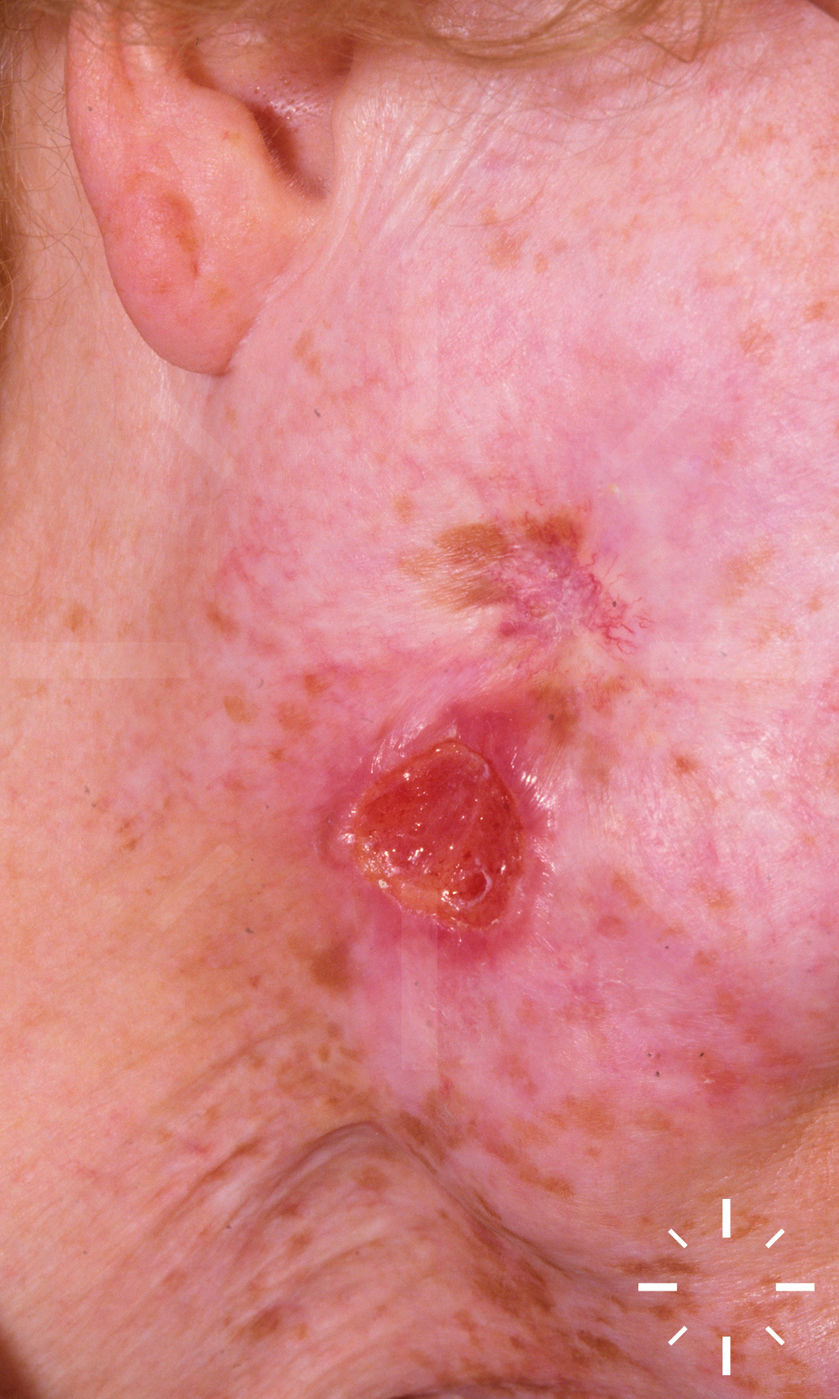
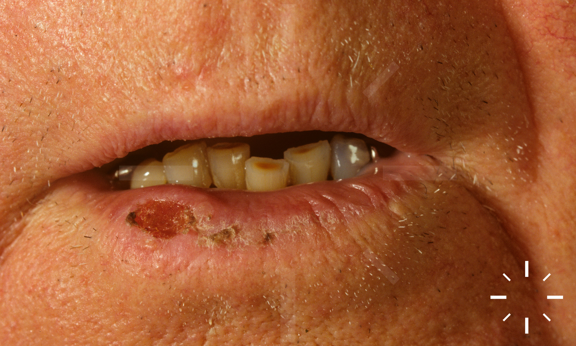
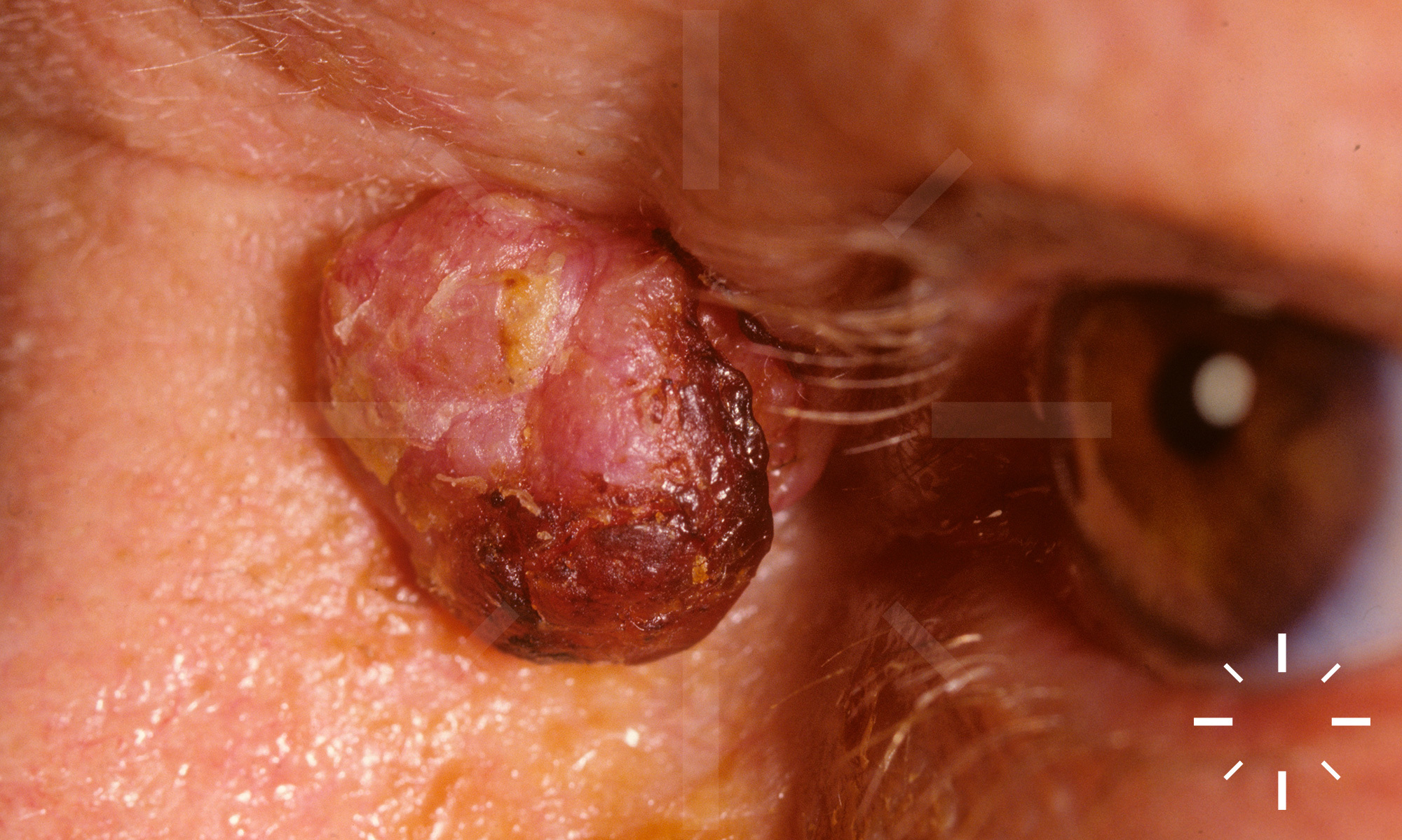
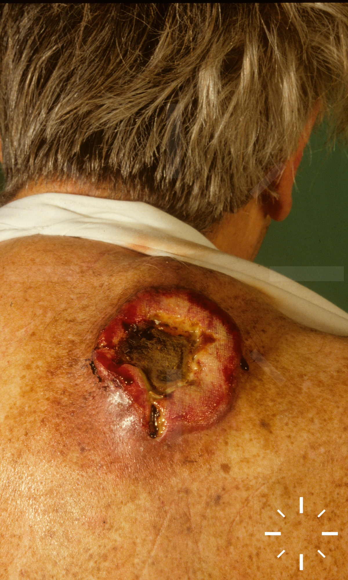
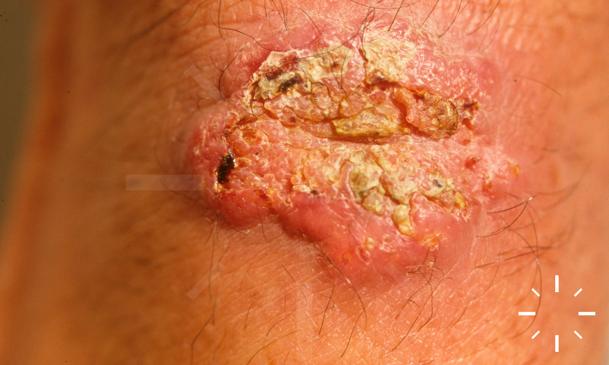
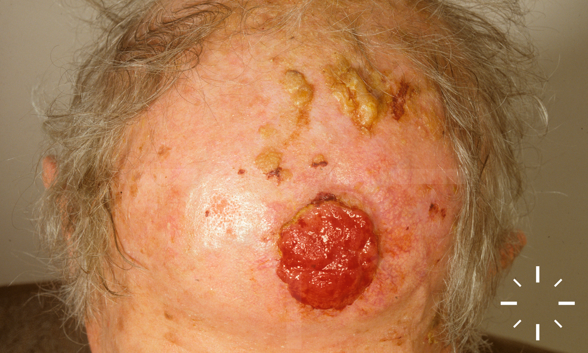
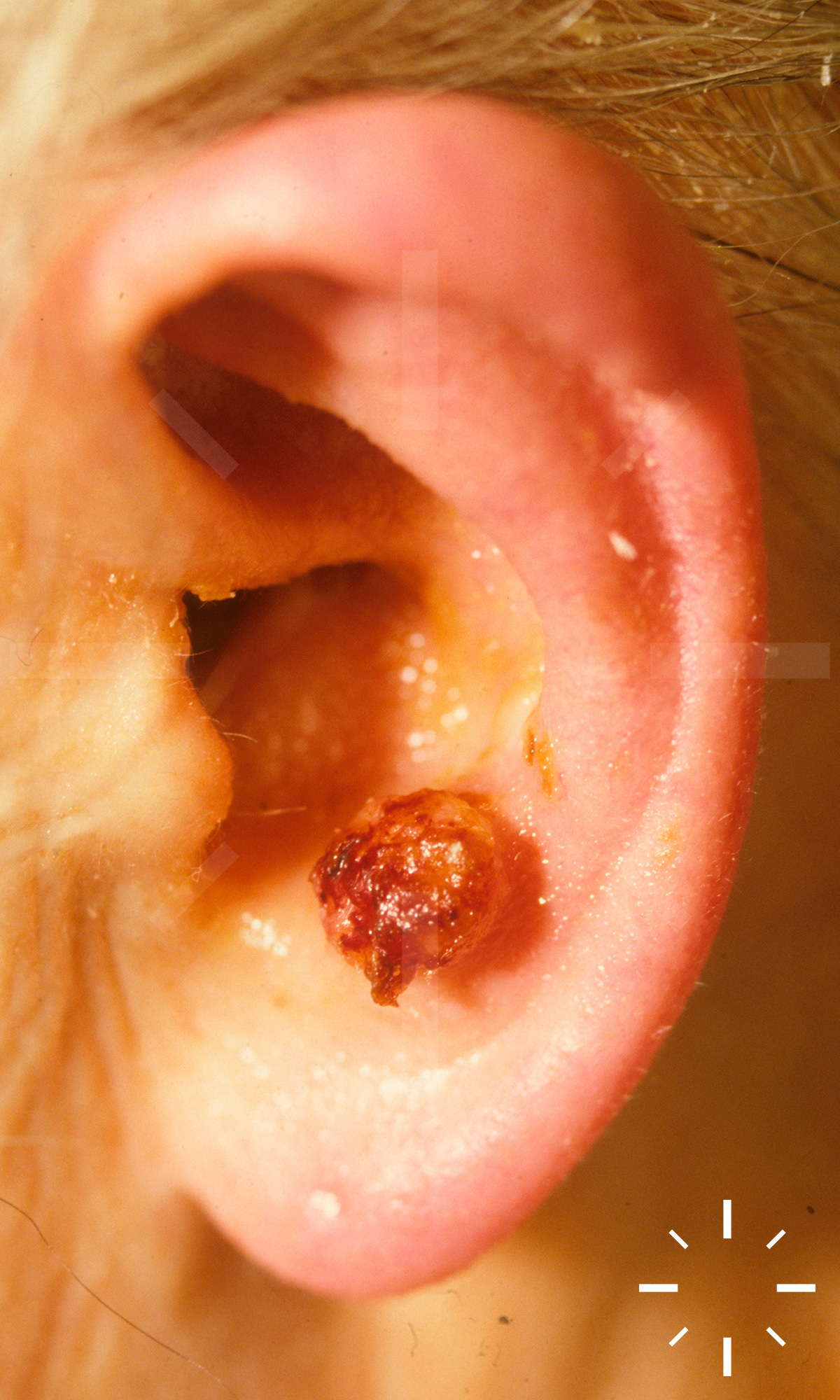
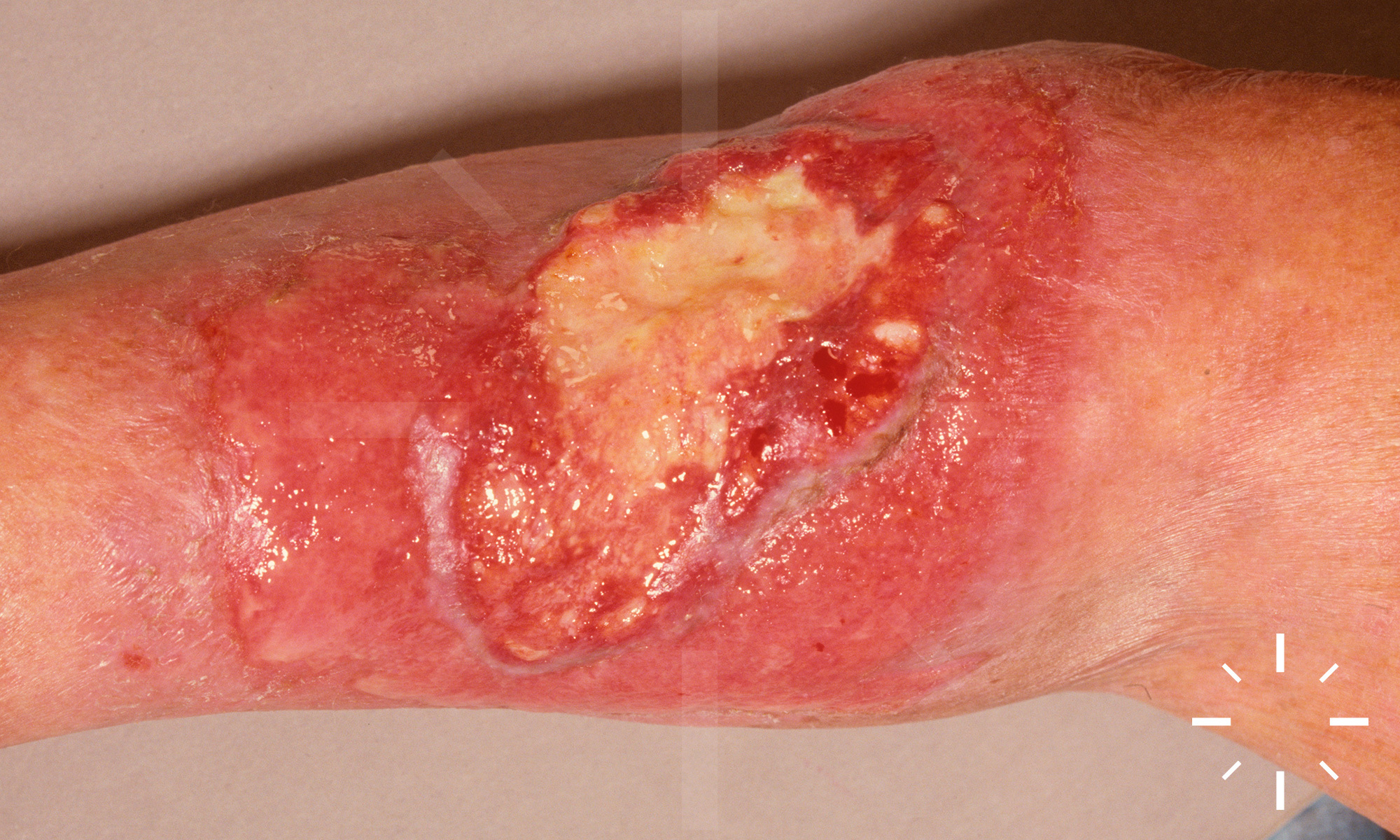
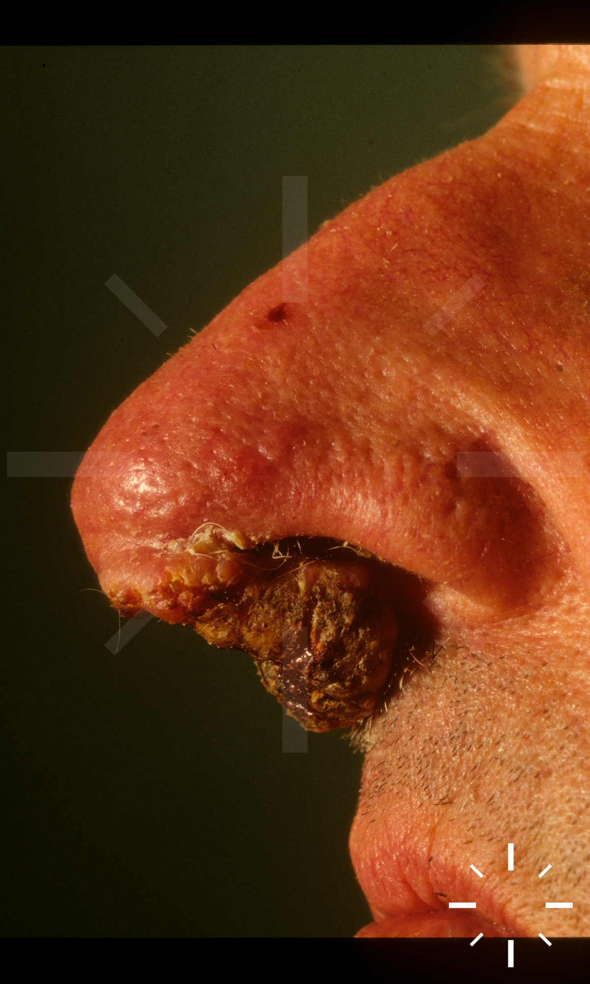
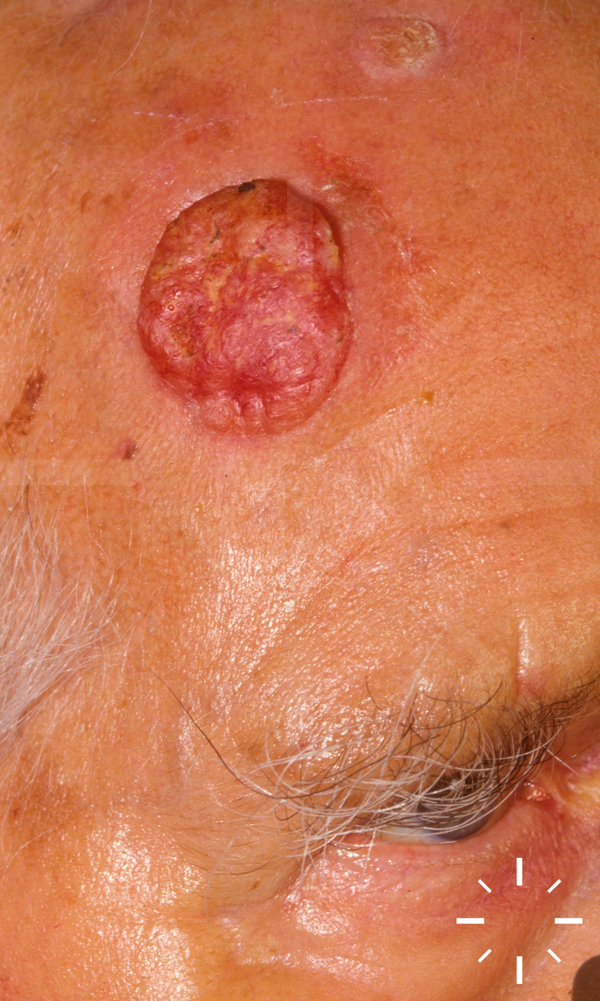
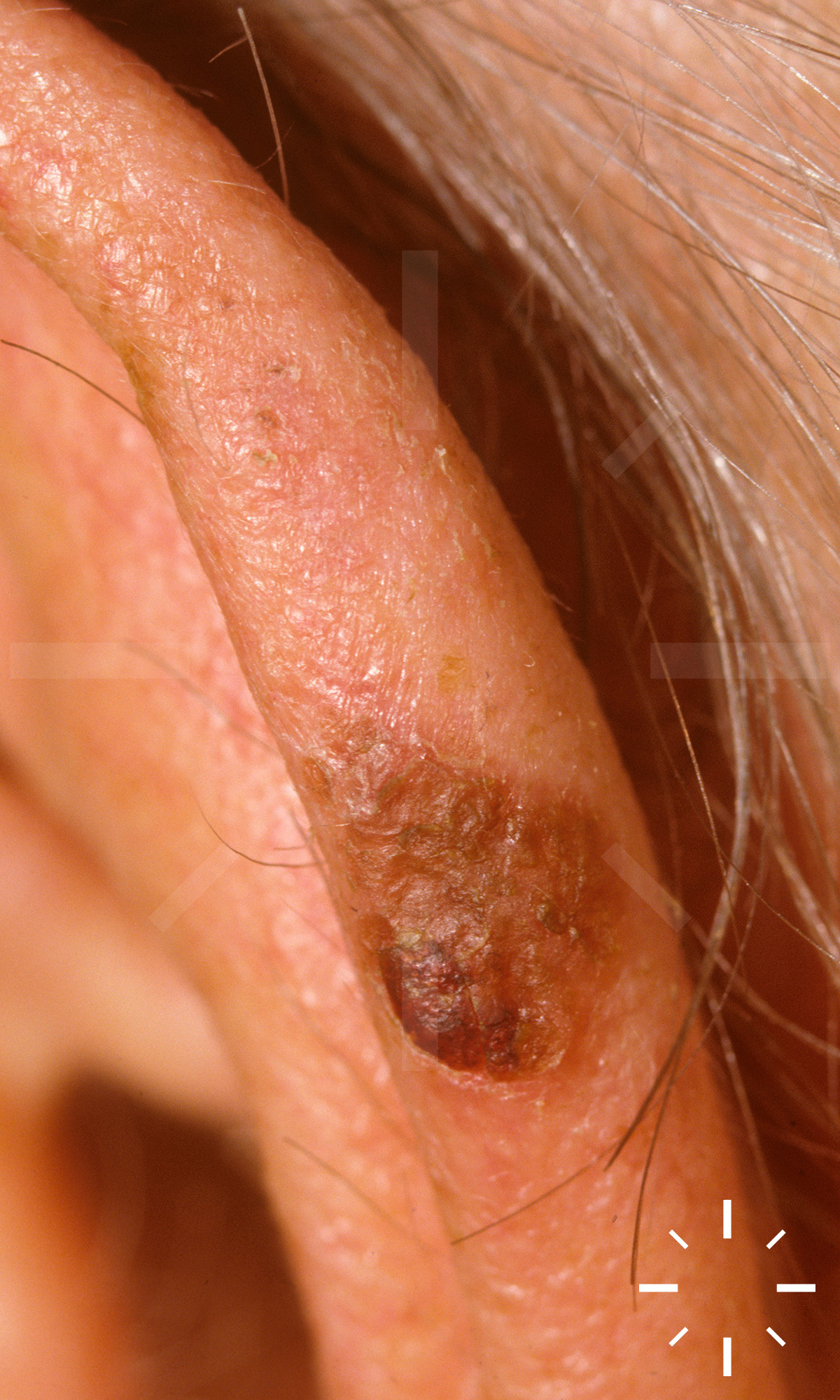
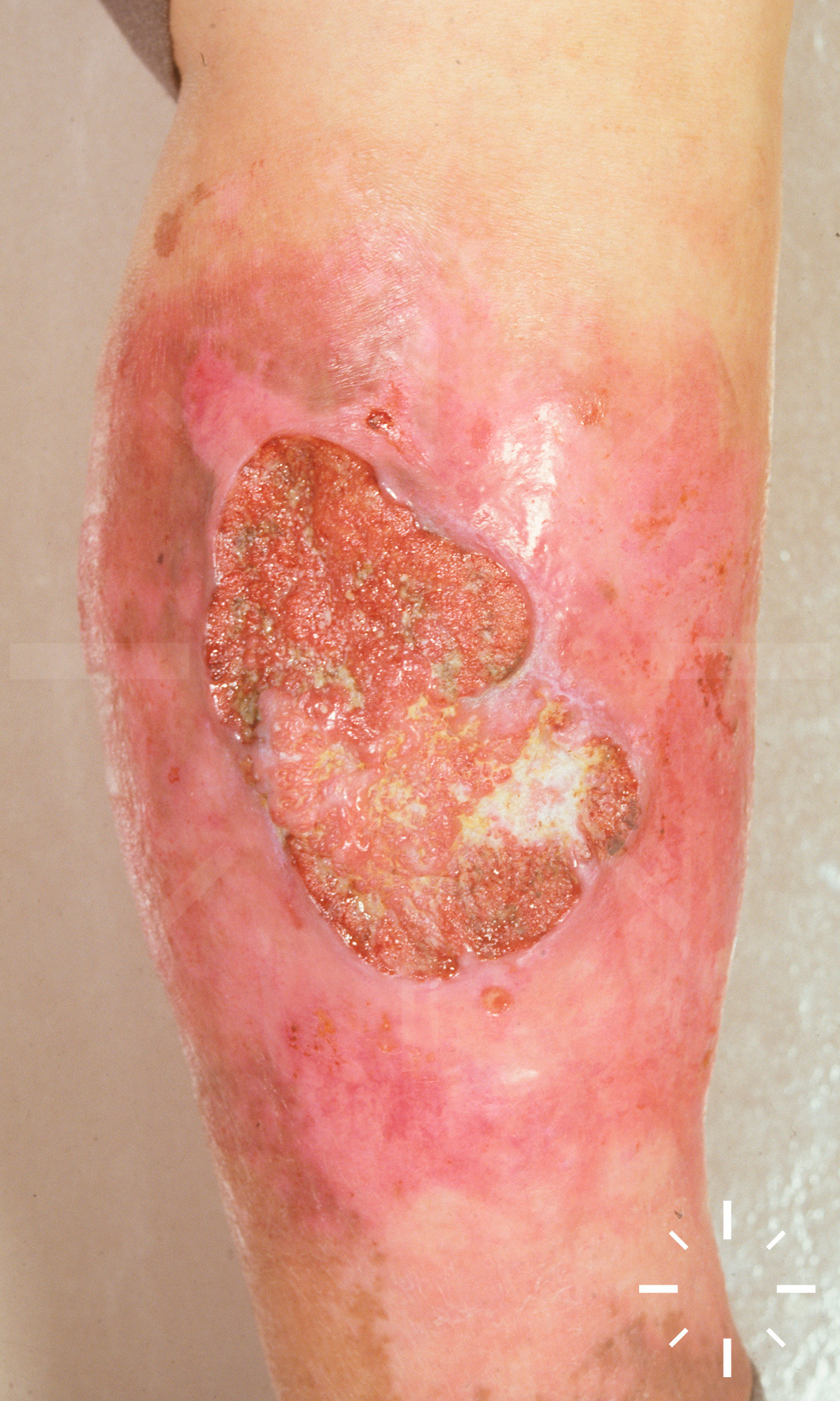
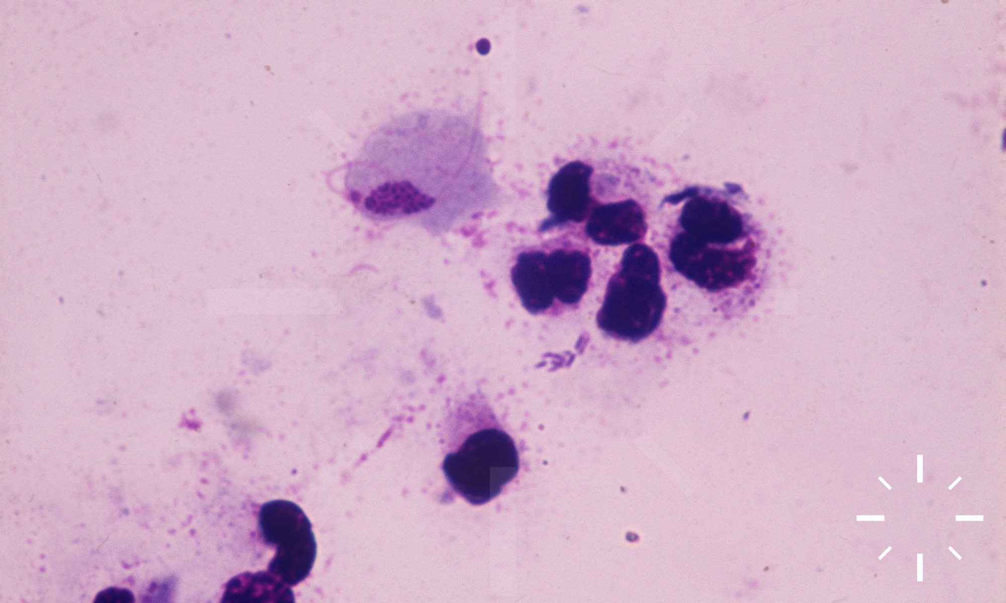
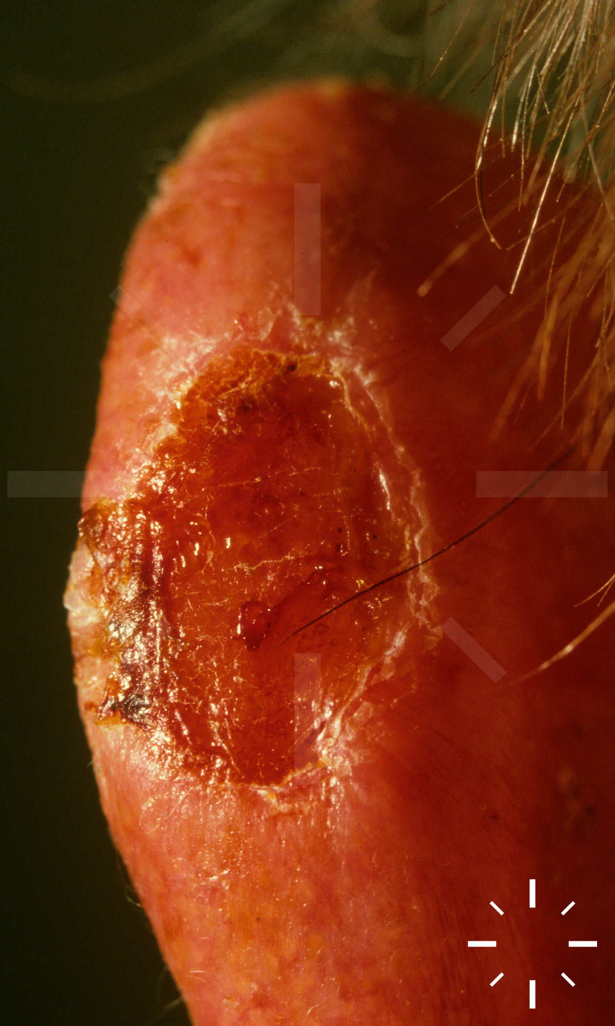
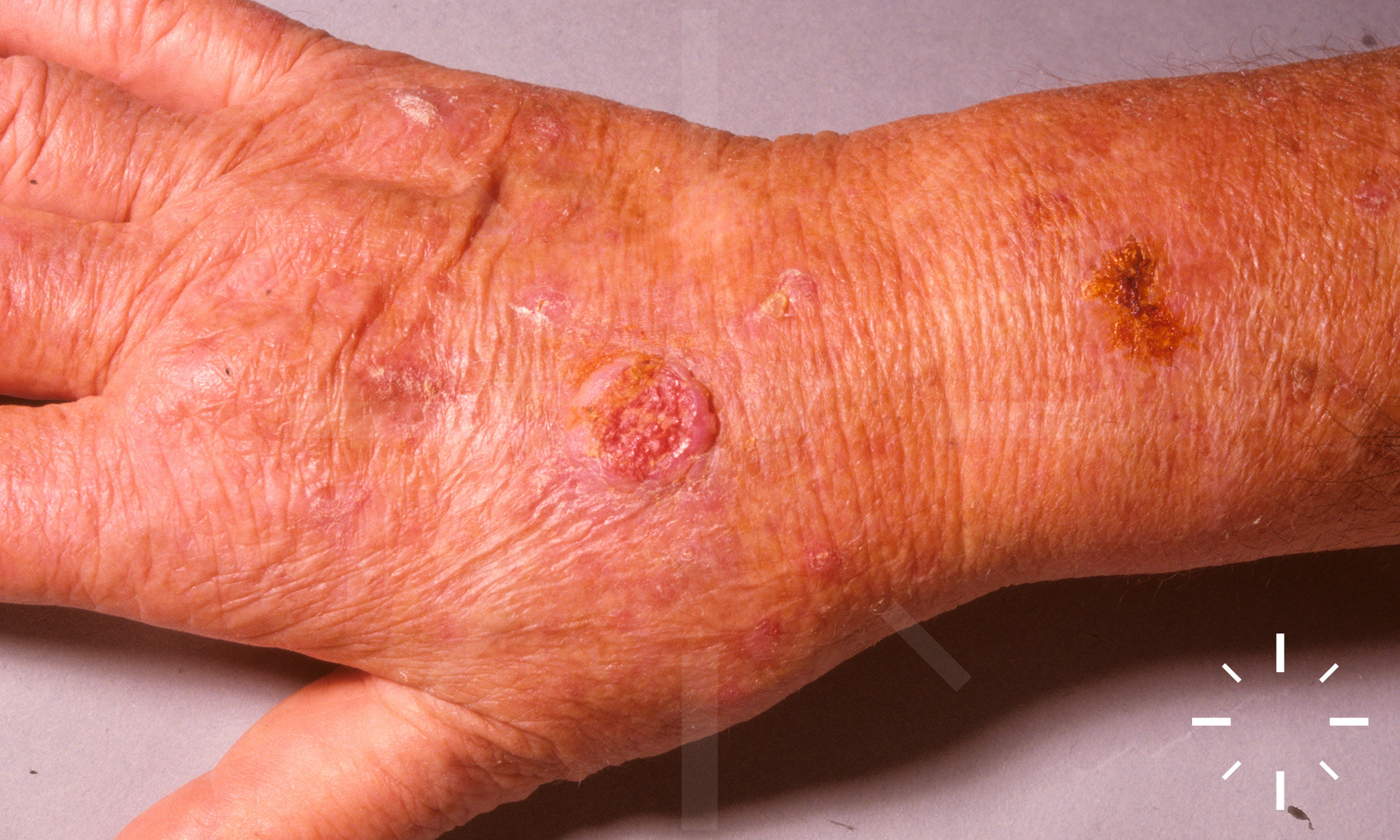
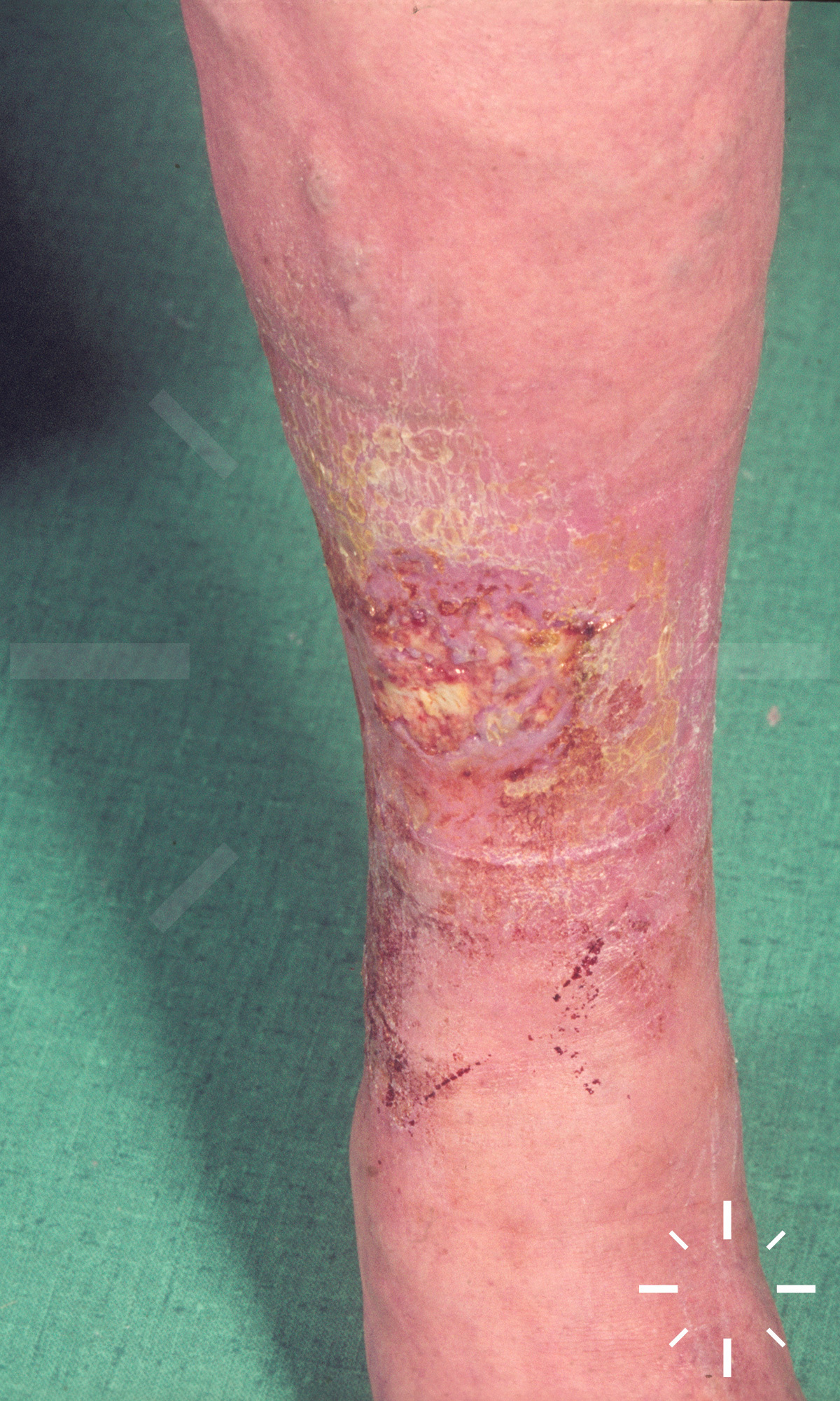
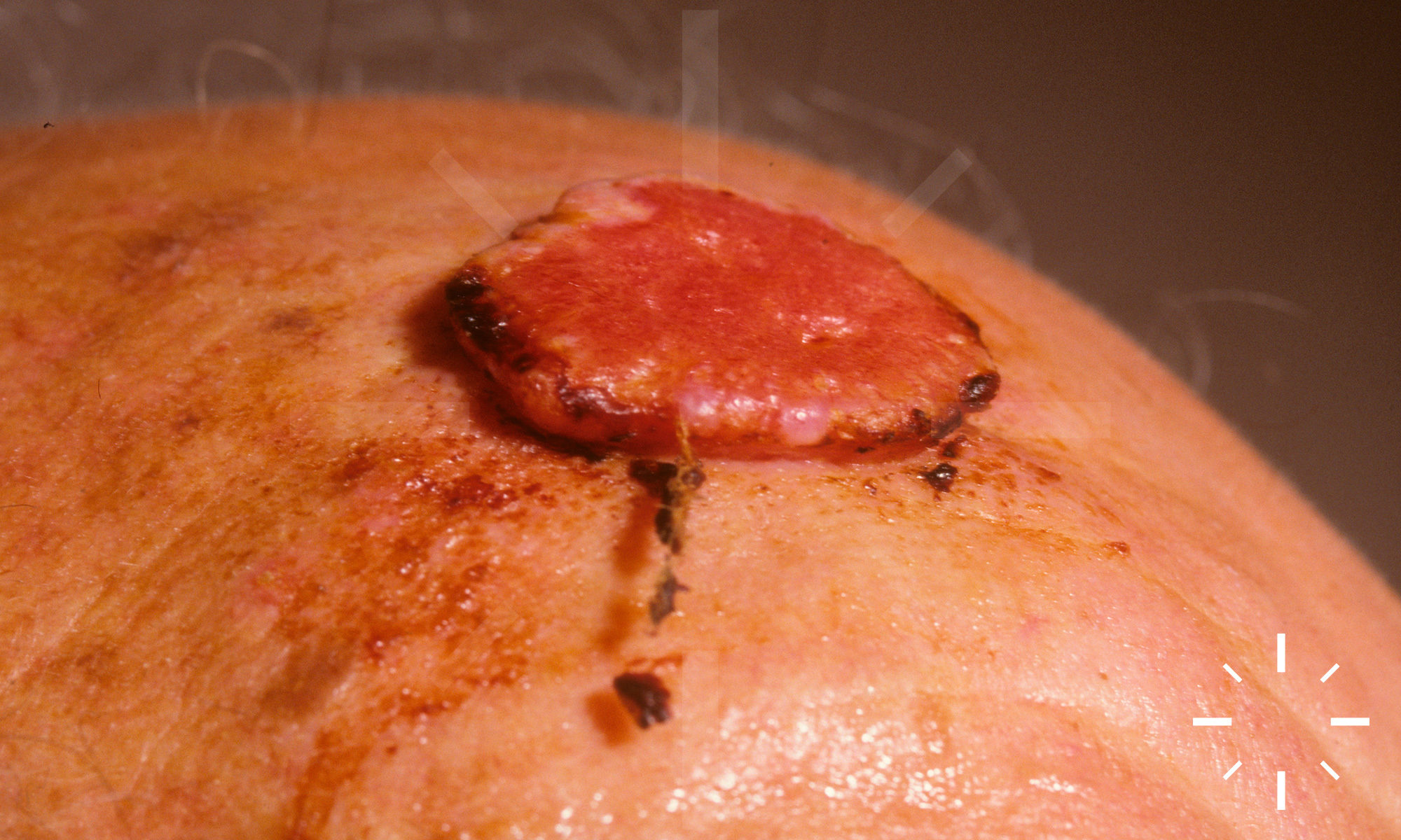
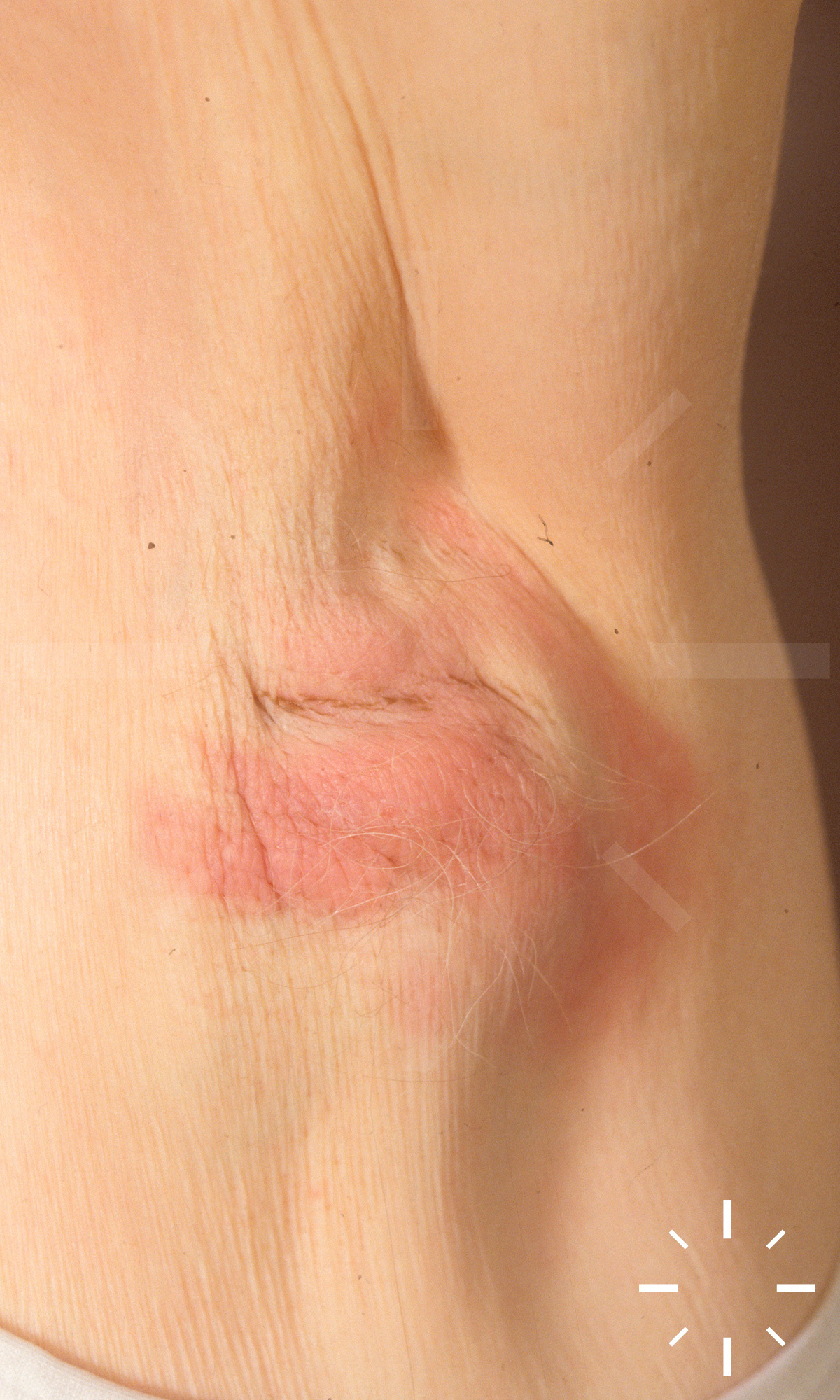
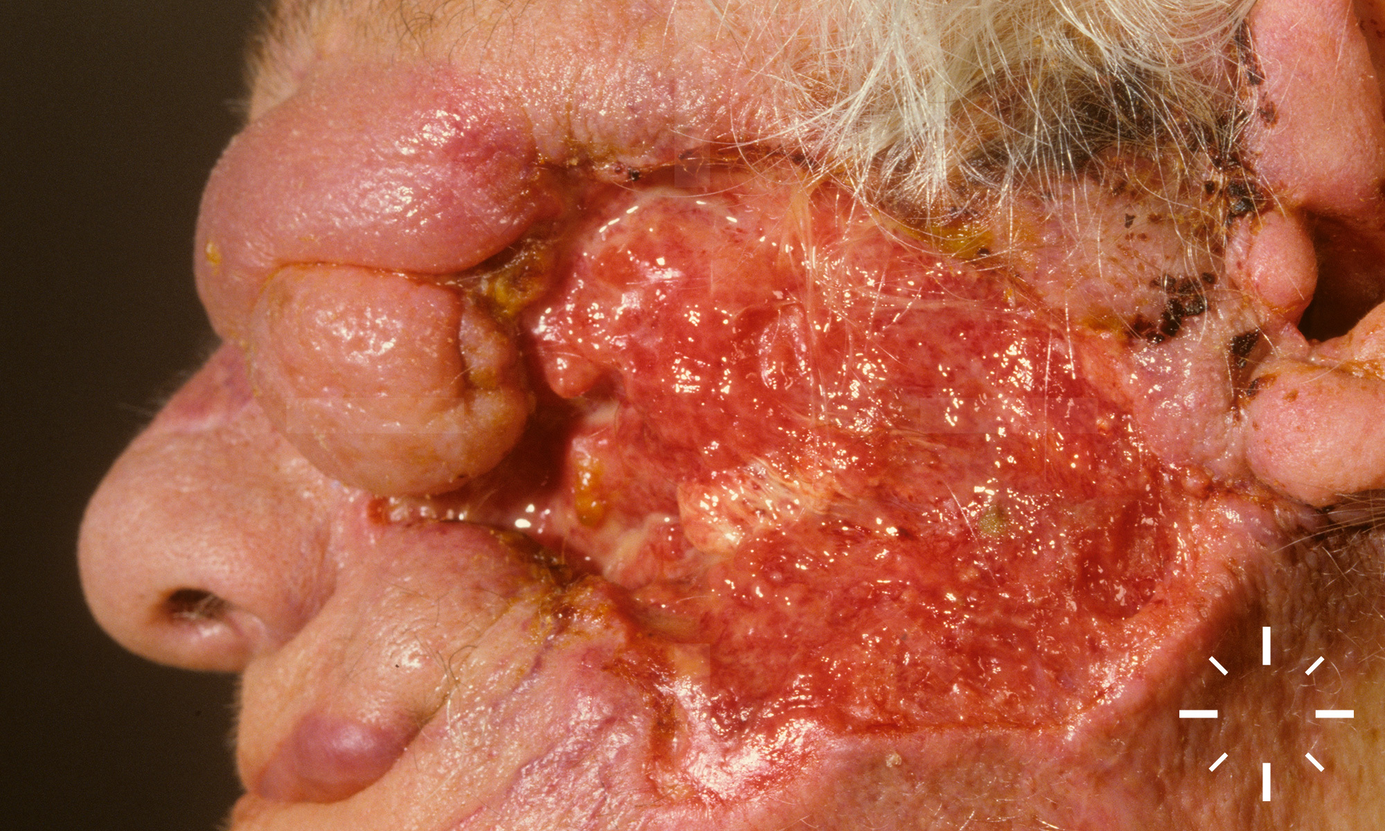
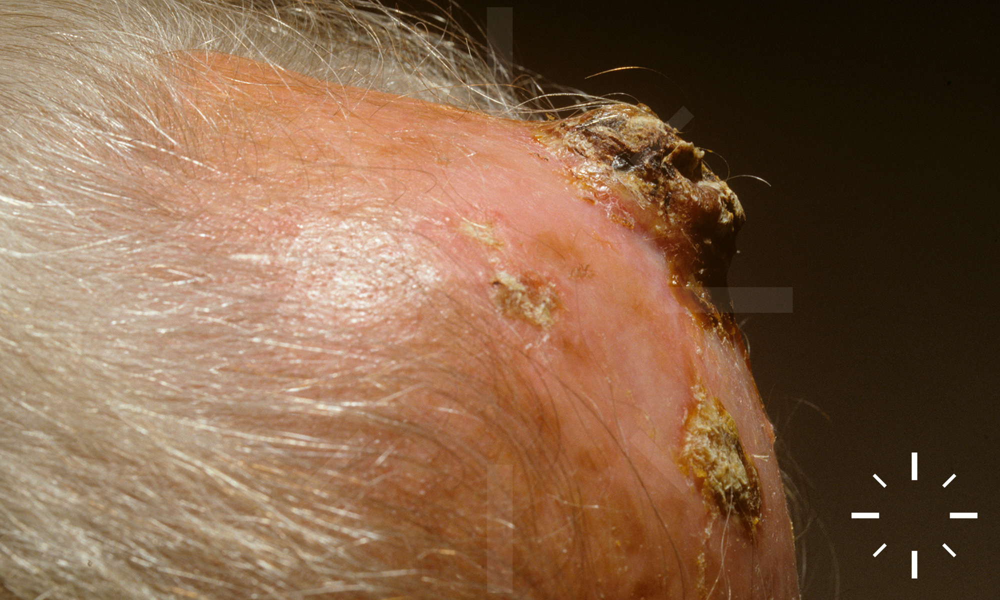
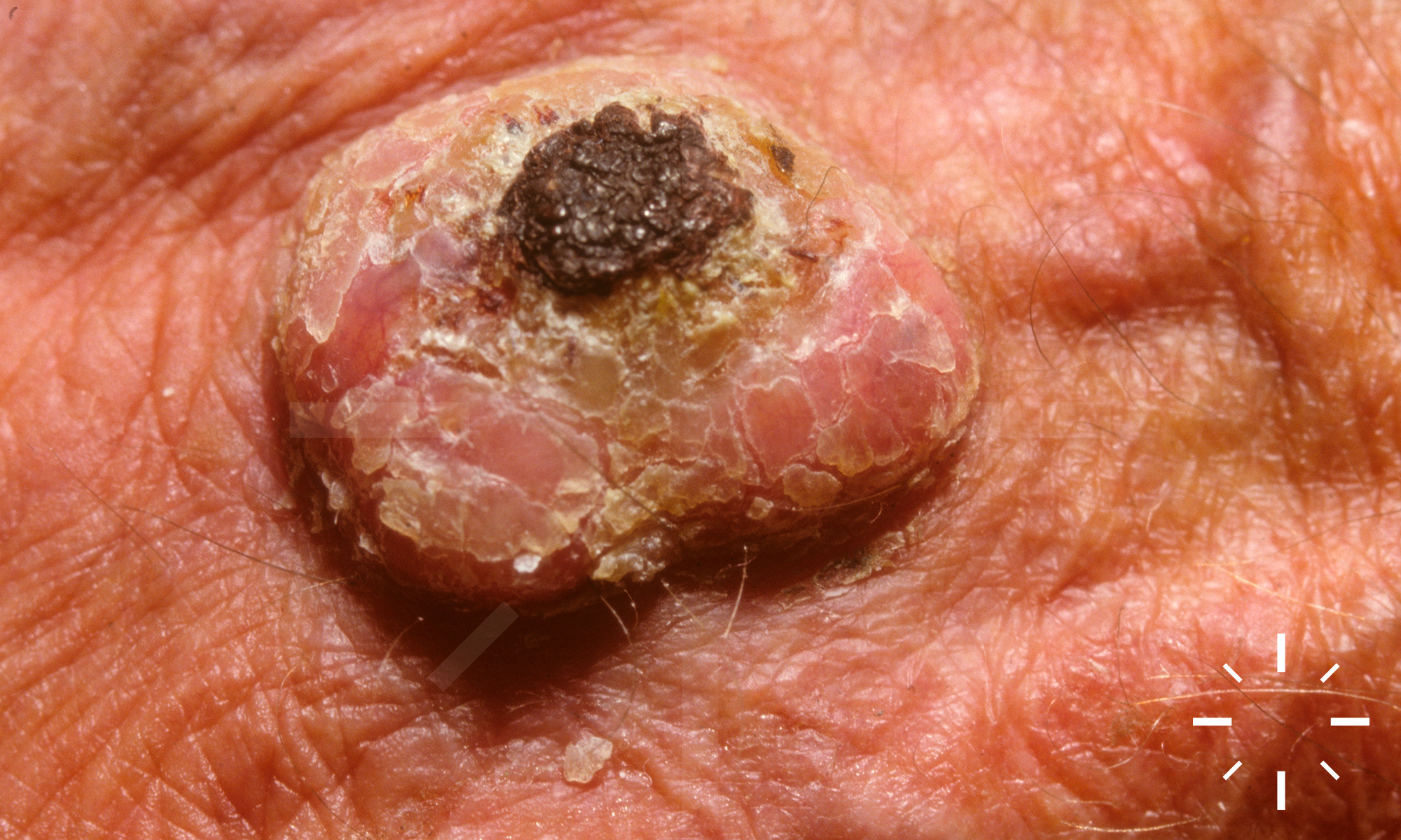
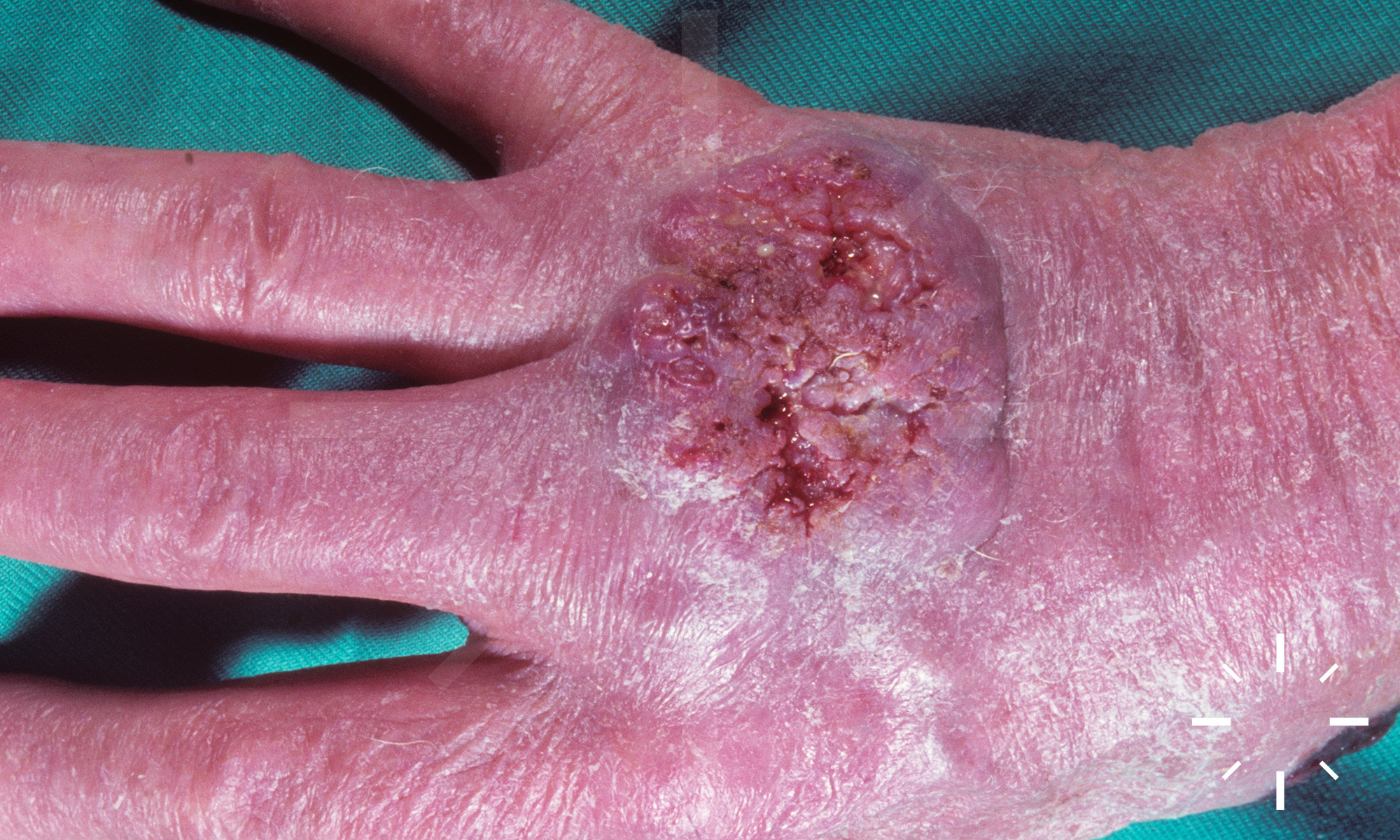
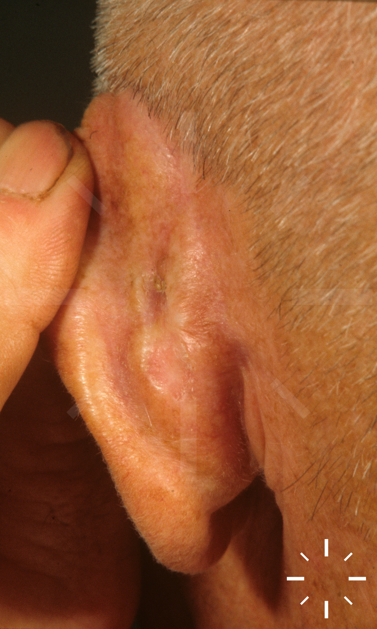
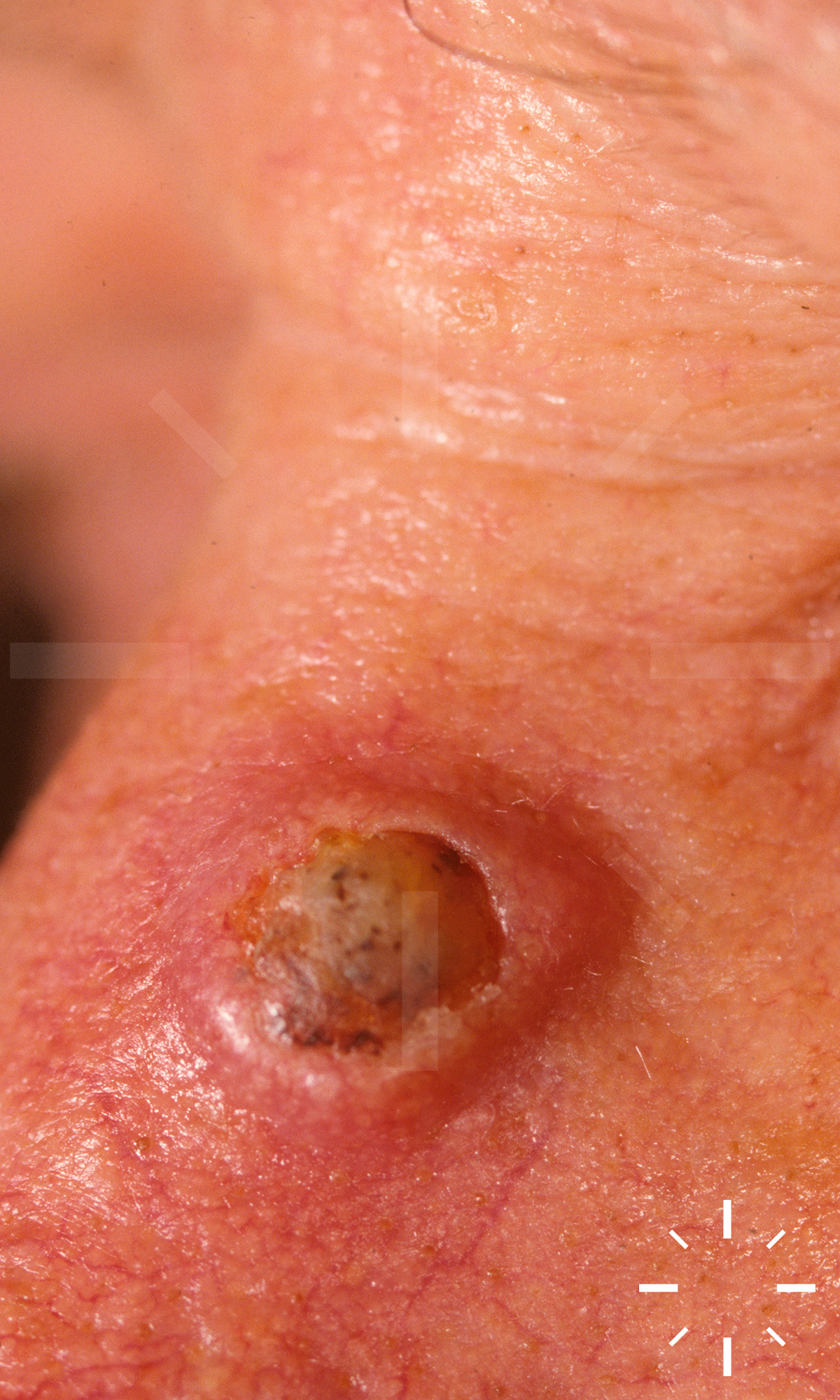
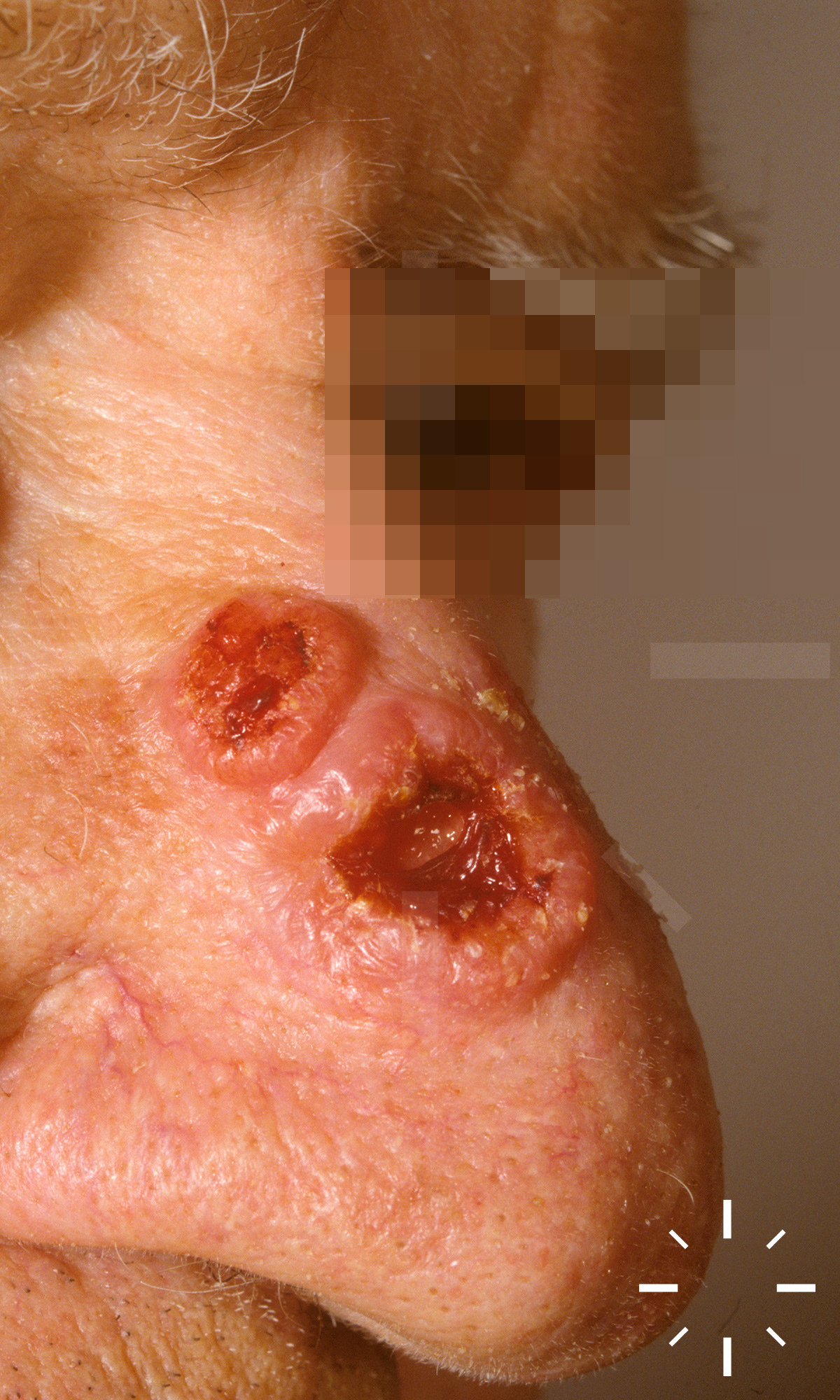
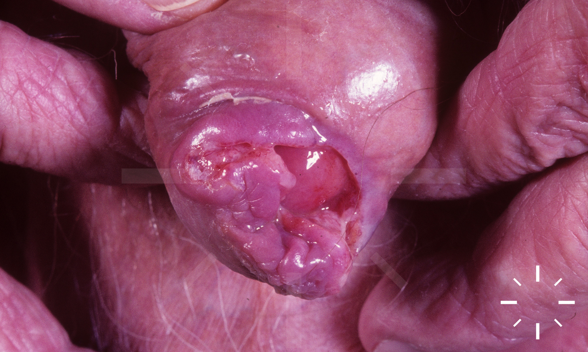
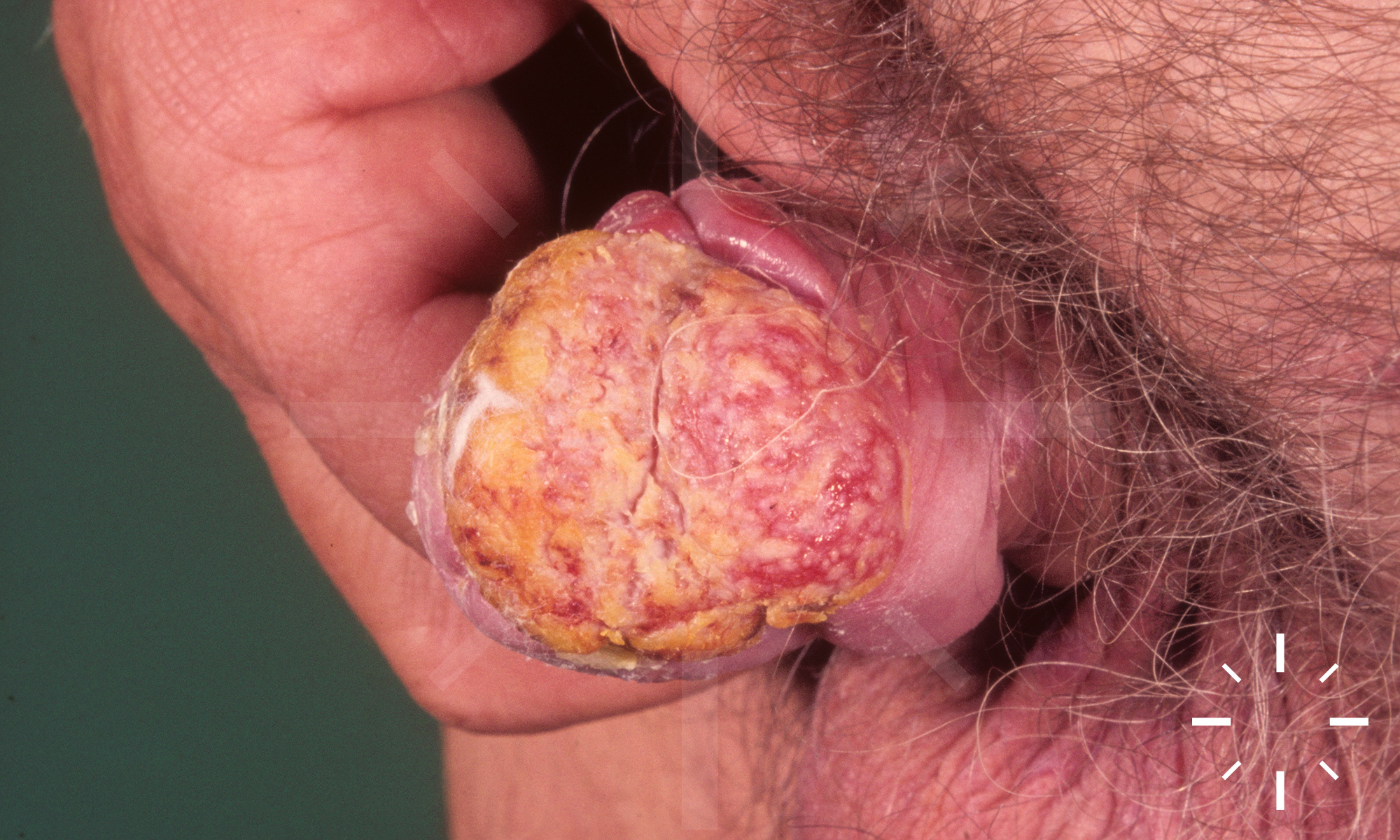
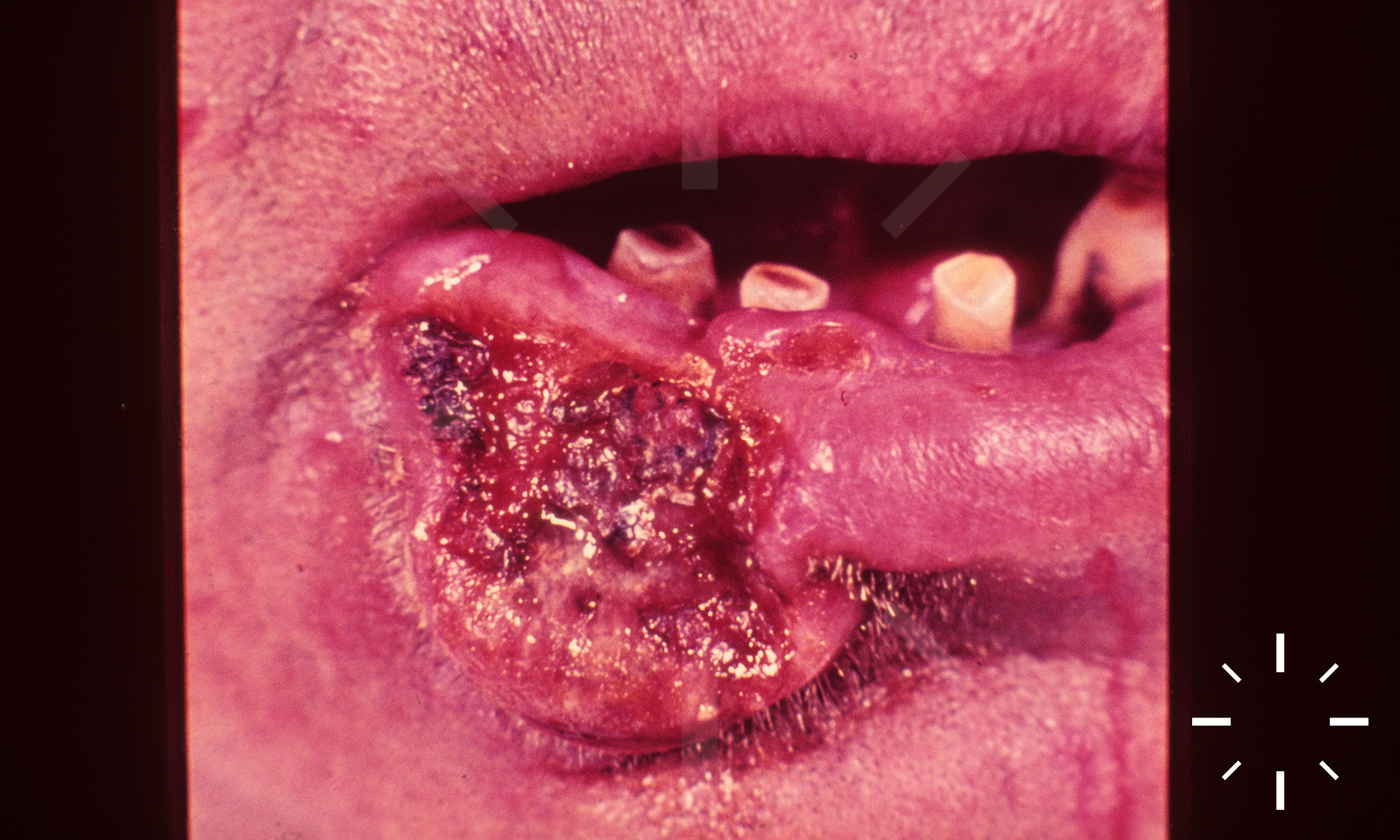
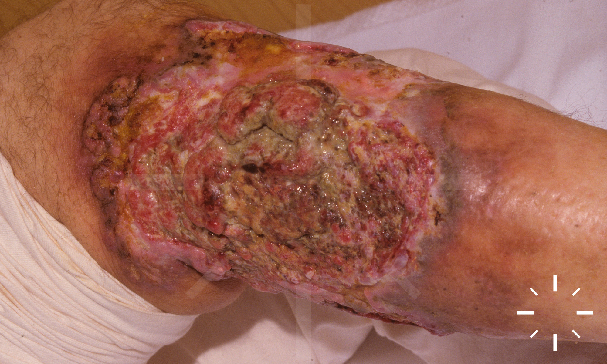
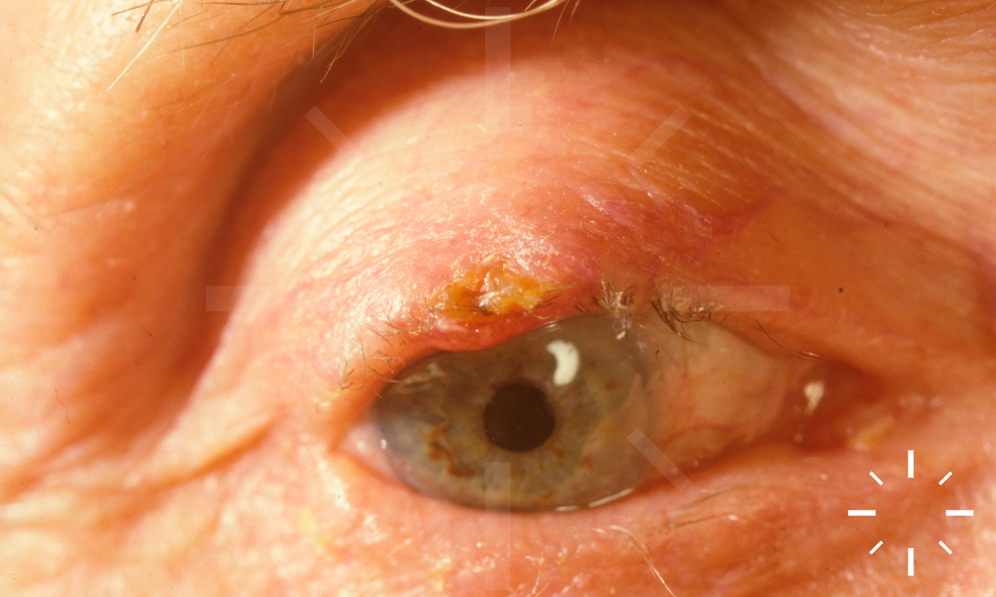
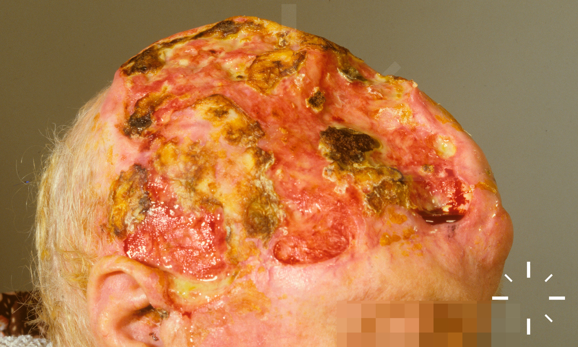
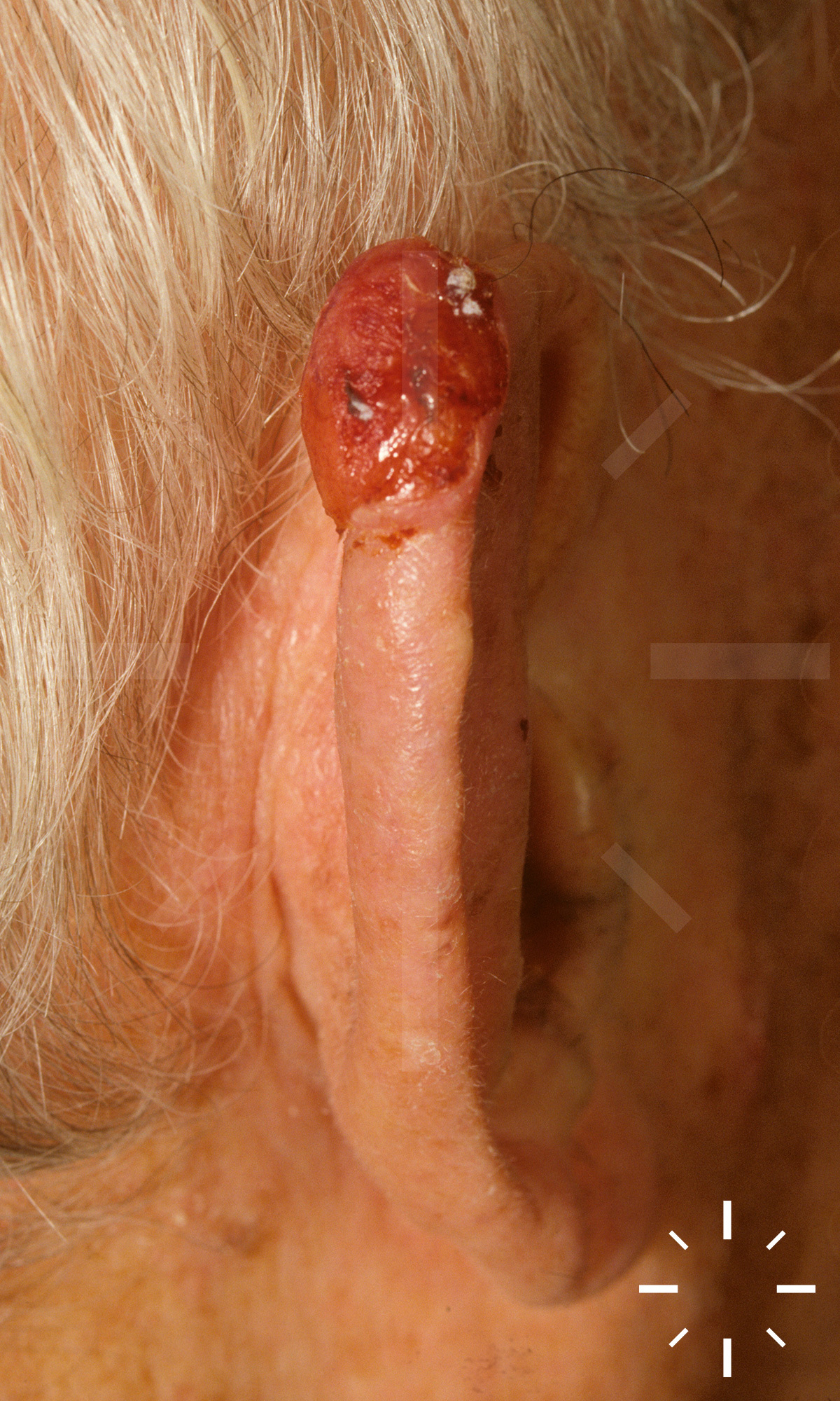
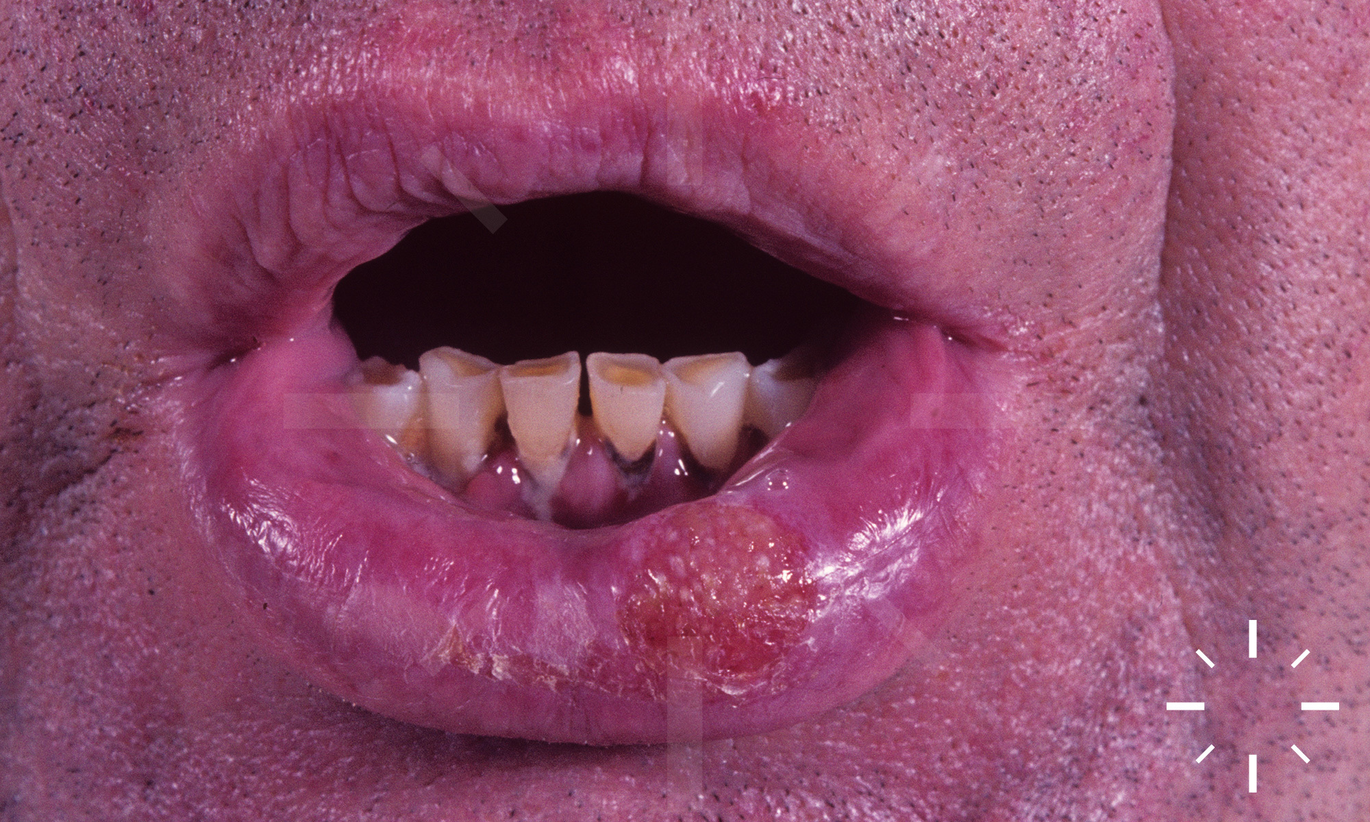
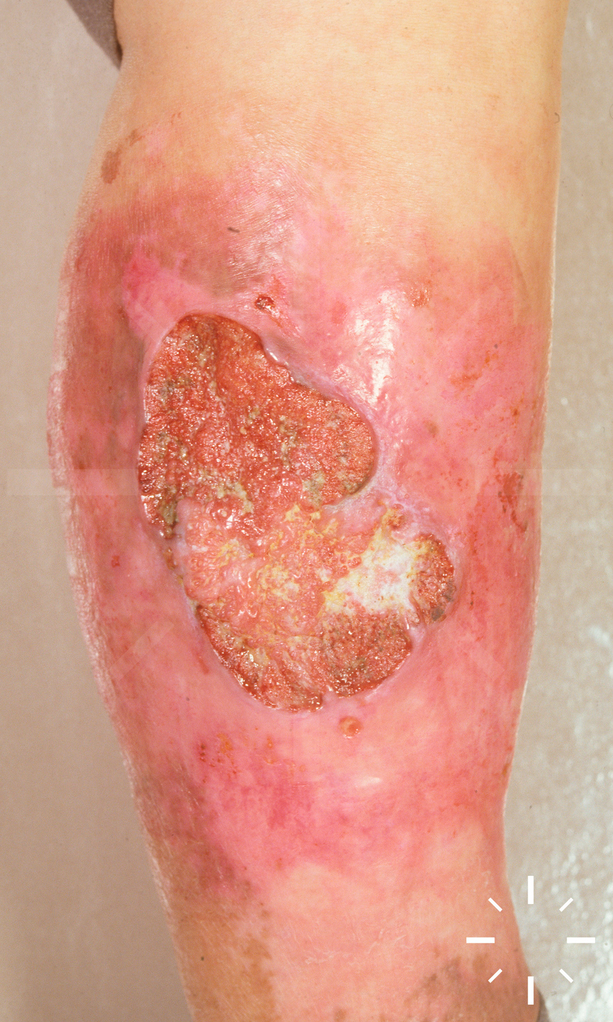
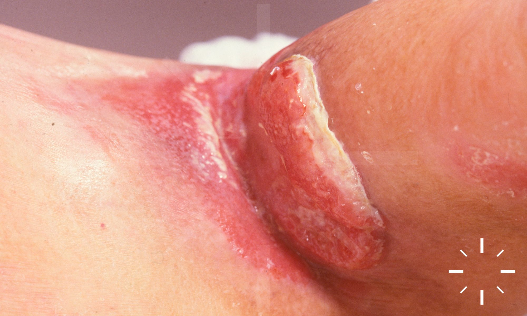
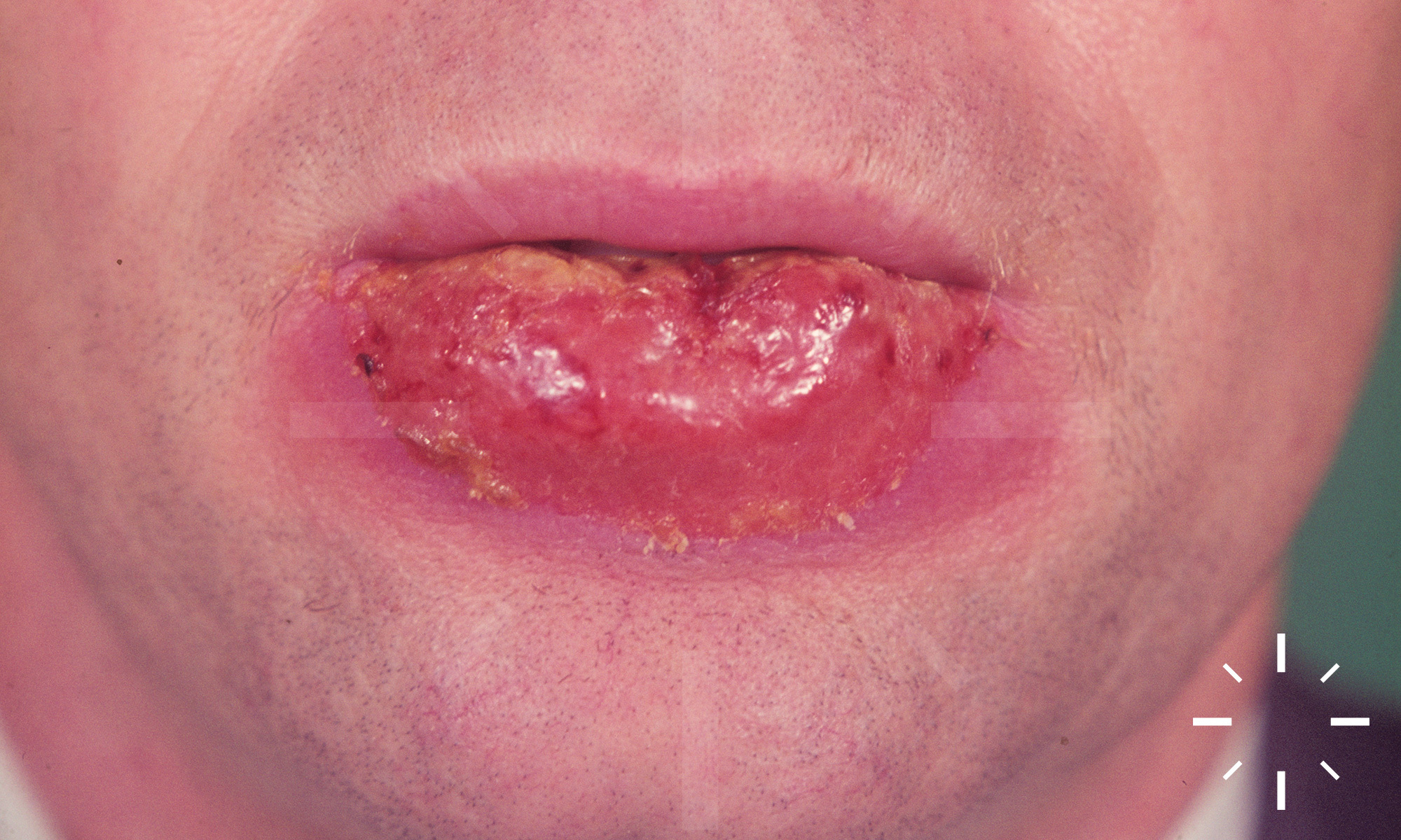
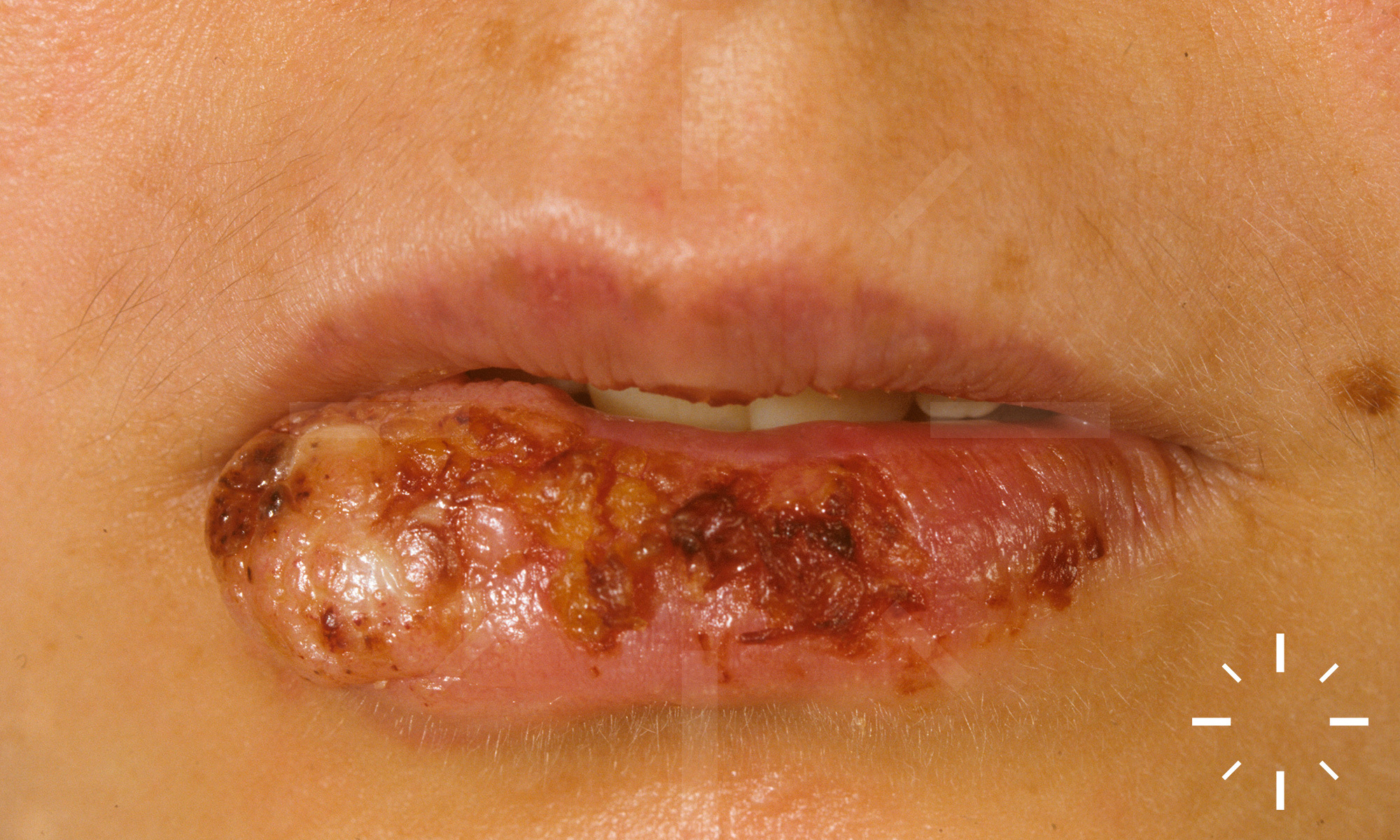
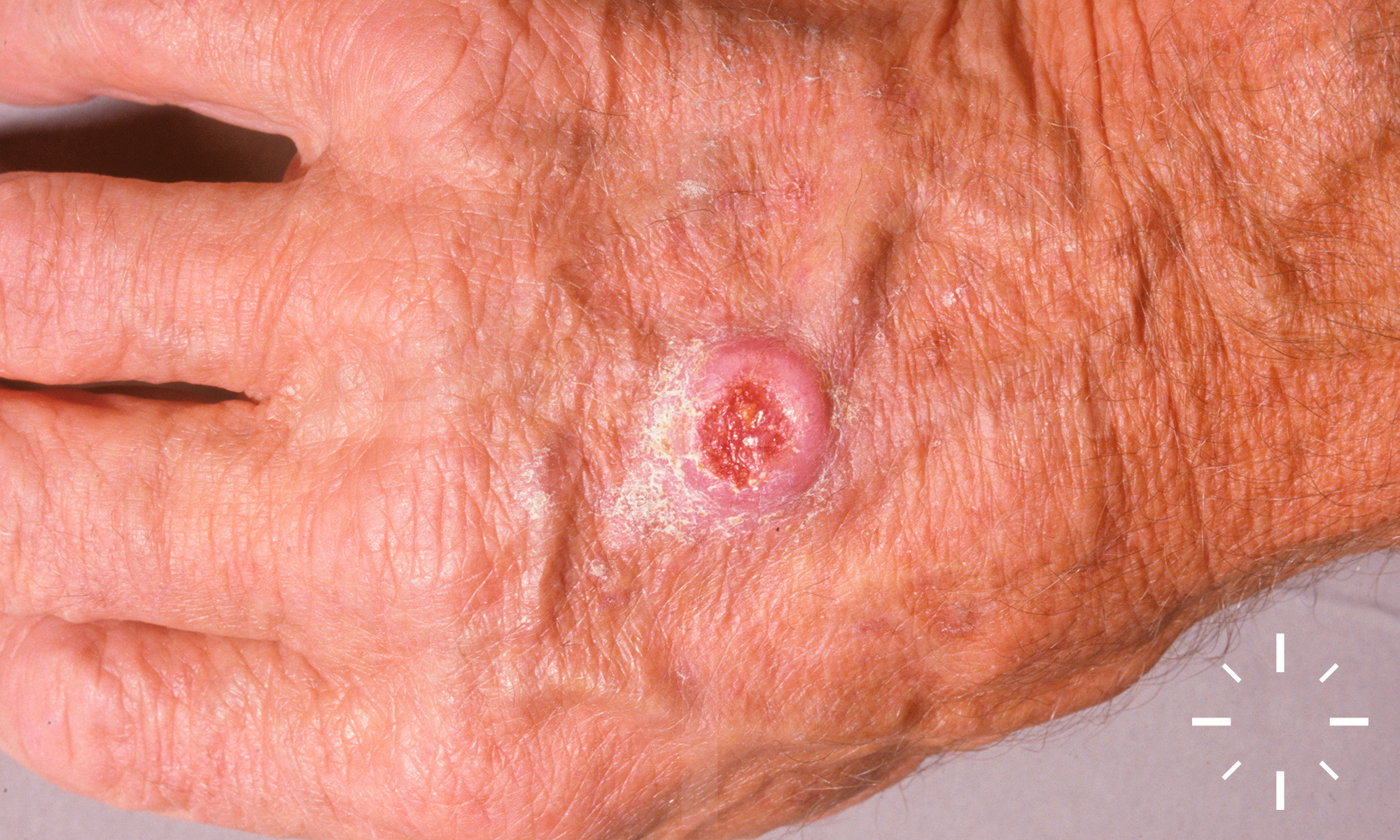
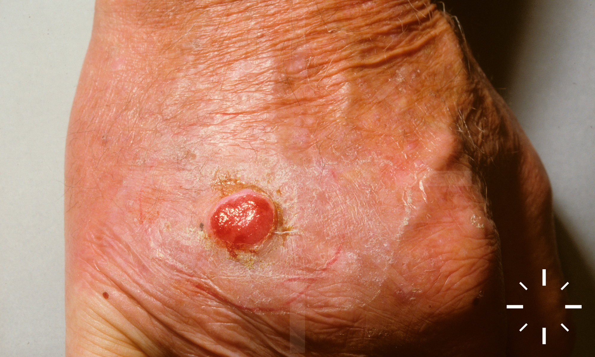
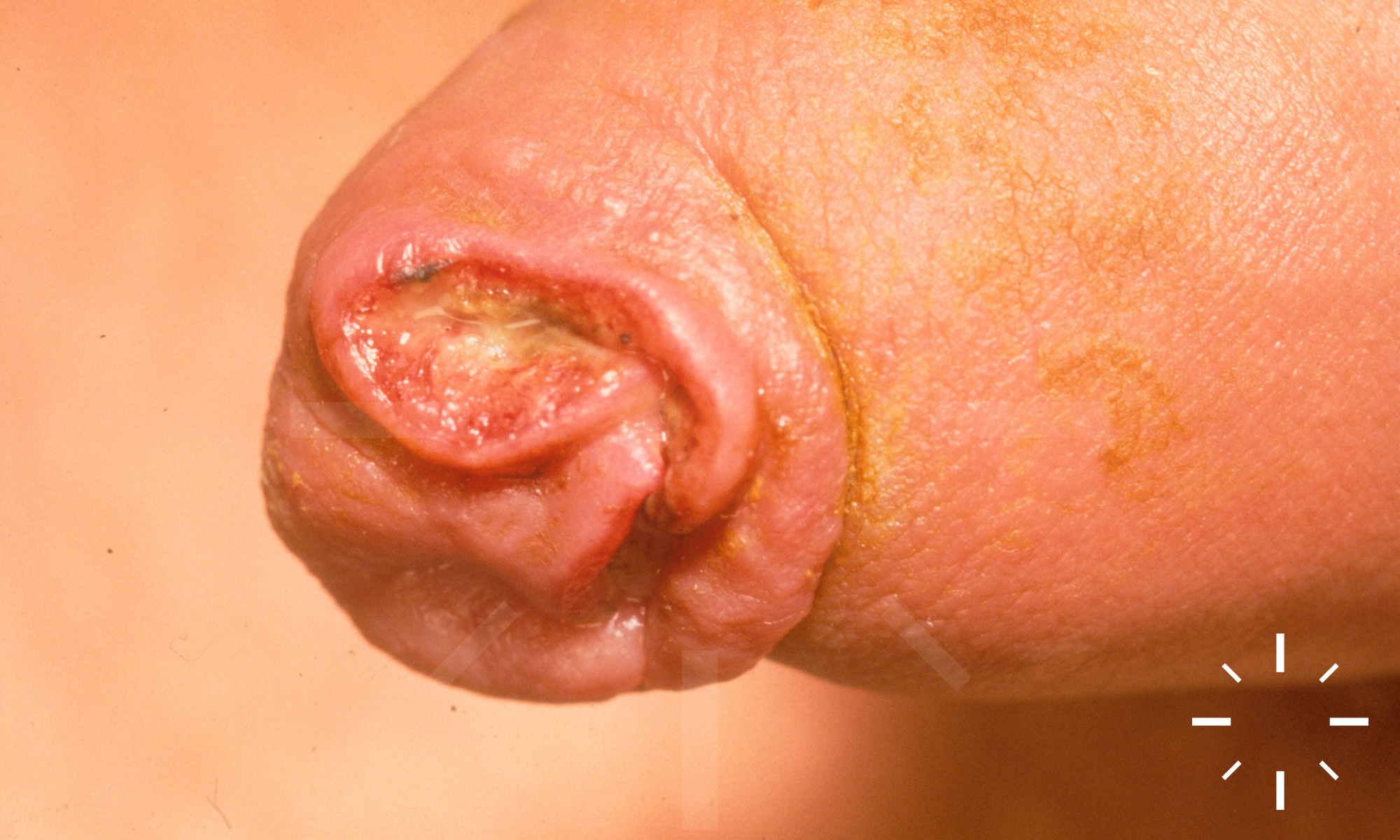
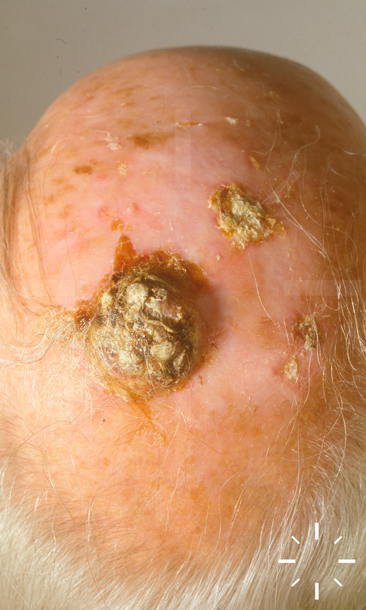
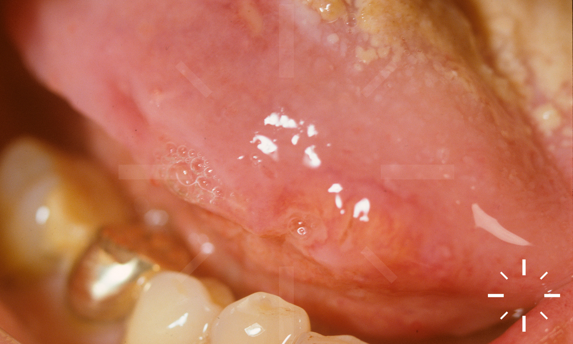
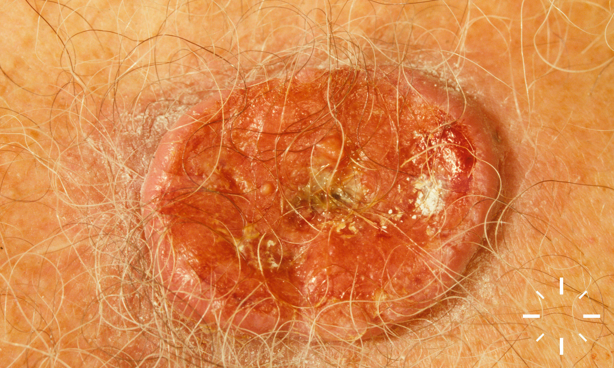
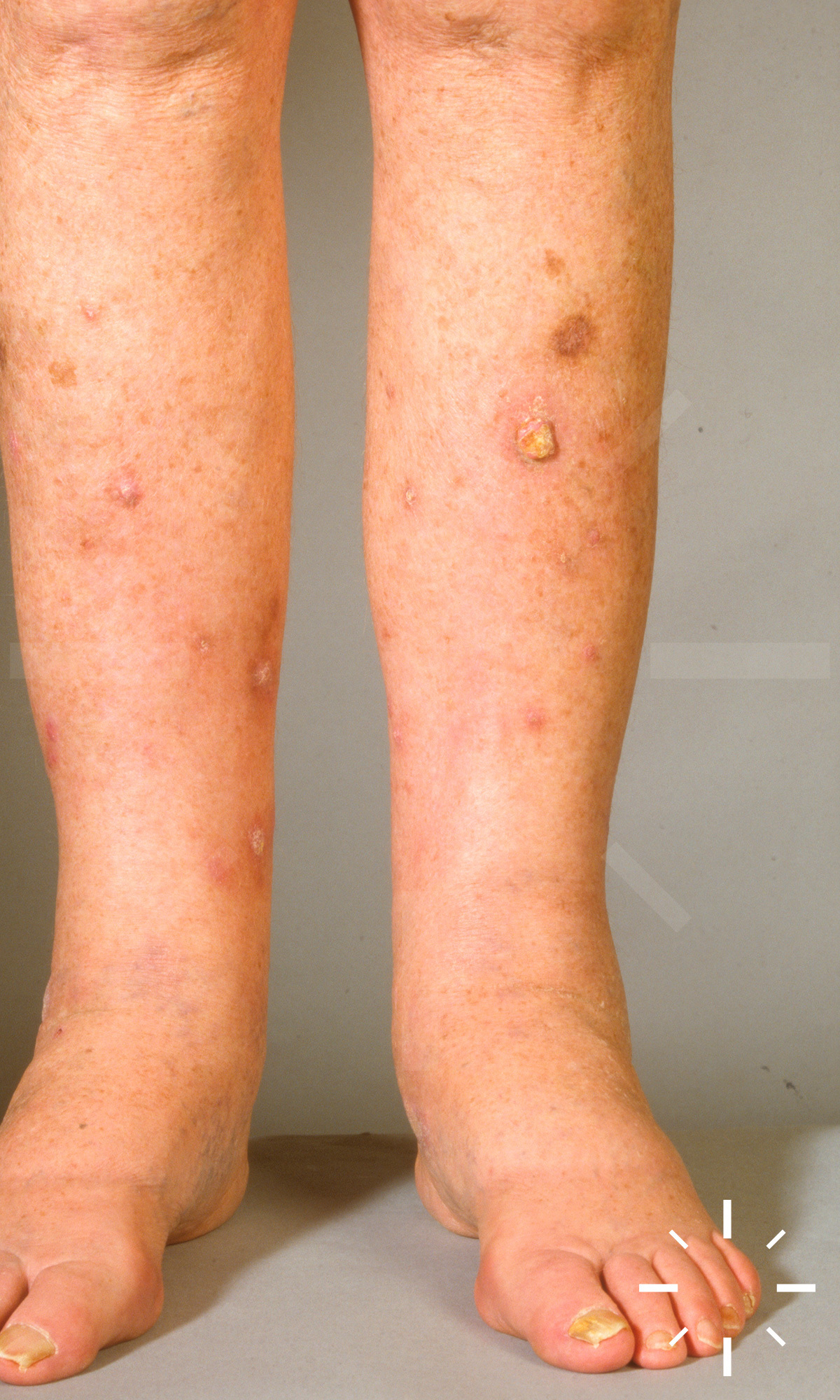
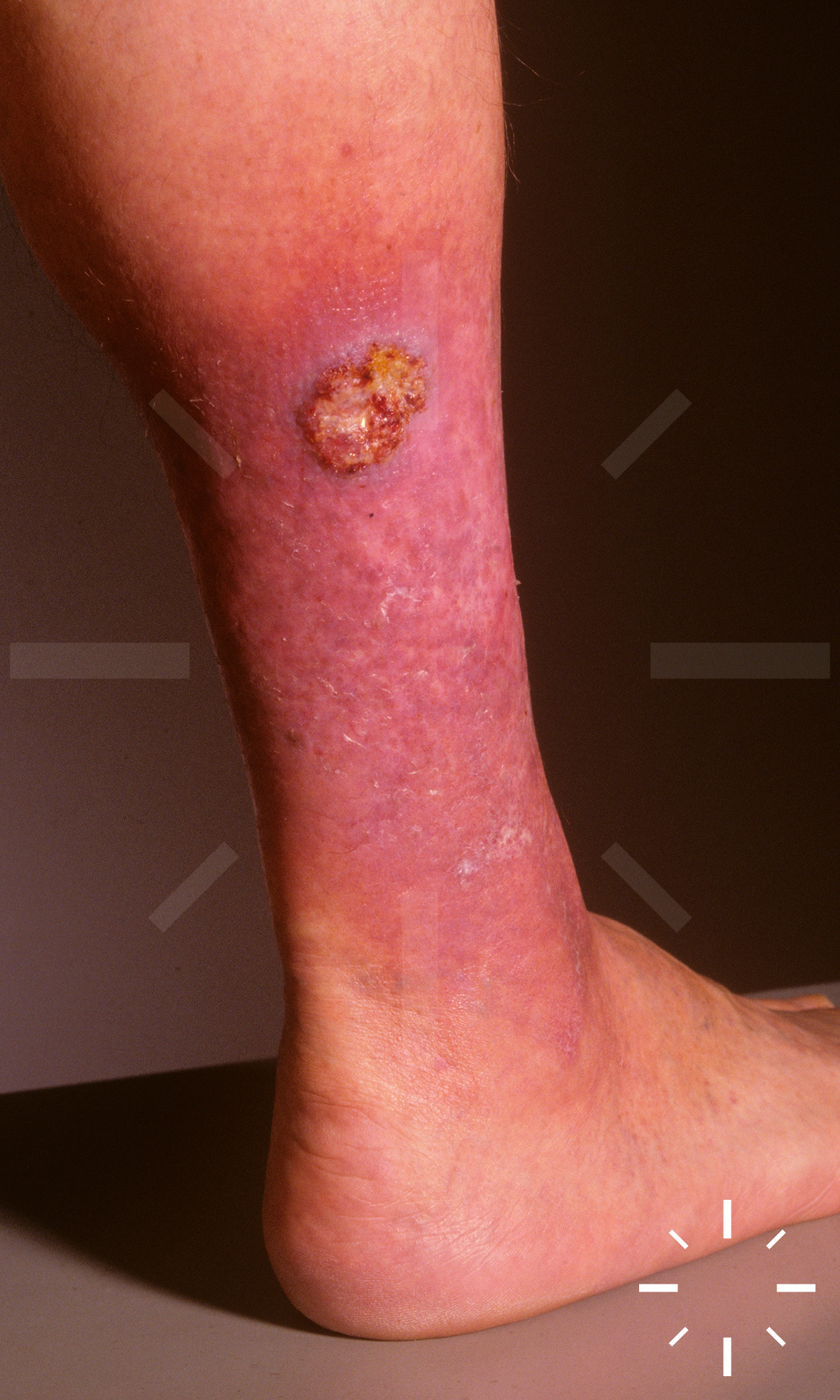
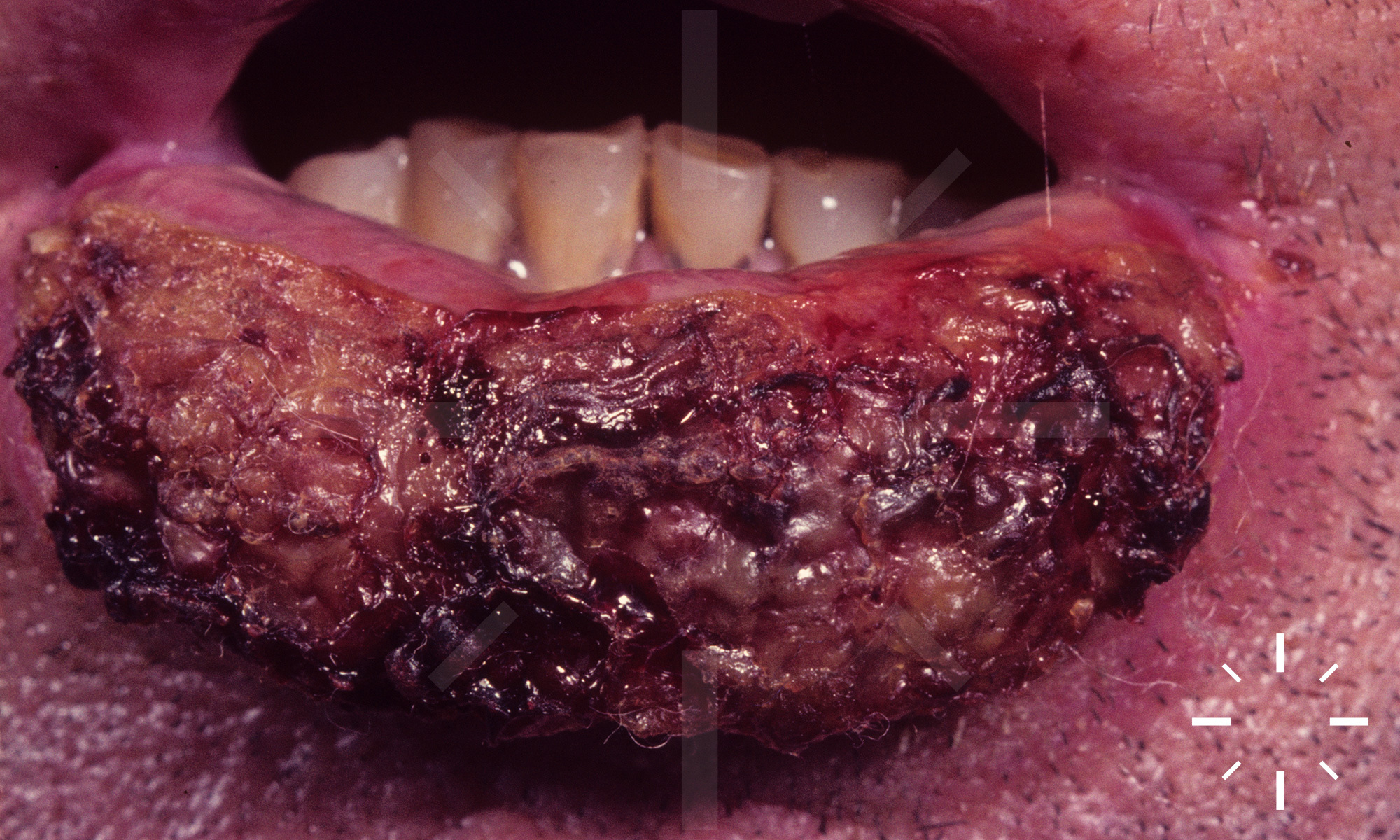
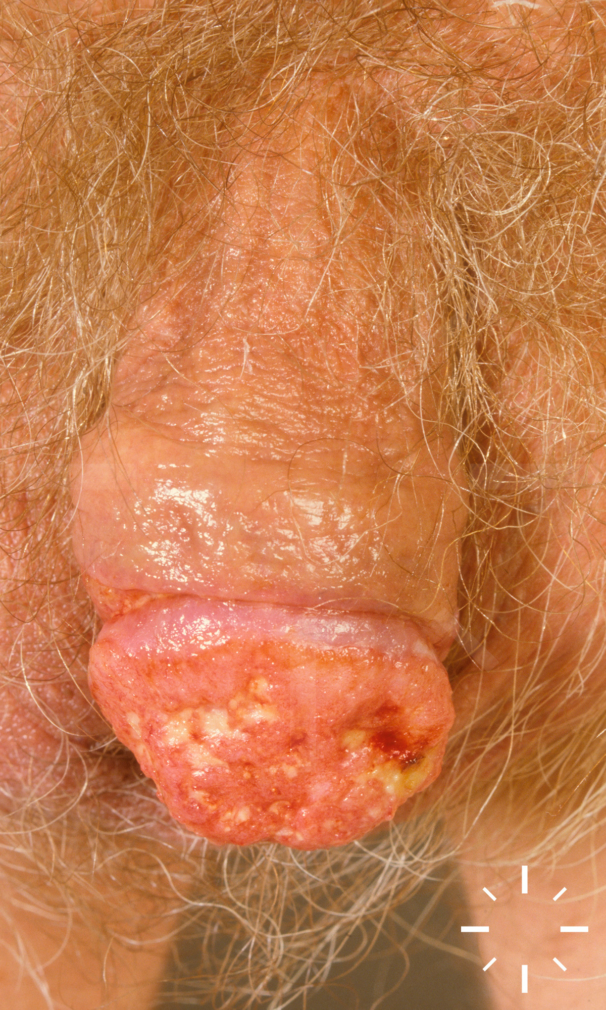
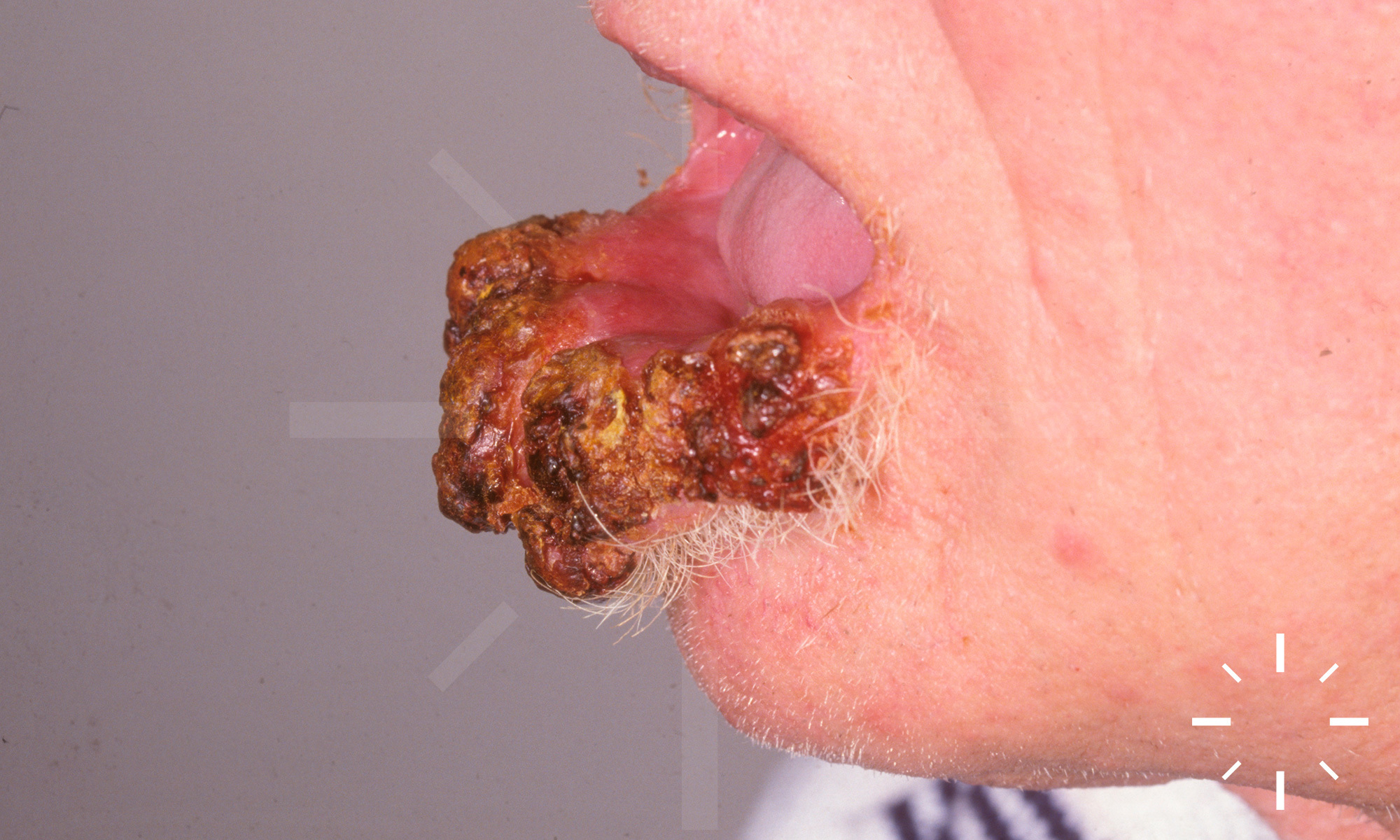
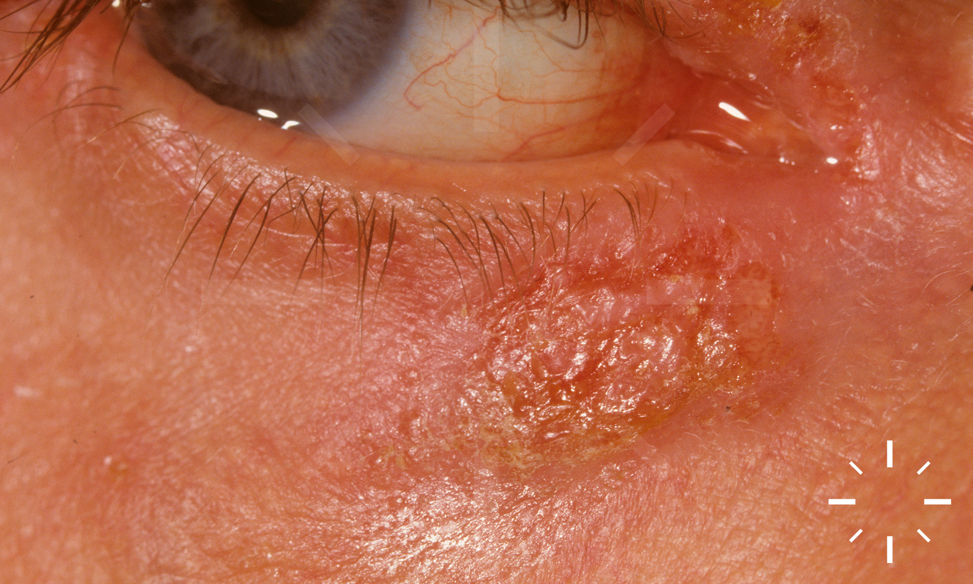
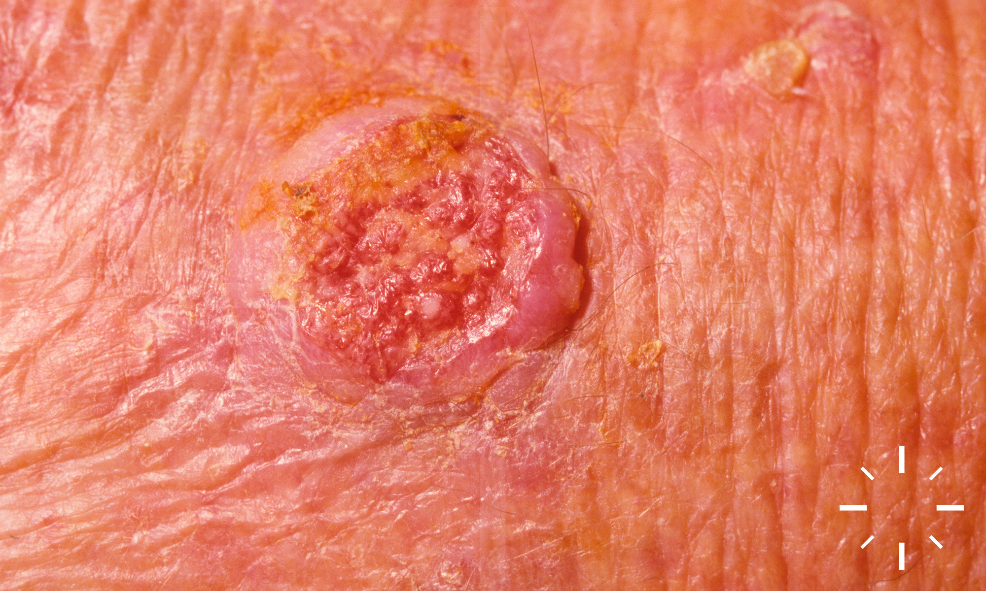
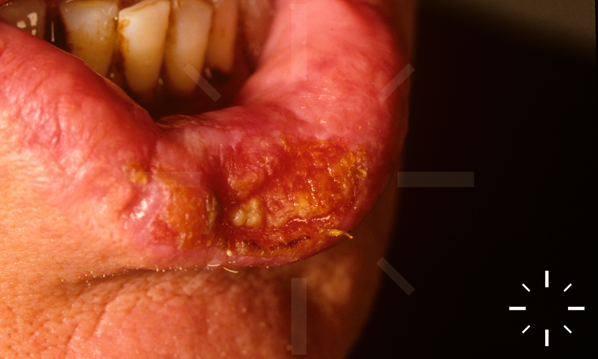
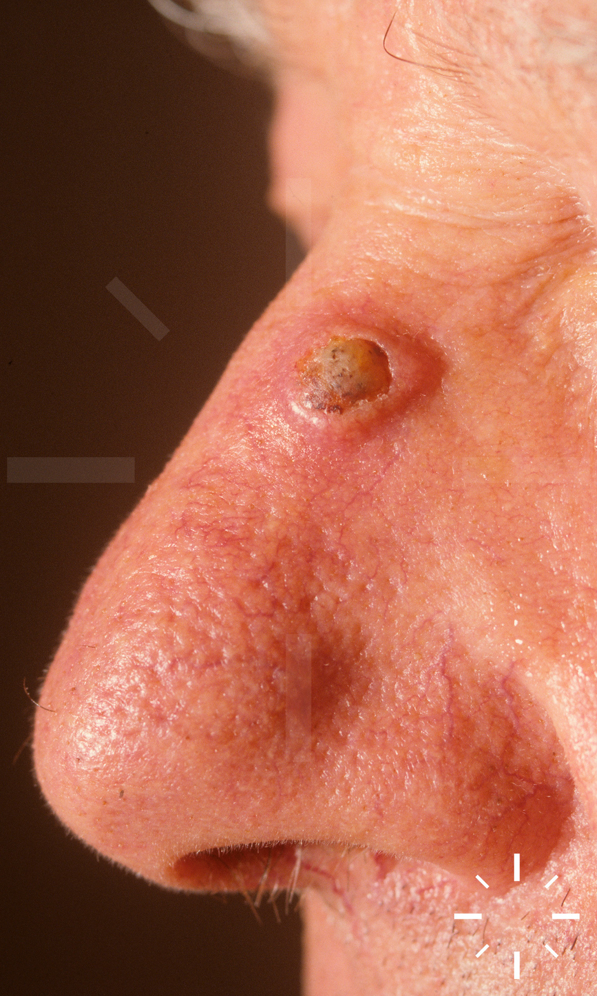
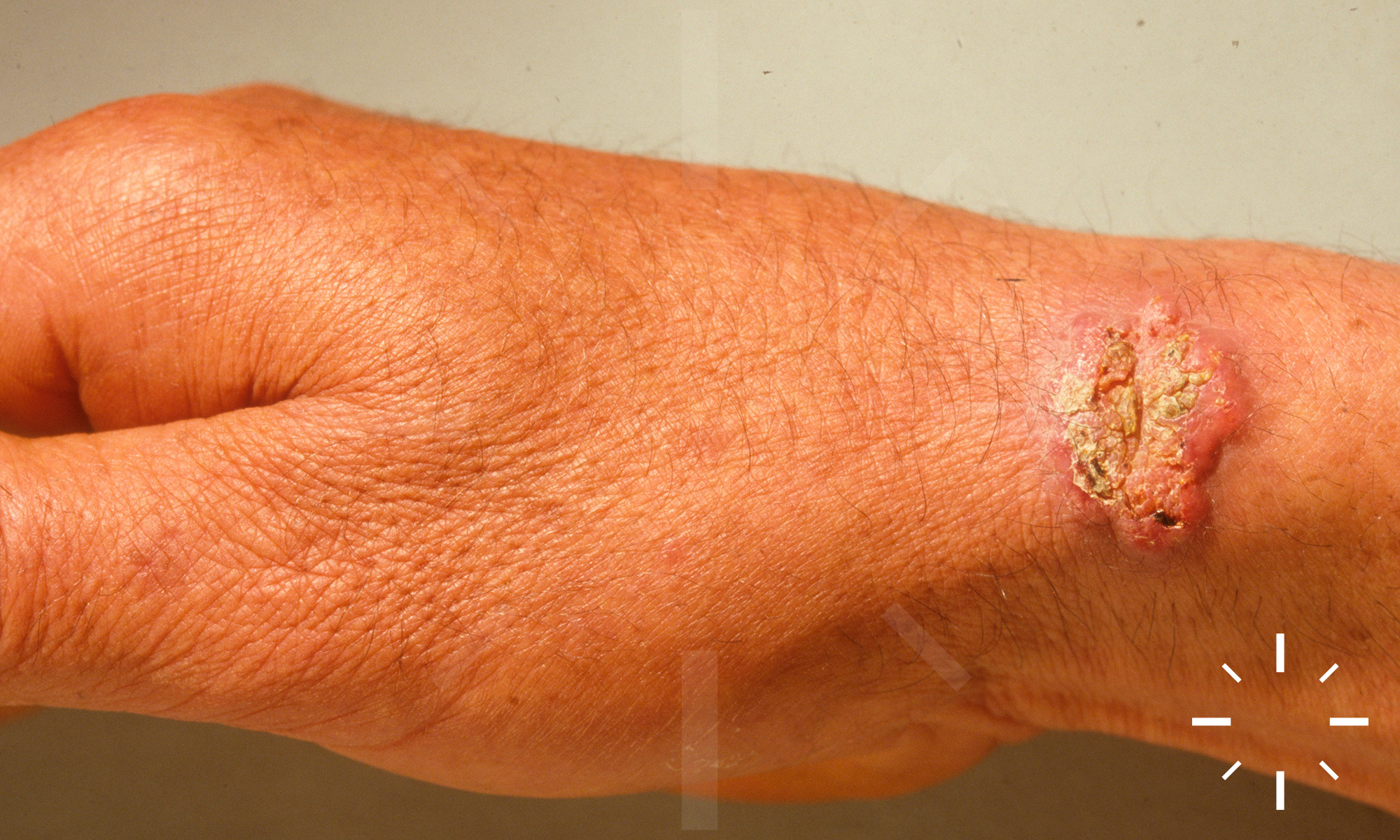
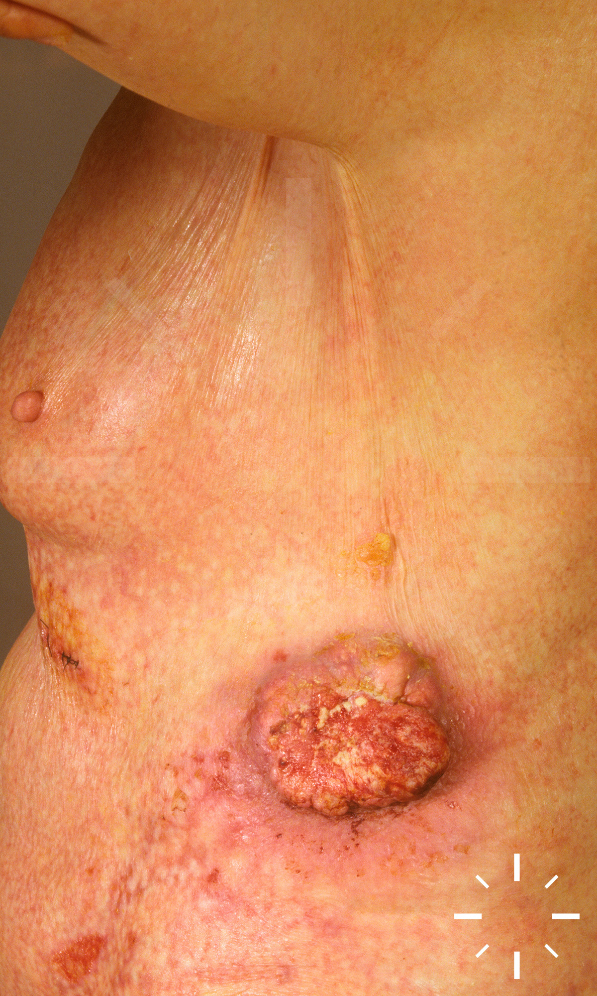
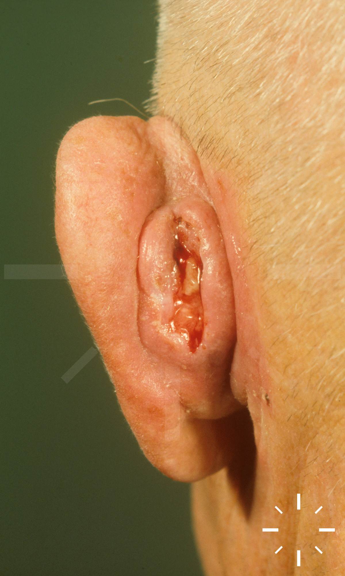
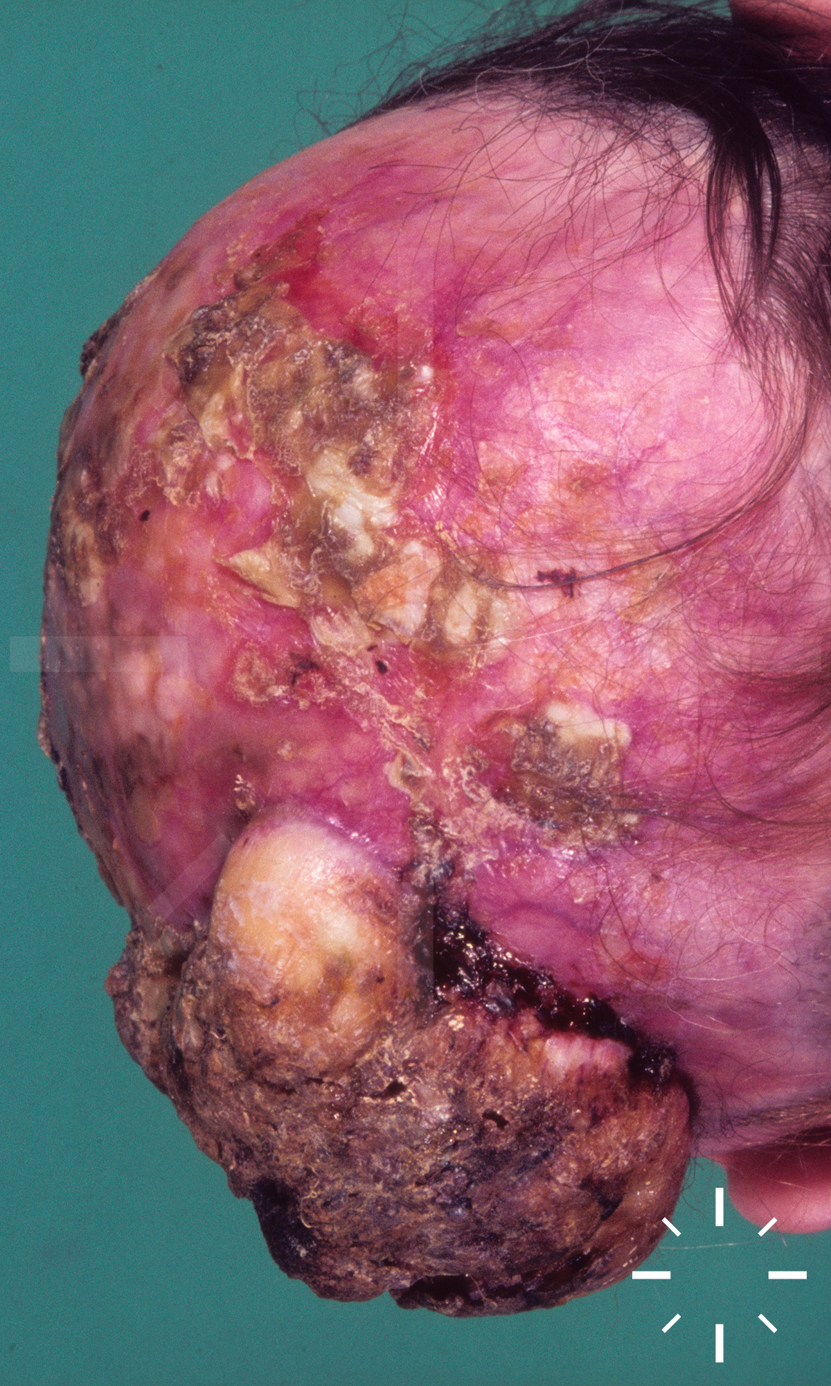
Spinocellular carcinoma

Last Updated: 2021-06-28

Author(s): Anzengruber F., Navarini A.

ICD11: -

Squamous cell carcinoma, spinalioma, epithelioma spinocellulare, carcinoma spinocellulare, cornifying squamous cell carcinoma of the skin, prickle cell carcinoma, spindle cell carcinoma, cornifying squamous cell carcinoma.

  • Epithelial carcinoma, which in rare cases can metastasise.

  • Second most common malignant skin tumour (Caucasian)
  • Incidence: 30/100,000 per year (north-south divide).
  • Average age: 70 years.
  • Men:Women = 2:1.
  • Spinocellular carcinomas account for about 20% of all non-melanoma skin cancer.
  • There has been a sharp increase in incidence in recent decades.

  • Histological classification:
  • Carcinoma in situ of the skin (KIN III)
  • Carcinoma in situ of the mucous membrane in penile, anal and vaginal carcinoma (PIN, AIN, VIN III)
  • Spinocellular carcinoma (classic type)
  • Acantholytic squamous cell carcinoma
  • Mucinsecreting squamous cell carcinoma
  • Desmoplastic squamous cell carcinoma
  • Small cell squamous cell carcinoma
  • Clear cell squamous cell carcinoma
  • Lymphoepithelioma-like carcinoma of the skin

  • In-situ carcinomas (actinic keratoses, Bowen's disease, Queyrat's erythroplasia, actinic cheilitis) are the precursors of SCC. However, squamous cell carcinomas can also develop from the begining. The etiopathogenesis is multifactorial.
  • This can be causal or predisposing:
  • Actinic keratoses.
  • Positive family history.
  • Light skin type.
  • Sun exposure (incl. solarium).
  • Ionising radiation.
  • High-risk human papilloma viruses.
  • Advanced age.
  • Chronic exposure to heat (erythema ab igne) or burns.
  • Chronic degenerative and chronic inflammatory skin changes.
  • Albinism.
  • Xeroderma pigmentosum.
  • Muir-Torre syndrome.
  • Epidermolysis bullosa hereditaria.
  • Scars (also Lupus vulgaris scars).
  • Atrophic lupus erythematosus foci.
  • Acrodermatitis chronica atrophicans.
  • Lower leg ulcer Marjolin ulcer (about 1:5000).
  • Acne inversa.
  • Lichen ruber erosivus mucosae.
  • Glossitis interstitialis syphilitica.
  • Lichen sclerosus et atrophicus of the vulva or penis.
  • Workers in oil refineries.
  • Coal tar industry.
  • Road construction with tar.
  • Soot (chimney-sweep crab).
  • Arsenic.
  • Immunosuppression (approx. 200 times higher risk).
  • Alcohol abuse, especially with high-proof spirits.
  • Smoking (tar distillates).

  • Leukoplakic, rough, painless, mostly exophytic, often hyperkeratotic, but sometimes also eroded plaques or nodules
  • In severe, horny hyperkeratosis one can speak of a Cornu cutaneum 
  • The verrucous carcinoma (Ackermann carcinoma) shows a plantar surface similar to warts
  • A metastasis occurs lymphogenically and later also hematogenically

  • Anamnesis with regard to predisposing factors.
  • Clinic.
  • Biopsy.
  • If the diagnosis is dermatopathologically confirmed, a full body inspection should be performed.
  • Palpation of the locoregional lymph nodes.
  • With tumour thickness of > 2mm à locoregional lymph node sonography.

  • 90% of all SCC is located in the head area
  • Rarer: oral mucosa, genital, toes, fingers

  • From the surface of the skin or the mucous membrane, finger-shaped, branching tumour strands infiltrate, destroying the basement membrane and growing deep into the tissue. 
  • The cells originate from the str. spinosum (ergo: spinocellular carcinoma). A cornified squamous epithelium with horny pearls (squamous eddies) is formed. Nuclear polymorphy, atypical mitoses, nuclear hyperchromasia, polyploidy, dyskeratosis, hyperkeratosis, cell atypia, mitoses, inflammatory infiltrate.
  • Immunohistological differentiation from basal cell carcinomas is carried out using Ber-EP4 markers.
  • Immunohistological differentiation from adnexal tumours is carried out using the cytokeratin marker CAM-5.2.

.

WHO classification as follows:

  • Spindle cell squamous cell carcinoma of the skin (aggressive behaviour).
  • Acantholytic (pseudoglandular) squamous cell carcinoma of the skin.
  • Verrucous squamous cell carcinoma of the skin (prognostically favourable).
  • Squamous cell carcinoma with horn formation.
  • Lymphoepithelioma-like squamous cell carcinoma of the skin.
  • The degree of differentiation according to Broders depends on the number of atypical, undifferentiated cells. 
  • Grade I <25%, grade II <50%, grade III <75%, grade IV >75%.
  • The higher the degree, the higher the metastatic tendency.

"High-risk spinocellular carcinoma" according to AJCC classification (2010):

  • Tumour thickness >2mm.
  • Penetration depth from Clark level 4.
  • Degree of de-differentiation from level III.
  • Perineural invasion.

  • Metastasis: ≤ 2 mm (0%), 2-6 mm (4%), > 6 mm (16%).
  • In case of metastasis the median survival time is 2 years.
  • Carcinomas of the penis, vulva and auricle tend to metastasise early. 
  • Aftercare: SOP currently being updated.

  1. Lim C, Tschuchnigg M, Lim J. Squamous cell carcinoma arising in an area of long-standing necrobiosis lipoidica. J Cutan Pathol 2006;33:581-3.
  2. Lin JH, Hsu M-L, Sheu HM, Lee J-Y. Coexistence of three variants of porokeratosis with multiple squamous cell carcinomas arising from lesions of giant hyperkeratotic porokeratosis. Journal of the European Academy of Dermatology and Venereology 2006;20:621-3.
  3. Morton C, Horn M, Leman J, et al. Comparison of Topical Methyl Aminolevulinate Photodynamic Therapy With Cryotherapy or Fluorouracil for Treatment of Squamous Cell Carcinoma In Situ. Arch Dermatol 2006;142.
  4. Patel GK, Goodwin R, Chawla M, et al. Imiquimod 5% cream monotherapy for cutaneous squamous cell carcinoma in situ (Bowen's disease): A randomized, double-blind, placebo-controlled trial. Journal of the American Academy of Dermatology 2006;54:1025-32.
  5. Veness MJ, Palme CE, Morgan GJ. High-risk cutaneous squamous cell carcinoma of the head and neck. Cancer 2006;106:2389-96.
  6. Ahlgrimm-Siess V, Hofmann-Wellenhof R, Zalaudek I, Cerroni L, Kerl H. Collision of Malignant Melanoma (Lentigo Maligna Type) with Squamous Cell Carcinoma in Solar-Damaged Skin of the Face. Dermatologic Surgery 2007;33:122-4.
  7. Bauman JE, Eaton KD, Martins RG. Treatment of Recurrent Squamous Cell Carcinoma of the Skin With Cetuximab. Arch Dermatol 2007;143.
  8. Dalle S, Depape L, Phan A, Balme B, Ronger-Savle S, Thomas L. Squamous cell carcinoma of the nail apparatus: clinicopathological study of 35 cases. Br J Dermatol 2007;156:871-4.
  9. Röwert-Huber J, Patel MJ, Forschner T, et al. Actinic keratosis is an early in situ squamous cell carcinoma: a proposal for reclassification. Br J Dermatol 2007;156:8-12.
  10. Veness MJ, Porceddu S, Palme CE, Morgan GJ. Cutaneous head and neck squamous cell carcinoma metastatic to parotid and cervical lymph nodes. Head Neck 2007;29:621-31.
  11. Brantsch KD, Meisner C, Schönfisch B, et al. Analysis of risk factors determining prognosis of cutaneous squamous-cell carcinoma: a prospective study. The Lancet Oncology 2008;9:713-20.
  12. Calzavara-Pinton PG, Venturini M, Sala R, et al. Methylaminolaevulinate-based photodynamic therapy of Bowen’s disease and squamous cell carcinoma. Br J Dermatol 2008;159:137-44.
  13. Garcia-Zuazaga J, Olbricht SM. Cutaneous Squamous Cell Carcinoma. Advances in Dermatology 2008;24:33-57.
  14. Geist DE, Garcia-Moliner M, Fitzek MM, Cho H, Rogers GS. Perineural Invasion of Cutaneous Squamous Cell Carcinoma and Basal Cell Carcinoma: Raising Awareness and Optimizing Management. Dermatologic Surgery 2008;34:1642-51.
  15. Arnold AW, Bruckner-Tuderman L, uuml, ger C, Itin PH. Cetuximab Therapy of Metastasizing Cutaneous Squamous Cell Carcinoma in a Patient with Severe Recessive Dystrophic Epidermolysis Bullosa. Dermatology 2009;219:80-3.
  16. de Koning MNC, Weissenborn SJ, Abeni D, et al. Prevalence and associated factors of betapapillomavirus infections in individuals without cutaneous squamous cell carcinoma. Journal of General Virology 2009;90:1611-21.
  17. Love WE, Bernhard JD, Bordeaux JS. Topical Imiquimod or Fluorouracil Therapy for Basal and Squamous Cell Carcinoma. Arch Dermatol 2009;145.
  18. Rishpon A, Kim N, Scope A, et al. Reflectance Confocal Microscopy Criteria for Squamous Cell Carcinomas and Actinic Keratoses. Arch Dermatol 2009;145.
  19. Turowski CB, Ross AS, Cusack CA. Human papillomavirus-associated squamous cell carcinoma of the nail bed in African-American patients. International Journal of Dermatology 2009;48:117-20.
  20. Cowen EW, Nguyen JC, Miller DD, et al. Chronic phototoxicity and aggressive squamous cell carcinoma of the skin in children and adults during treatment with voriconazole. Journal of the American Academy of Dermatology 2010;62:31-7.
  21. Lavogiez C, Delaporte E, Darras-Vercambre S, et al. Clinicopathological Study of 13 Cases of Squamous Cell Carcinoma Complicating Hidradenitis Suppurativa. Dermatology 2010;220:147-53.
  22. Gormley RH, Groft CM, Miller CJ, Kovarik CL. Digital squamous cell carcinoma and association with diverse high-risk human papillomavirus types. Journal of the American Academy of Dermatology 2011;64:981-5.
  23. Koba S, Inoue T, Okawa T, et al. Merkel cell carcinoma with cytokeratin 20-negative and thyroid transcription factor-1-positive immunostaining admixed with squamous cell carcinoma. Journal of Dermatological Science 2011;64:77-9.
  24. Ueda Y, Enomoto T, Kimura T, Yoshino K, Fujita M, Kimura T. Two Distinct Pathways to Development of Squamous Cell Carcinoma of the Vulva. Journal of Skin Cancer 2011;2011:1-7.
  25. Hood AF, Farmer ER. Data Needed for Management of Cutaneous Squamous Cell Carcinoma. JAMA Dermatol 2013;149:41.
  26. Iarrobino A, Messina JL, Kudchadkar R, Sondak VK. Emergence of a squamous cell carcinoma phenotype following treatment of metastatic basal cell carcinoma with vismodegib. Journal of the American Academy of Dermatology 2013;69:e33-e4.
  27. Majores M, Bierhoff E. [Actinic keratosis, Bowen's disease, keratoacanthoma and squamous cell carcinoma of the skin]. Pathologe 2015;36:16-29.
  28. Rowan SD, Hu SL, Brotzman JS, Redding SW, Rankin KV, Vigneswaran N. Knowledge Assessment of the Dental Community in Texas on the Role of Human Papilloma Virus in Oropharyngeal Cancer. Tex Dent J 2015;132:528-36.
  29. Deen K, Burdon-Jones D. Imiquimod in the treatment of penile intraepithelial neoplasia: An update. Australas J Dermatol 2016.
  30. Breuninger, H. (2016). Plattenepithelkarzinom der Haut. Awmf.org. Retrieved 30 May 2016, from http://www.awmf.org/leitlinien/detail/ll/032-022.html
  31. Carthier, T. (2016). Treatment and prognosis of cutaneous squamous cell carcinoma. Uptodate.com. Retrieved 30 May 2016, from http://www.uptodate.com/contents/treatment-and-prognosis-of-cutaneous-squamous-cell-carcinoma?source=search_result&search=plattenepithelkarzinom+der+haut&selectedTitle=5%7E150
  32. Lim, J. (2016). Clinical features and diagnosis of cutaneous squamous cell carcinoma (SCC). Uptodate.com. Retrieved 30 May 2016, from http://www.uptodate.com/contents/clinical-features-and-diagnosis-of-cutaneous-squamous-cell-carcinoma-scc?source=search_result&search=plattenepithelkarzinom+der+haut&selectedTitle=3%7E150
  33. Padillla, S. (2016). Epidemiology and risk factors for cutaneous squamous cell carcinoma. Uptodate.com. Retrieved 30 May 2016, from http://www.uptodate.com/contents/epidemiology-and-risk-factors-for-cutaneous-squamous-cell-carcinoma?source=search_result&search=plattenepithelkarzinom+der+haut&selectedTitle=4%7E150